

doi: 10.1016/j.jseint.2024.02.004.
eCollection 2024 Jul.
Arthroscopic humeral head defect filling with osteochondral autografts transplantation for near-track Hill-Sachs lesions
Affiliations
- PMID: 39035648
- PMCID: PMC11258713
- DOI: 10.1016/j.jseint.2024.02.004
Item in Clipboard
Arthroscopic humeral head defect filling with osteochondral autografts transplantation for near-track Hill-Sachs lesions
JSES Int.
.
No abstract available
Keywords: Autologous ostechondral transplantation; Bipolar bone loss; Gleno-humeral instability; Hill sachs; Near-track; On-track.
Figures
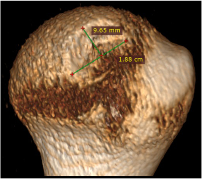
Preoperative assessment of HSL size. Through 3D-CT reconstruction of the HH, the number and size of grafts needed to fill the defect can be planned. Only the articular part of the HS is considered. HSL, Hill-Sachs lesion; HH, humeral head; HS, Hill-Sachs; CT, computed tomography; 3D, three-dimensional.
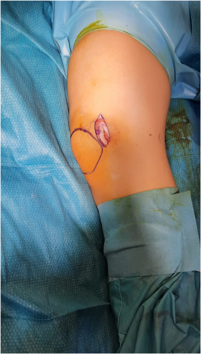
Lateral decubitus, left knee (ipsilateral to the affected shoulder): Through a lateral mini-arthrotomy, the proximal lateral aspect of the femoral trochlea (donor site) is exposed. The donor harvester is positioned perpendicular to the donor surface and impacted to a depth of approximately 15 mm.

Arthroscopic view through anterior-superior portal of left shoulder with patient in lateral decubitus. (A) Through the cannula placed in the posterior portal, the recipient harvester is positioned perpendicular to the osteochondral defect and impacted to a depth of 10-13 mm, then the harvester is removed, creating the bone socket; (B) A graduated alignment rod is used to measure the final recipient socket depth and check the correct insertion angle.

Arthroscopic view through anterior-superior portal of left shoulder with patient in lateral decubitus. The graft, taken from the knee still inside the donor harvester through the posterior portal is inserted and press-fitted perpendicularly into the recipient socket.

Arthroscopic view through anterior-superior portal of left shoulder with patient in lateral decubitus. Thanks to a specific plastic tamp in the posterior portal, final graft placement is performed at the level of the cartilage edge.
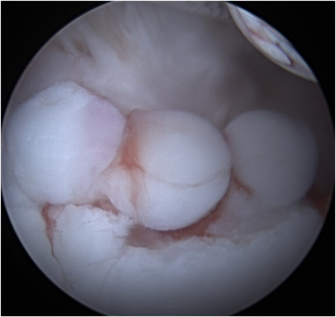
Arthroscopic view through posterior portal of left shoulder with patient in lateral decubitus. Final result at the end of the procedures, with the HSL filled by multiple grafts. HSL, Hill-Sachs lesion.
Similar articles
-
Defining Critical Humeral Bone Loss: Inferior Craniocaudal Hill-Sachs Extension as Predictor of Recurrent Instability After Primary Arthroscopic Bankart Repair.Am J Sports Med. 2024 Jan;52(1):181-189. doi: 10.1177/03635465231209443. Am J Sports Med. 2024. PMID: 38164666
-
The effect of a combined glenoid and Hill-Sachs defect on glenohumeral stability: a biomechanical cadaveric study using 3-dimensional modeling of 142 patients.Am J Sports Med. 2015 Jun;43(6):1422-9. doi: 10.1177/0363546515574677. Epub 2015 Mar 20. Am J Sports Med. 2015. PMID: 25794869
-
[Incidence, Morphology and Clinical Significance of Hill-Sachs Lesions in Shoulder Instability - CT Scan Evaluation of the Group of Patients].Acta Chir Orthop Traumatol Cech. 2021;88(6):434-441. Acta Chir Orthop Traumatol Cech. 2021. PMID: 34998447 Czech.
-
Osseous Defects Seen in Patients with Anterior Shoulder Instability.Clin Orthop Surg. 2015 Dec;7(4):425-9. doi: 10.4055/cios.2015.7.4.425. Epub 2015 Nov 13. Clin Orthop Surg. 2015. PMID: 26640623 Free PMC article. Review.
-
When to Abandon the Arthroscopic Bankart Repair: A Systematic Review.Sports Health. 2020 Sep/Oct;12(5):425-430. doi: 10.1177/1941738120940676. Epub 2020 Jul 27. Sports Health. 2020. PMID: 32716726 Free PMC article.
References
-
- Burkhart S.S., Danaceau S.M. Articular arc length mismatch as a cause of failed bankart repair. Arthroscopy. 2000;16:740–744. - PubMed
LinkOut - more resources
Full Text Sources