. 2022 Oct 1;20(3):A376-A386.
doi: 10.59390/JCRT2250. eCollection 2022 Spring.

Recording from Snail Motor Nerves to Investigate Central Pattern Generation

Affiliations

Recording from Snail Motor Nerves to Investigate Central Pattern Generation

Robert A Wyttenbach et al. J Undergrad Neurosci Educ. .

Abstract

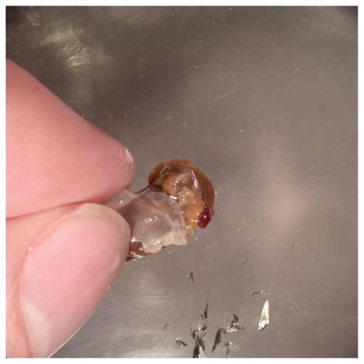
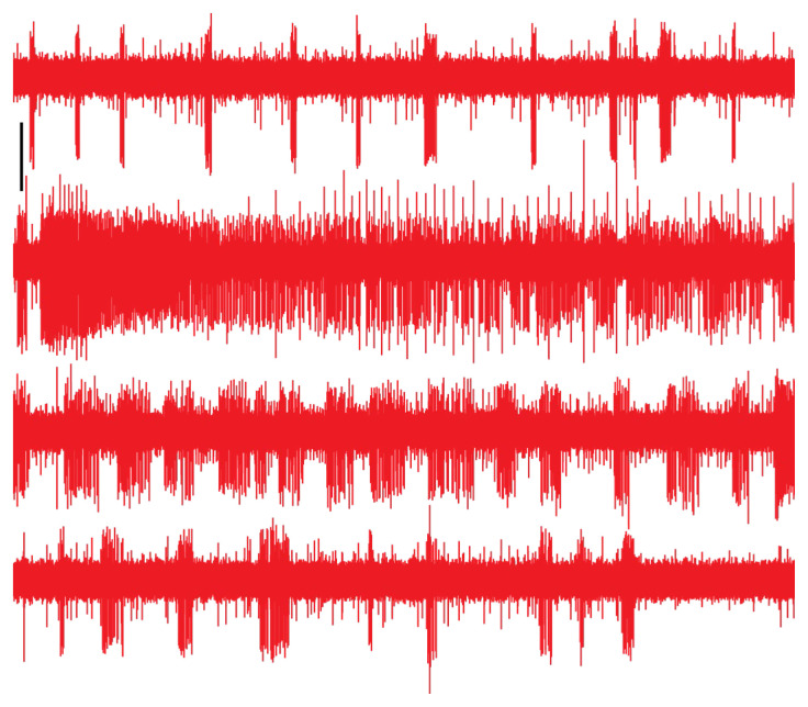
Feeding in pond snails has long been a model system for central pattern generation and its modulation. The pattern is generated by a small set of neurons in the buccal ganglia, which innervate the buccal mass, esophagus, and salivary glands. In this exercise, students observe feeding behavior and then record and quantify rhythmic motor activity and its response to feeding stimulants and neuromodulators. In a standard three-hour class period, students do a dissection, record from several nerves, and perform experimental manipulations such as adding feeding stimulants, serotonin, or dopamine to the preparation. Depending on the course goals, data can be presented qualitatively or cyclic measurements and spike-rate analysis can be done. This exercise leads to discussion of neural circuitry and intrinsic properties that support pattern generation for rhythmic activities such as feeding, locomotion, and respiration.

Keywords: CPG; Helisoma; Lymnaea; Planorbis; buccal; dopamine; extracellular; feeding; neuromodulation; pond snail; serotonin.


Figures

Video 1
Video 1
Feeding Behavior. As a snail moves, it frequently grazes on the substrate. This video shows parts of the triphasic motor pattern. As the mouth opens, the odontophore protrudes backwards. As the odontophore moves forward again, the radula scrapes the surface, loosening food particles. Finally, the mouth closes as the odontophore pushes food back to the esophagus. The rhythm is variable and may include long pauses.
Video 2
Video 2
Removing the Shell. Cut the shell for about one full turn along its spiral. Break the shell away as you proceed. Try not to cut deeply into the snail’s body, but do not worry if body fluid leaks. If the shell breaks apart completely, you may need help orienting the body later.
Video 3
Video 3
Exposing the Ganglia. Pin the snail in a dish, fold back the mantle, and cut through the skin to expose the body cavity. Find the circumesophageal ganglia and esophagus (remove other organs or move them aside as needed). Cut the esophagus behind the ring of ganglia and then pull it through the ring. Tilt the buccal mass forward and secure it with a pin through the esophagus.
Video 4
Video 4
Nerve Recording. Pull saline into the suction electrode, move it onto a nerve, and apply suction. This example shows recording from the right LBN and right PBN. Both have bursts of activity that change over time.
Figure 1
Figure 1
One feeding cycle in Helisoma (left) and Lymnaea (right). Yellow circles in the upper images show areas magnified below. The odontophore protrudes back as the mouth opens, the odontophore moves forward such that the radula scrapes the surface, and the mouth closes as the odontophore pushes food back into the esophagus. Each cycle shown here took 1.4 s. These are frames from Video 1, in which snails were feeding on the side of the aquarium.
Figure 2
Figure 2
Dissection. A. Remove the shell by cutting around its edge (Helisoma, left) or in the middle of the shell opening (Lymnaea, right). Continue the cut for at least one full turn of the shell and break off the shell fragments to expose the body. The remaining steps show Helisoma; the procedure is the same for Lymnaea. B. Hold the snail down with three pins. Add saline now or after the next step. C. Pull the mantle back and pin it, then cut the body cavity open. D. Pin the body walls down, then cut the esophagus and pull it through the circumesophageal ganglia. E. Tilt the buccal mass forward and secure it with a pin through the esophagus.
Figure 3
Figure 3
Completed dissection. With the buccal mass tilted forward, the buccal ganglia and nerves should be visible. ET esophageal trunk; PBN posterior buccal nerve; LBN lateral buccal nerve; CBC cerebro-buccal connective; VBN ventral buccal nerve. electrode tip, so only a slight suction may be needed. If the LBN, VBN, and CBC are often joined for some distance before splitting into three distinct branches.
Figure 4
Figure 4
Helisoma buccal ganglia and nerves. After dissection, ganglia will be oriented with the caudal side visible and ET nerves pointed away from the body. Many of the neurons have known functions and axonal projections. For example, recording from PBN should show a burst from neuron 6; this would precede dense bursts from 27 and 19, both in VBN (data from Murphy, 2001; see Table 1, Figures 3–6, and associated text).
Figure 5
Figure 5
Cyclic measurements. A. Measure the burst period, burst duration, and number of spikes per burst (if spikes are distinct). Do this for several bursts and report mean and standard deviation. From the means, calculate duty cycle = duration/period (as a percentage). B. When more than one spike size has bursts with the same period, measure the duration, spike count, and duty cycle of each. From the delay between the two distinct bursts, calculate phase difference = delay/period (as a percentage). C. If there are independent burst patterns, measure the period, duration, duty cycle, and spike count of each.
Figure 6
Figure 6
Dopamine modifies a rhythm. A. This Helisoma PBN initially had regular bursts of 27±7.5 spikes, lasting 0.58±0.10 s. Omitting the “missing” burst, the period was 7.1±0.7 s, for an 8% duty cycle. B. Five minutes after dopamine was added (final concentration 10−4 M), bursts were less frequent but longer and denser. Omitting the sparse spikes leading up to each burst: 180±25 spikes/burst, 4.6±1.1 s burst duration, 11.3±1.2 s period, 41% duty cycle. Vertical scale bars are 20 μV. Spike times came from the analysis tools in Wyttenbach et al. (2014).
Figure 7
Figure 7
Burst phases. A. An unidentified nerve in Lymnaea had bursts from two neurons. Bursts of large spikes consistently had two spikes and a regular period (7.0±1.1 s) and duration (0.27±0.01 s). Bursts of small spikes were less consistent, with 29±4.4 spikes and 1.3±0.3 s durations. These bursts followed the start of a large-spike burst by 0.90±0.28 s, lagging the large-burst period by 13±4%. B. The area outlined in blue. C. Instantaneous spike rate declined exponentially (time constant 0.26 s, R2 = 0.95) during the burst in (B). Vertical scale bars for traces are 20 μV.
Figure 8
Figure 8
Serotonin disrupts a rhythm. A. In the Lymnaea nerve shown in Figure 7, serotonin was added at the arrowhead (final concentration 10−4 M). Within 10 s, it activated large axons, shut down the larger rhythmically active axon, and increased tonic activity of the small axon. B. One-second pieces of the 20-s areas highlighted in (A), showing activity before (orange) and after (green and blue) serotonin was added. C. Histograms of spike sizes in the 20-s areas (2 μV bins), showing that increased activity in the small axon persists for several minutes.
Figure 9
Figure 9
Serotonin modifies a rhythm. A. An unidentified nerve in Helisoma had occasional bursts in two axons. B. Two minutes later, 40 s after serotonin was added (final concentration 10−4 M), the larger axon stopped firing and the smaller axon fired regular bursts with a 1.75 s period. During serotonin application, the signal-to-noise ratio decreased slightly, so spikes are smaller. In each case, the recording (red, scale bar 40 μV) was rectified and integrated and then smoothed (blue, scale bar 10 nV·s; 1 s triangular window) to clearly show cyclic behavior. Calculation was done by ADinstruments LabChart 8.1.
Figure 10
Figure 10
Lettuce juice modifies a rhythm. Nerve ET in Helisoma had activity in multiple neurons, with one of them producing bursts. Each row is 30 s, continuing from top to bottom for a total of 120 s. Lettuce juice was added near the end of the top row. Within a few seconds, additional neurons started firing (larger spikes) and the rate and duty cycle of the bursting neuron increased (second row). Over the next 30 s, activity died down and reverted to the original pattern. Vertical scale bar is 20 μV.

Similar articles

References

    1. Benjamin PR. Lymnaea . Scholarpedia. 2008;3:4124.
    1. Benjamin PR, Rose RM. Central generation of bursting in the feeding system of the snail, Lymnaea stagnalis. J Exp Biol. 1979;80:93–118. doi: 10.1242/jeb.80.1.93. - DOI - PubMed
    1. Booth JRH, Sane V, Gather MC, Pulver SR. Inexpensive methods for live imaging of central pattern generator activity in the Drosophila larval motor system. J Undergrad Neurosci Educ. 2020;19:A124–A133. Available at https://pubmed.ncbi.nlm.nih.gov/33880100/ - PMC - PubMed
    1. Dillon RT colleagues. The Freshwater Gastropods of North America Volume 1: Atlantic drainages, Georgia through Pennsylvania. Charleston, SC: FWGNA Press; 2019. Available at https://www.fwgna.org/
    1. Elliott CJ, Susswein AJ. Comparative neuroethology of feeding control in molluscs. J Exp Biol. 2002;205:877–896. doi: 10.1242/jeb.205.7.877. - DOI - PubMed

LinkOut - more resources