. 2024;12(1):2345165.
doi: 10.1080/21681163.2024.2345165. Epub 2024 May 1.

Adaptive Image Segmentation Reveals Substantial Cortical Bone Remodeling During Early Fracture Repair

Affiliations

Adaptive Image Segmentation Reveals Substantial Cortical Bone Remodeling During Early Fracture Repair

Alireza Ariyanfar et al. Comput Methods Biomech Biomed Eng Imaging Vis. 2024.

Abstract

The goal of this study was to develop an image analysis algorithm for quantifying the effects of remodeling on cortical bone during early fracture healing. An adaptive thresholding technique with boundary curvature and tortuosity control was developed to automatically identify the endocortical and pericortical boundaries in the presence of high-gradient bone mineral density (BMD) near the healing zone. The algorithm successfully segmented more than 47,000 microCT images from 12 healing ovine osteotomies and intact contralateral tibiae. Resampling techniques were used to achieve data dimensionality reduction on the segmented images, allowing characterization of radial and axial distributions of cortical BMD. Local (transverse slice) and total (whole bone) remodeling scores were produced. These surrogate measures of cortical remodeling derived from BMD revealed that cortical changes were detectable throughout the region covered by callus and that the localized loss of cortical BMD was highest near the osteotomy. Total remodeling score was moderately and significantly correlated with callus volume and mineral composition (r > 0.64, p < 0.05), suggesting that the cortex may be a source of mineral needed to build callus.

Keywords: adaptive thresholding; bone fracture healing; cortical remodeling; micro-computed tomography; ovine osteotomy.


Conflict of interest statement

Declaration of Interest Statement The authors have no conflicts of interest relevant to this work.

Figures

Figure 1.
Figure 1.
Quantitative analysis of remodeling in an operated ovine tibia relative to the intact contralateral tibia from the same animal required axial registration of CT slices from two separate scans. Scans were aligned by matching the level of the nutrient artery canal (annotated NAC) in the two scans. In this example animal, the axial offset is hoff =135 slices. Numbers on each image refer to the slice position in image stack, with white text for intact and red text for operated.
Figure 2.
Figure 2.
Representative example of the boundary detection procedure for a single slice of an operated limb, segmented using non-adaptive thresholding. The raw slice image (a) was de-noised by applying a Gaussian filter, with a structural similarity index measure (SSIM) of 0.98 with respect to the raw image (b). In this example, preliminary pericortical (c), endocortical (d), and callus (e) boundaries were defined by binarizing with thresholds τperi =7,000, τendo =5950, and τcall = 4,250 to differentiate higher density bone from the combined region of all mineralized tissue including callus. The preliminary pericortical boundary (c) defined by thresholding was not correct due to cortical remodeling, so a convexity constraint was enforced to produce a more physiological shape (f). Boundaries were then uniformly resampled (g) and smoothed with a Savitsky-Golay filter (h), which were subsequently cleaned up to remove redundant points (i). Wherever the corrected pericortical boundary was larger than the detected callus boundary, the pericortical radius was collapsed to the detected callus radius (j). In this example, no further change was needed because callus was thicker than 15 pixels along most radial lines (k).
Figure 3.
Figure 3.
Examples of paths over which circumferential line average (ρ¯r), maximum circumferential line average (P), and radial line average (ρ¯θ) BMD were calculated.
Figure 4.
Figure 4.
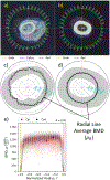
Comparison of paired slices from operated and contralateral limbs located just proximal to the osteotomy where substantial callus is present. a/b) Images after boundary detection and resampling in 1-degree circumferential increments. c/d) Radial line average BMD (ρ¯θ) superimposed over the cortical cross-section shape in each image for reference. e) Point cloud of BMD (ρ) distribution in each cross section. Representative densities for operated (Pop) and contralateral (Pco) slice images are indicated by the large dots. Considerable cortical remodeling was measured for this slice (R = 10%).
Figure 5.
Figure 5.
Comparison of paired slices from operated and contralateral limbs located ~68 mm proximal to the centre of osteotomy where little callus is present. a/b) Images after boundary detection and resampling in 1-degree circumferential increments. c/d) Radial line average BMD (ρ¯θ) superimposed over the cortical cross-section shape in each image for reference. e) Point cloud of BMD (ρ) distribution in each cross section. Representative densities for operated (Pop) and contralateral (Pco) slice images are indicated by the large dots. Moderate cortical remodeling was measured for this slice (R = 5%).
Figure 6.
Figure 6.
Global differences between the operated and contralateral limb of one animal were evaluated based on the changes of a) normalized representative densities (P¯) in each limb and b) remodeling index (R) with characteristic measures obtained from its scatter.
Figure 7.
Figure 7.
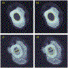
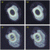
Successful detection of endocortical boundaries in different slice images within one operated limb required adaptive endocortical thresholding (τendo). a/b) For a slice located ~11 mm distal to the osteotomy center with no endosteal callus, the endocortical boundary was properly detected with τendo = 4,250, but improperly detected with τendo = 6,650 (correct threshold in d). c/d) For a slice located 4 mm proximal to the osteotomy center with endosteal callus, the endocortical was incorrectly detected with τendo = 4,250 (correct threshold in a), but properly detected with τendo = 6,650.
Figure 8.
Figure 8.
Detection of pericortical boundaries in the same slice images of Figure 7. a/b) Pericortical boundary was properly detected with τperi = 7,600, but was improperly dilated with τperi = 7,000 (correct threshold in d). c/d) Pericortical boundary was severely eroded with τperi = 7,600 (correct threshold in a), but correctly follows pericortical wall with τperi = 7,000.
Figure 9.
Figure 9.
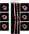
Noteworthy pattern of extreme remodeling in the Sheep L. Remodeling at the outer wall of cortical bone (slice #1500) formed a ring of low BMD at the outer wall (#1638) extending inward to the core (#1783) and reaching the inner wall partially (#1883) before occupying an irregular region of cortex (#2032 and #2132).

Similar articles

Cited by

References

    1. Augat P, Hollensteiner M, von Rüden C. 2021. The role of mechanical stimulation in the enhancement of bone healing. Injury. 52:S78–S83. 10.1016/j.injury.2020.10.009 - DOI - PubMed
    1. Becker M, Witzel C, Kierdorf U, Frölich K, Kierdorf H. 2020. Ontogenetic changes of tissue compartmentalization and bone type distribution in the humerus of Soay sheep. Journal of Anatomy. 237(2):334–354. 10.1111/joa.13194 - DOI - PMC - PubMed
    1. Bissinger O, Götz C, Wolff K-D, Hapfelmeier A, Prodinger PM, Tischer T. 2017. Fully automated segmentation of callus by micro-CT compared to biomechanics. Journal of Orthopaedic Surgery and Research. 12(1):108. 10.1186/s13018-017-0609-9 - DOI - PMC - PubMed
    1. Buie HR, Campbell GM, Klinck RJ, MacNeil JA, Boyd SK. 2007. Automatic segmentation of cortical and trabecular compartments based on a dual threshold technique for in vivo micro-CT bone analysis. Bone. 41(4):505–515. 10.1016/j.bone.2007.07.007 - DOI - PubMed
    1. Claes L, Recknagel S, Ignatius A. 2012. Fracture healing under healthy and inflammatory conditions. Nature Reviews Rheumatology. 8(3):133–143. 10.1038/nrrheum.2012.1 - DOI - PubMed

LinkOut - more resources