. 2024 Jul 22;20(7):e1012335.
doi: 10.1371/journal.ppat.1012335. eCollection 2024 Jul.

Highly restrictive and directional penetration of the blood cerebral spinal fluid barrier by JCPyV

Affiliations

Highly restrictive and directional penetration of the blood cerebral spinal fluid barrier by JCPyV

Bethany A O'Hara et al. PLoS Pathog. .

Abstract

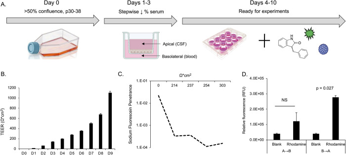
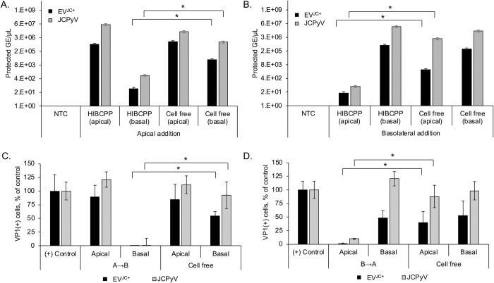
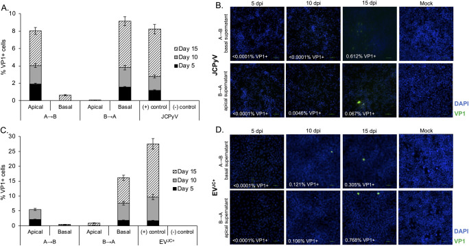
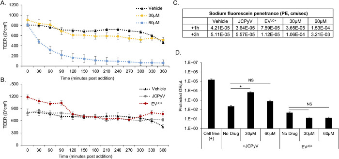
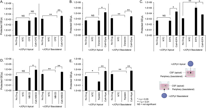
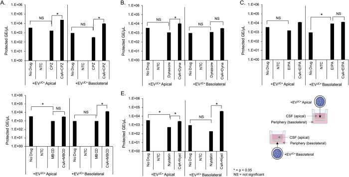
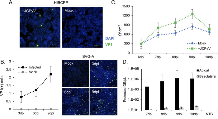
The human polyomavirus JCPyV is an opportunistic pathogen that infects greater than 60% of the world's population. The virus establishes a persistent and asymptomatic infection in the urogenital system but can cause a fatal demyelinating disease in immunosuppressed or immunomodulated patients following invasion of the CNS. The mechanisms responsible for JCPyV invasion into CNS tissues are not known but direct invasion from the blood to the cerebral spinal fluid via the choroid plexus has been hypothesized. To study the potential of the choroid plexus as a site of neuroinvasion, we used an adult human choroid plexus epithelial cell line to model the blood-cerebrospinal fluid (B-CSF) barrier in a transwell system. We found that these cells formed a highly restrictive barrier to virus penetration either as free virus or as virus associated with extracellular vesicles (EVJC+). The restriction was not absolute and small amounts of virus or EVJC+ penetrated and were able to establish foci of infection in primary astrocytes. Disruption of the barrier with capsaicin did not increase virus or EVJC+ penetration leading us to hypothesize that virus and EVJC+ were highly cell-associated and crossed the barrier by an active process. An inhibitor of macropinocytosis increased virus penetration from the basolateral (blood side) to the apical side (CSF side). In contrast, inhibitors of clathrin and raft dependent transcytosis reduced virus transport from the basolateral to the apical side of the barrier. None of the drugs inhibited apical to basolateral transport suggesting directionality. Pretreatment with cyclosporin A, an inhibitor of P-gp, MRP2 and BCRP multidrug resistance transporters, restored viral penetration in cells treated with raft and clathrin dependent transcytosis inhibitors. Because choroid plexus epithelial cells are known to be susceptible to JCPyV infection both in vitro and in vivo we also examined the release of infectious virus from the barrier. We found that virus was preferentially released from the cells into the apical (CSF) chamber. These data show clearly that there are two mechanisms of penetration, direct transcytosis which is capable of seeding the CSF with small amounts of virus, and infection followed by directional release of infectious virions into the CSF compartment.


Conflict of interest statement

The authors have declared that no competing interests exist.

Figures

Fig 1
Fig 1. Establishment and characterization of a choroid plexus barrier system.
A) Barrier development protocol. Cells are maintained at high density (>50% confluence) and used for experiments between passages 30–38. Cells are seeded to transwell inserts and follow a stepwise serum reduction protocol with daily monitoring until ready for experiments. Transwell cultures persist with sufficiently high TEER values for up to ten days. B) Representation of TEER values over time. TEER increases steadily over time up to day 9. C) Penetration of sodium fluorescein (SF), a small molecule tracer dye. SF was used to measure the rate of penetrance (cm/sec) from the apical to basal chamber over one hour. As TEER increases, penetrance decreases, indicating that a restrictive barrier has formed. D) Bidirectional transport of rhodamine 123. Relative fluorescence (RFU) was measured in the apical and basal chambers after 3 hours. Rhodamine is preferentially transported in the basolateral to apical direction (B to A) rather than apical to basolateral (A to B) indicating the barrier is polarized. Error bars in panels B and D represent the standard deviation between three independent experiments, in triplicate. Images in Fig 1A, were created with BioRender.com under license to Brown University.
Fig 2
Fig 2. HIBCPP barrier cultures significantly restrict JCPyV and EVJC+.
A) Barriers were set up on transwell inserts as described. 310/ml genomes of JCPyV or EVJC+ were added to the apical cell surface of HIBCPP barrier cultures and viral genomes were quantified by qPCR from both apical and basal chamber supernatants after 48h. B) JCPyV or EVJC+ were added to the basolateral cell surface of HIBCPP barrier cultures and viral genomes were quantified from both apical and basal chamber supernatants after 48h. C-D) Apical and basal supernatants from panels A and B were used to infect SVG-A cells and scored for VP1 at 3 days post infection. The majority of JCPyV or EVJC+ is restricted to the chamber in which its added. * = p < 0.05. NTC = no template control. (B to A), basolateral to apical direction. (A to B) apical to basolateral direction. Infection with purified JCPyV or EVJC+ was used as the positive (+) control, to compare the spread of infection. Error bars represent the standard deviation between three independent experiments, in triplicate.
Fig 3
Fig 3. Barrier-infiltrating JCPyV and EVJC+ establish infected foci in primary human astrocytes following multiple rounds of replication.
Supernatants from the barrier infiltration experiment shown in Fig 3 were used to reinfect primary human astrocytes (NHA) and allowed to replicate for 15 days. Apical and basal supernatants were collected after 48h incubation with virus or EVJC+ and used to infect NHA. The infection was monitored for viral spread over time. A) VP1 expression in NHA following barrier infiltration by JCPyV at days 5, 10 and 15 post infection. VP1+ cells increase over time. B) Clusters of infected cells are apparent by day 15. C) VP1 expression in NHA following barrier infiltration by EVJC+ at days 5, 10 and 15 post infection. VP1+ cells increase over time. D) Clusters of infected cells are apparent by day 15. DAPI (total cell count) is shown in blue and VP1 (infected cell count) is shown in green. Mock = SVG-A cells incubated with supernatant from naïve HIBCPP cells from day 7, and stained for VP1 at 15dpi. Virus is detectable at day 15 by both indirect immunofluorescence and qPCR, and is able to spread over time. (B to A), basolateral to apical direction. (A to B) apical to basolateral direction. Infection with purified JCPyV was used as the positive (+) control, to compare spread. Mock infection with viral diluent alone (DMEM/F12 media) was used as the negative (-) control. Error bars represent the standard deviation between three independent experiments, in triplicate.
Fig 4
Fig 4. Intercellular disruption of HIBCPP barriers does not increase penetration of JCPyV or EVJC+.
A) Following initial TEER readings, vehicle control (DMSO) or 30μM and 60μM capsaicin were added to barrier cultures. TEER readings were taken every 30 minutes, starting at time zero, for 6 hours. Capsaicin disrupted intercellular barrier integrity in a dose dependent manner. B) Following initial TEER readings, JCPyV, vehicle control, or EVJC+ were added to barrier cultures. TEER readings were taken every 30 minutes, starting at time zero, for 6 hours. Addition of JCPyV, vehicle or EVJC+ did not disrupt barrier integrity. C) Sodium fluorescein penetrance was calculated following 1 hour and 3 hour exposure to capsaicin, JCPyV, EVJC+ or vehicle. As expected, PE increases following barrier incubation with capsaicin. D) Quantification of viral penetration by qPCR after capsaicin treatment. NS = not significant, * = p < 0.05. Error bars represent the standard deviation between three independent experiments, in triplicate.
Fig 5
Fig 5. The effect of transcytosis inhibitors on barrier penetration by JCPyV.
HIBCPP cells were cultured on transwell inserts as described and treated with transcytosis inhibitors alone, or cyclosporin A for one hour followed by inhibitors. 310/ml genome copies of purified JCPyV was added to either the apical or basal chamber for an additional 24h. A) qPCR quantification of JCPyV barrier penetration following treatment with 100μM chlorpromazine. B) qPCR quantification of JCPyV barrier penetration following treatment with 100μM dynasore. C) qPCR quantification of JCPyV barrier penetration following treatment with 100μM EIPA. D) qPCR quantification of JCPyV barrier penetration following treatment with 5mM methyl-beta-cyclodextran (MßCD). E) qPCR quantification of JCPyV barrier penetration following treatment with 5μM nystatin. Cyclosporin A (CsA) restored virus penetration from the basolateral to apical chambers to equivalent with the untreated control (no drug, panels A-E). NS = not significant, * = p < 0.05, ** = p < 0.01. NTC = no template control. Error bars represent the standard deviation between three independent experiments, in triplicate. Images in Fig 5E were created with BioRender.com under license to Brown University.
Fig 6
Fig 6. The effect of transcytosis inhibitors on barrier penetration by EVJC+.
HIBCPP cells were cultured on transwell inserts as described and treated with transcytosis inhibitors alone, or cyclosporin A for one hour followed by inhibitors. 310/ml genome copies of EVJC+ was added to either the apical or basal chamber for an additional 24h. A) qPCR quantification of EVJC+ barrier penetration following treatment with 100μMchlorpromazine. B) qPCR quantification of EVJC+ barrier penetration following treatment with 100μM dynasore. C) qPCR quantification of EVJC+ barrier penetration following treatment with 100μM EIPA. D) qPCR quantification of EVJC+ barrier penetration following treatment with 5mM methyl-beta-cyclodextran (MßCD). E) qPCR quantification of EVJC+ barrier penetration following treatment with 5μM nystatin. Inhibitors did not block basal to apical penetration of EVJC+. NS = not significant, * = p < 0.05. NTC = no template control. Error bars represent the standard deviation between three independent experiments, in triplicate. Images in Fig 6E were created with BioRender.com under license to Brown University.
Fig 7
Fig 7. Release of infectious virions from JCPyV+ HIBCPP barriers.
A) HIBCPP monolayers were infected with JCPyV for 2 hours. Three days post infection, infected and mock infected cells were plated to transwell culture dishes and 96 well plates. HIBCPP cells were stained for VP1 at 7dpi to verify the presence of a productive infection. VP1+ cells are shown in green; DAPI was used to calculate a total cell count and is shown in blue. Scale bar = 100μm. B) Media collected from day 7 was used to infect SVG-A cultures in a growth assay over 15 days. HIBCPP cells release infectious virions capable of establishing an infection and spreading in target cells. VP1+ cells are shown in green; DAPI was used to generate a total cell count and is shown in blue. Scale bar = 100μm. C) TEER was monitored daily and is shown as Ω*cm2. Values were calculated by subtracting the TEER reading of cell free control wells from that of the sample wells, and multiplying by the transwell surface area. D) A sample of media from the apical and basal chambers was collected daily and analyzed for protected viral genome content by qPCR. Virus is preferentially released into the apical chamber. Error bars represent the standard deviation between three independent experiments, in triplicate. Mock = mock infected cells, negative control; DPI = days post infection; NTC = no template control.

Similar articles

Cited by

References

    1. Assetta B, Atwood WJ. The biology of JC polyomavirus. Biol Chem. 2017;398(8):839–55. Epub 2017/05/12. doi: 10.1515/hsz-2016-0345 . - DOI - PubMed
    1. Haley SA, Atwood WJ. Progressive Multifocal Leukoencephalopathy: Endemic Viruses and Lethal Brain Disease. Annu Rev Virol. 2017;4(1):349–67. Epub 2017/06/24. doi: 10.1146/annurev-virology-101416-041439 . - DOI - PubMed
    1. Berger JR, Hartung HP. Commentary: Progressive multifocal leukoencephalopathy genetic risk variants for pharmacovigilance of immunosuppressant therapies. Front Neurol. 2023;14:1146027. Epub 2023/04/04. doi: 10.3389/fneur.2023.1146027 ; PubMed Central PMCID: PMC10062523. - DOI - PMC - PubMed
    1. Cortese I, Beck ES, Al-Louzi O, Ohayon J, Andrada F, Osuorah I, et al.. BK virus-specific T cells for immunotherapy of progressive multifocal leukoencephalopathy: an open-label, single-cohort pilot study. Lancet Neurol. 2021;20(8):639–52. Epub 2021/07/25. doi: 10.1016/S1474-4422(21)00174-5 ; PubMed Central PMCID: PMC8395368. - DOI - PMC - PubMed
    1. Cortese I, Muranski P, Enose-Akahata Y, Ha SK, Smith B, Monaco M, et al.. Pembrolizumab Treatment for Progressive Multifocal Leukoencephalopathy. N Engl J Med. 2019;380(17):1597–605. Epub 2019/04/11. doi: 10.1056/NEJMoa1815039 . - DOI - PubMed

Substances

LinkOut - more resources