. 2024 Jul 22;22(7):e3002074.
doi: 10.1371/journal.pbio.3002074. eCollection 2024 Jul.

miR-203 secreted in extracellular vesicles mediates the communication between neural crest and placode cells required for trigeminal ganglia formation

Affiliations

miR-203 secreted in extracellular vesicles mediates the communication between neural crest and placode cells required for trigeminal ganglia formation

Yanel E Bernardi et al. PLoS Biol. .

Abstract

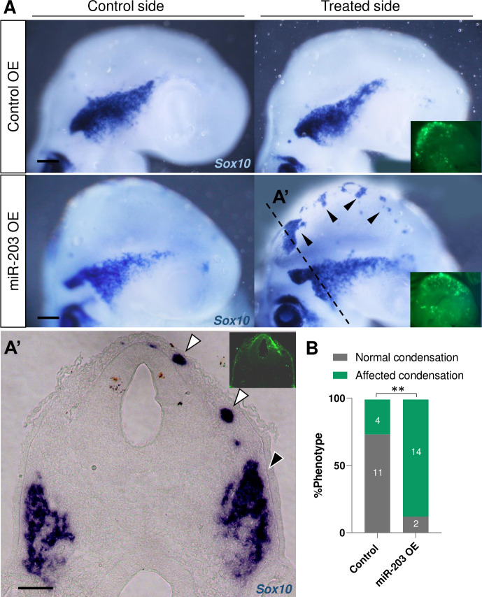
While interactions between neural crest and placode cells are critical for the proper formation of the trigeminal ganglion, the mechanisms underlying this process remain largely uncharacterized. Here, by using chick embryos, we show that the microRNA (miR)-203, whose epigenetic repression is required for neural crest migration, is reactivated in coalescing and condensing trigeminal ganglion cells. Overexpression of miR-203 induces ectopic coalescence of neural crest cells and increases ganglion size. By employing cell-specific electroporations for either miR-203 sponging or genomic editing using CRISPR/Cas9, we elucidated that neural crest cells serve as the source, while placode cells serve as the site of action for miR-203 in trigeminal ganglion condensation. Demonstrating intercellular communication, overexpression of miR-203 in the neural crest in vitro or in vivo represses an miR-responsive sensor in placode cells. Moreover, neural crest-secreted extracellular vesicles (EVs), visualized using pHluorin-CD63 vector, become incorporated into the cytoplasm of placode cells. Finally, RT-PCR analysis shows that small EVs isolated from condensing trigeminal ganglia are selectively loaded with miR-203. Together, our findings reveal a critical role in vivo for neural crest-placode communication mediated by sEVs and their selective microRNA cargo for proper trigeminal ganglion formation.


Conflict of interest statement

The authors have declared that no competing interests exist.

Figures

Fig 1
Fig 1. miR-203 expression is activated during trigeminal ganglion formation.
(A) Schematic diagrams of a cross-section through the cranial NT illustrating the migration (B, B’) of NC and their coalescence (C, C’) and condensation (D, D’) with placode cells during TG formation in chicken embryos. Scale bars: 100 μm. Transverse section of miR-203 after in situ hybridization using an LNA-DIG-labeled probe followed by immunostaining for HNK1 (neural crest marker in red) and Tuj1 (placodal marker in green) at HH13 (B), HH16 (C), and HH20 (D). Scale bars: 50 μm. While miR-203 is absent from migrating NC cells, its expression reinitiates at the time of ganglion coalescence and remains present in the condensed ganglion. Red and green arrowheads in C’ denoted early coalescing NC and placode cells expressing miR-203, respectively. NC, neural crest; NT, neural tube; TG, trigeminal ganglion.
Fig 2
Fig 2. Overexpression of miR-203 promotes ectopic and premature NC condensation and enhanced aggregation into the trigeminal ganglion.
(A) In situ hybridization for Sox10 in HH15-16 embryos unilaterally electroporated (treated side showing green fluorescence in insets) with miR-203 (miR-203 OE) or empty control (Control OE) overexpression vectors. (A’) Cross section of miR-203 OE embryo at the level shown in A (dotted line) reveals an ectopic Sox10+ group of cells (white arrowhead) and a denser TG (black arrowhead) on the treated side. Scale bars: 100 μm. (B) Quantification of embryos showing a phenotype (normal versus affected condensation having ectopic condensation and/or denser TG) on Control and miR-203 OE. Numbers in the graph represent the numbers of analyzed embryos. **P = 0.001 by contingency table followed by a χ2 test. TG, trigeminal ganglion.
Fig 3
Fig 3. miR-203 is produced in NC but it places of action are placode cells to secure normal trigeminal ganglion condensation.
Electroporation schemes for region-specific transfections (see also S2 Fig). Embryos at HH9 were unilaterally electroporated (treated side showing green fluorescence in insets) in the NT or ectodermal placodes with miR-203 sponge (n = 12) or scrambled sponge (n = 12) plasmids (A, B); or with miR-203 gRNAs + Cas9 (n = 10) or Scrambled gRNAs + Cas9 (NT n = 8; ectodermal placode n = 11) (C, D) (see also S1 Fig and S1 Data). Trigeminal ganglia condensation was analyzed by ISH for Sox10 at HH17-18. Scale bars: 100 μm. Scatter plots represent the quantification of trigeminal ganglia areas (treated side versus control side). ****P < 0.0001, **P = 0.0085 by two-tailed unpaired t test. ns, non-significant differences. Values are means ± SD. NC, neural crest; NT, neural tube.
Fig 4
Fig 4. Extracellular vesicles are actively released by NC cells in the vicinity of placode cells during TG condensation.
TEM from dissected trigeminal ganglia at HH17 shows that cells (identify each with different colors) have many direct cell-to-cell contacts to each other (A), the presence of MVBs releasing exosomes-like structures into the extracellular space (ExS) (B), and a shedding vesicle protruding from PM (C). Bars: 2 μm (A), 200 nm (B and C). (D) Diagram of pHluo-CD63-mScarlet functional assay. (E) Embryos electroporated in the NT with pHluo-CD63-mScarlet at HH9- and visualized at stage HH16 exhibited GFP+/mScarlet+ fluorescence in the condensing trigeminal ganglion area (dotted line); in contrast, cells located in the midbrain are enrich in mScarlet+ (white arrowheads). Scale bar: 100 μm. TG, trigeminal ganglion; E, eye. (F) Migratory neural crest cells electroporated with pHluo-CD63-mScarlet exhibit intraluminal MVB (pHluo-/mScarlet+), extracellular/releasing EVs (pHluo+/mScarlet+) and deposit trails behind the cells. Scale bar: 20 μm. (G) Transverse section through the TG showing NC cells electroporated with pHluo-CD63-mScarlet and placode cells immunostained for Tuj1 (magenta). Scale bar: 10 μm. (G’) Zoom of box in G shows a placode cells (Tuj1+) with pHluo puncta incorporated into their cytoplasm (white arrowheads). Scale bar: 20 μm. EV, extracellular vesicle; MVB, multivesicular body; NC, neural crest; PM, plasma membrane; TEM, transmission electron microscopy.
Fig 5
Fig 5. Extracellular vesicles produced by NC cells are engulfed by placode cells in explant co-cultures.
(A) Scheme of a chicken embryo at HH9- with NT electroporated with a pHluo (pseudocolored in yellow) and placode cells electroporated with membrane RFP (pseudocolored in magenta), respectively. The electroporated dorsal NT (mostly containing NC cells) and ectodermal trigeminal placode cells were dissected at HH9+ and HH10-, respectively, and co-cultured to examine their interactions. (B) NC cells (yellow) exhibiting trails (possibly migrasomes or retraction fibers) and cytonemes-like structures in very close contact with placode cells (magenta). (B’) Higher magnification of box in B showing the cytonemes from NC cells contacting placode cells. See also S1 Movie. (C) Placode cells showing vesicular structures like macropinosomes in their membrane (white arrowheads). See also z-stack in S2 Movie. (D, D’) NC cells expressing pHluo were surrounded by numerous extracellular pHluo+ puncta (possibly EVs deposits) and trails (possibly migrasomes or retraction fibers). Co-cultured placodal cells showing intra-cytoplasmic vesicular structures containing pHluo+ puncta (white arrowheads). See also S3 and S4 Movies. (E) Placode cell co-cultured with NC cells expressing pHluo produce filopodia that move toward the EVs deposits and engulf them. (E’) Zoom of box in E showing the time-lapse showing placode cells engulfing a pHluo+ puncta (white arrowhead). See also S5 Movie. (F) 3D reconstruction of placode protrusion observed in (E) demonstrating the internalized pHluo+ puncta (white arrowhead). See also S6 Movie. Scale bars: 10 μm. EV, extracellular vesicle; NC, neural crest; NT, neural tube.
Fig 6
Fig 6. miR-203 generated in NC cells inhibits a sensor target electroporated in placode cells both in vivo and ex vivo.
(A) Scheme of chick embryo with NT electroporated with miR-203 or empty control overexpressing vectors (with cytoplasmic EGFP) and placode cells electroporated with a dual-colored sensor vector (pSdmiR-203) containing 2 copies of complementary sequences to the mature miR-203. pCAG, Chick β-actin promoter; d4EGFPn nuclear-localized destabilized EGFP with a half-life of 4 h; mRFPn, nuclear-localized monomeric red fluorescent protein. Box of the condensing trigeminal ganglion indicates area of enlarged inset exemplifying the transfer of miR-203 from NC (green) to placode cells (orange because of the expression of both EGFP and RFP), thus affecting the EGFP translation (red cell). (B) Transverse section of HH17 embryos immunostained for Tuj1 (magenta), to identify the trigeminal ganglia with coalescing NC (electroporated with miR-203 OE vector) and placode (electroporated with the dual-colored sensor vector; see white arrowheads) cells. (B’) Zoom of box in B identifying migratory NC cells with cytoplasmic EGFP+ (black arrowhead) and placode cells with nuclear EGFP+/RFP+ (white arrowhead) or only RFP+ (dotted circles). Scale bar: 100 μm. See also S5A Fig for DAPI staining. (C) Time-lapse of co-cultured placode ectodermal cells (electroporated with the dual-colored sensor vector) and dorsal NT (electroporated with miR-203 OE) explants. Placodal cells (PC1-6) interact with NC cells (NC1-2) where the EGFP channel was pseudocolored (red>white>blue) to visualize the EGFP decay in placode cells over time. See also S5B Fig for control and S7 Movie. (D) Scatter plot from in vivo experiments analyzing the EGFP/RFP intensity for individual placode cells reaching the condensing area at HH17 from miR-203 OE or Control electroporated embryos (cells counted in 2–5 sections per embryo, n = 4 embryos from 2 independent electroporations; see S1 Data). ****P < 0.0001 calculated using unpaired Student’s t test. Values are means ± SD. (E) Scatter plot from ex vivo co-cultured experiment analyzing the EGFP/RFP normalized intensity for individual placode cells interacting with NC cells from miR-203 OE or Control electroporated embryos (100 cells, n = 4 co-cultured explants for each treatment from 2 independent experiments; see S1 Data). ****P < 0.0001 calculated using unpaired Student’s t test. Values are means ± SD. NC, neural crest; NT, neural tube.
Fig 7
Fig 7. Endogenous miR-203 is selectively loaded into sEVs produced during TG coalescence.
(A) Graphs show particle concentration, size, and mode size (value are mode ± SD) using NTA from enriched sEVs samples (sEV1 and sEV2) isolated from dissected trigeminal ganglia at HH17 (see S1 Data). (B) TEM image showing isolated exosomes after sEVs purified from dissected trigeminal ganglia. Scale bar: 100 nm. (C) RT-PCR from 2 independent whole dissected TG (TG1 and TG2) and isolated sEVs from condensing TG (sEV1 and sEV2) raised against the endogenous miR-203 (containing the EXOmotif) and miR-34a-5p (containing the CELLmotif). NTA, nanoparticle tracking analysis; sEV, small extracellular vesicle; TEM, transmission electron microscopy; TG, trigeminal ganglion.

Update of

References

    1. Breau MA, Schneider-Maunoury S. Cranial placodes: models for exploring the multi-facets of cell adhesion in epithelial rearrangement, collective migration and neuronal movements. Dev Biol. 2015;401:25–36. doi: 10.1016/j.ydbio.2014.12.012 - DOI - PubMed
    1. Saint-Jeannet JP, Moody SA. Establishing the pre-placodal region and breaking it into placodes with distinct identities. Dev Biol. 2014;389:13–27. doi: 10.1016/j.ydbio.2014.02.011 - DOI - PMC - PubMed
    1. Crane J, Trainor P. Neural crest stem and progenitor cells. Annu Rev Cell Dev Biol. 2006;22:267–286. doi: 10.1146/annurev.cellbio.22.010305.103814 - DOI - PubMed
    1. Graham A, Shimeld SM. The origin and evolution of the ectodermal placodes. J Anat. 2013;222:32. doi: 10.1111/j.1469-7580.2012.01506.x - DOI - PMC - PubMed
    1. Patthey C, Schlosser G, Shimeld SM. The evolutionary history of vertebrate cranial placodes–I: Cell type evolution. Dev Biol. 2014;389:82–97. doi: 10.1016/j.ydbio.2014.01.017 - DOI - PubMed

MeSH terms

LinkOut - more resources