. 2024 Sep;31(9-10):455-466.
doi: 10.1038/s41434-024-00466-w. Epub 2024 Jul 22.

Characterization of brain transduction capability of a BBB-penetrant AAV vector in mice, rats and macaques reveals differences in expression profiles

Affiliations

Characterization of brain transduction capability of a BBB-penetrant AAV vector in mice, rats and macaques reveals differences in expression profiles

María Bunuales et al. Gene Ther. 2024 Sep.

Abstract

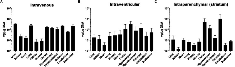
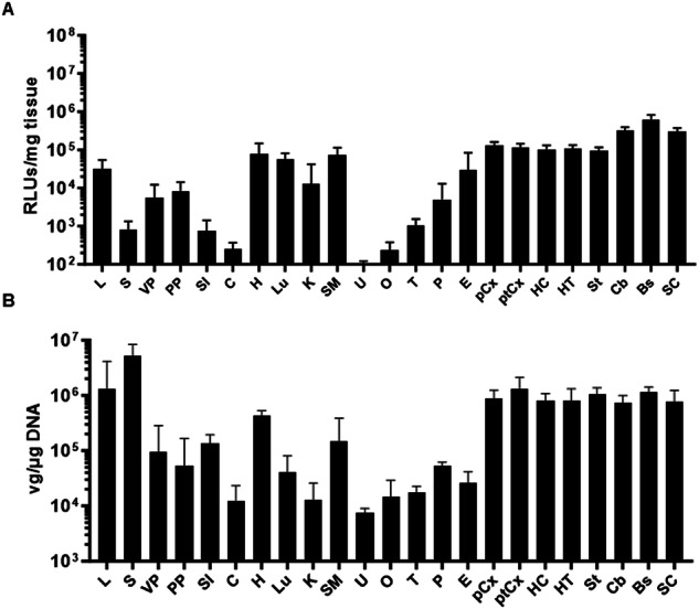
Different screening methods are being developed to generate adeno-associated viral vectors (AAV) with the ability to bypass the blood-brain barrier (BBB) upon intravenous administration. Recently, the AAV9P31 stood out as the most efficient version among a library of peptide-displaying capsids selected in C57BL/6 mice using RNA-driven biopanning. In this work we have characterized in detail its biodistribution in different mouse strains (C57BL/6 and Balb/c), as well as in Sprague Dawley rats and non-human primates (Macaca fascicularis). Using GFP and NanoLuc reporter genes, we confirmed homogeneous infection and transgene expression across the CNS of mice injected intravenously with AAV9P31. A more restricted pattern was observed upon either intracerebroventricular or intraparenchymal injection. Following intravenous delivery, region- and cell-specific differential patterns of transduction were observed in the mouse brain, including a preferential transduction of astrocytes and neurons in the cerebral cortex and striatum, whereas neurons were the only transduced cell type in subcortical locations across the hippocampus, thalamus, hypothalamus, mesencephalon, brainstem and cerebellum. Furthermore, transduced microglial cells were never found in any CNS location. Peripheral organs transduced upon intravenous administration included lung, liver, peritoneum, heart and skeletal muscle. However, a comparable performance of AAV9P31 to bypass the BBB in rats and macaques was not observed, although a more limited neuronal transduction was found in the brainstem of rats upon intravenous delivery. Finally, intracerebroventricular delivery in macaques resulted in neuronal transduction in cortical, subcortical structures and cerebellum following a patchy pattern. In conclusion, the widespread CNS transduction obtained in mice upon intravenous delivery of AAV9P31 represents a powerful tool for modeling a wide variety of neurological disorders as well as an appealing choice for the evaluation of gene therapy-based therapeutics.


Conflict of interest statement

The authors declare no competing interests.

Figures

Fig. 1
Fig. 1. Biodistribution of AAV9P31-CMV-GFP following different routes of administration in C57BL6/J mice.
Different groups of 7 weeks-old C57BL6/J mice (3 females and 3 males) received the AAV9P31-CMV-GFP vector by i.v. (retro-orbital) injection at 2 × 1013 vg/kg (A), or by i.c.v. injection at 1.2 × 1011 vg (B), or by unilateral intrastriatal injection at 6 × 1010 vg (C). One month later, mice were sacrificed and viral genomes were quantified in the indicated CNS structures and peripheral organs by qPCR.
Fig. 2
Fig. 2. Detection of transduced cells upon administration of AAV9P31-CMV-GFP following different routes in C57BL6/J mice.
Different groups of 7 weeks-old C57BL6/J mice (3 females and 3 males) received the AAV9P31-CMV-GFP vector by i.v. (retro-orbital) injection at 2 × 1013 vg/kg (A), or by i.c.v. injection at 1.2 × 1011 vg (B), or by unilateral intrastriatal injection at 6 × 1010 vg (C). One month later, mice were sacrificed and tissue samples were processed for detection of GFP by immunohistochemistry. Pictures show representative images of the indicated tissues. A Low-power photomicrographs taken from three different rostrocaudal levels of the CNS illustrating the brain-wide expression of GFP. B At the level of the cerebral cortex, a predominant transduction of astrocytes was constantly observed, together with a more moderate expression being found in both pyramidal and non-pyramidal neurons. Image taken from the primary somatosensory cortex. C Cellular phenotypes expressing GFP in the hippocampal formation are limited to neurons (image taken from the CA1 field). D The striatum is the brain territory showing the weakest GFP expression, mainly comprising scattered astrocytes and medium-sized spiny neurons. E Basal forebrain neurons were strongly transduced with GFP. F, G Different types of neurons in the cerebellum were transduced with GFP, comprising neurons in the molecular, Purkinje and granular layers of the cerebellar cortex. Purkinje neurons were randomly transduced, most often showing a Golgi-like stain (inset in G). Scale bars are 3.0 mm in (A); 200 µm in (B, D, E); 100 µm in (C); 250 µm in (F) and 50 µm in (G).
Fig. 3
Fig. 3. Cellular phenotypes expressing GFP in the cerebral cortex upon i.v. delivery of AAV9P31-CMV-GFP in mice.
C57BL6/J mice (3 females and 3 males) received the AAV9P31-CMV-GFP vector by i.v. (retro-orbital) injection at 2 × 1013 vg/kg. One month later, mice were sacrificed and brain samples were processed for immunofluorescence using antibodies against GFP (green) and the indicated cell markers: NeuN for neurons (blue) and GFAP for astrocytes (purple). A-A” A random transduction of astrocytes was found throughout the cerebral cortex. B-B” Expression of GFP spreads within the whole cytoplasm of transduced astrocytes, even reaching the most distal and thinnest astrocytic processes. C, D” Pyramidal neurons were also randomly transduced, sometimes showing Golgi-like morphologies. A representative example of a GFP+ layer II-III pyramidal neuron and surrounding astrocytes is shown in (C-C”’), whereas (D-D”’) illustrate a GFP+ layer V pyramidal neuron as well as a neighboring transduced astrocyte. Scale bars are 20 μm in (A-A” & D-D”); 10 μm in (B-B”) and 40 μm in (C-C”).
Fig. 4
Fig. 4. Cellular phenotypes expressing GFP in subcortical locations upon i.v. delivery of AAV9P31-GFP in mice.
C57BL6/J mice (3 females and 3 males) received the AAV9P31-CMV-GFP vector by i.v. (retro-orbital) injection at 2 × 1013 vg/kg. One month later, mice were sacrificed and brain samples were processed for immunofluorescence using antibodies against GFP (green) and the indicated cell markers: NeuN for neurons (blue) and GFAP for astrocytes (purple). In all analyzed subcortical territories, neurons were found to be the only cellular phenotype transduced with GFP, as shown for the hippocampal formation (A-A”’; image taken from the CA1 field); lateral hypothalamic nuclei (C-C”’), substantia nigra pars compacta (D-D”’), locus coeruleus (E-E”’), as well as in motor and sensory brainstem nuclei such as the facial nerve (F-F”’), and inferior olive (G-G”’) nuclei, respectively. The same pattern of exclusive neuronal transduction was found in the cerebellar cortex, where only Purkinje and granule neurons were GFP+ (H-H”’). The striatum was found to be the only subcortical structure with few GFP+ astrocytes (B-B”). Scale bars are 40 μm in all panels.
Fig. 5
Fig. 5. Biodistribution and kinetics of transgene expression in C57BL/6 J mice treated with i.v. injection of AAV9P31-CMV-NL.
A–C Seven weeks-old C57BL/6 J mice (3 females and 3 males) received the AAV9P31-CMV-NL vector by i.v. (retro-orbital) injection at 2 × 1013 vg/kg. One month later, mice received an intraperitoneal injection of the NanoLuc substrate (furimazine) and light emission was visualized in a luminometer (A). The pictures show a representative image of the dorsal and ventral views. Mice were then sacrificed for dissection of different CNS structures and peripheral organs. One portion of the tissue samples was used for quantification of luciferase activity ex vivo (B), and other portion was processed DNA isolation and quantification of viral genomes by qPCR (C). The same dose of vector and route of administration was used to treat 2 weeks-old C57BL/6 J mice (n = 6). Longitudinal detection (D) and quantification (E) of NanoLuc expression was performed by BLI at the indicated times after vector administration. In the BLI pictures, maximal and minimal light emission are represented by red and blue pseudocolors, respectively. Quantification corresponds to total light emission (photons/s) from ROIs located in the head and abdominal areas.
Fig. 6
Fig. 6. Biodistribution and transgene expression in Balb/c mice treated with i.v. injection of AAV9P31-CMV-NL.
Seven weeks-old Balb/c mice (3 females and 3 males) received the AAV9P31-CMV-NL vector by i.v. (retro-orbital) injection at 2 × 1013 vg/kg. One month later, mice were sacrificed for dissection of different CNS structures and peripheral organs. One portion of the tissue samples was used for quantification of luciferase activity ex vivo (A), and other portion was processed DNA isolation and quantification of viral genomes by qPCR (B).
Fig. 7
Fig. 7. Immunoperoxidase detection of GFP transduced cells upon i.v. delivery of AAV9P31-CMV-GFP in rats.
Seven weeks-old rats (one female and one male) received the AAV9P31-CMV-GFP vector by i.v. (retro-orbital) injection at 2 × 1013 vg/kg. One month later, rats were sacrificed and tissue samples were processed for detection of GFP by immunohistochemistry. A-A’ Low-power photomicrograph and inset taken from the primary somatosensory cortex showing just a few, scattered astrocytes expressing GFP across the whole rostrocaudal extent of the cerebral cortex. B–D’ GFP+ neurons were unexpectedly found in different locations of the caudal mesencephalon and brainstem such as the dorsal raphe nucleus (B-B’), the abducens nucleus (C-C’) and the inferior olive (D-D’). GFP+ neurons were only found in these discrete locations of the rat brain, together with few scattered neurons in different brainstem nuclei. Scale bars are 100 µm in panel (A); 25 µm in (B’); 1000 µm in (B, D, E); and 200 µm in (B’, C’, D’).
Fig. 8
Fig. 8. Immunohistochemical analysis of transgene expression in tissues from macaques treated with AAV9P31 vectors by different routes.
Macaca fascicularis (n = 2, male and female) were treated simultaneously with vectors AAV9P31-CMV-GFP (2 × 1013 vg/kg), AAV9P31-CMV-tdT (4 × 1013 vg) and AAV9P31-CMV-NL (2 × 1011 vg) following i.v., i.c.v. and intraparenchymal injections (substantia nigra), respectively. Animals were sacrificed one month after injection, and the expression of the respective transgenes was analyzed by IHQ. In the case of NanoLuc (AAV9P31-CMV-NL), the antibody was directed against the N-terminal HA tag fused to the protein. A-A” The systemic delivery of AAV9P31-CMV-GFP resulted in an almost complete lack of CNS transduction. GFP+ astrocytes were only found in the white matter area located above the putamen nucleus through the tract of the intraputaminal delivery of NL (HA-tagged) vector. For orientation purposes, the boundaries of the caudate and putamen nuclei, as well as both segments of the globus pallidus are delineated. B, C The intraparenchymal delivery resulted in local HA+ neurons and astrocytes at the level of the injected putamen nucleus (B’). AAV9P31 shared similar retrograde spreading properties of the native AAV9 capsid, therefore leading to transduction of GPe neurons projecting to the putamen (HA+ arkypallidal neurons; B”), as well as neurons located in the left substantia nigra pars compacta (C’-C”) innervating the left putamen nucleus through the nigrostriatal pathway. D–F Widespread neuronal transduction in the cerebral cortex, subcortical structures and the cerebellum upon i.c.v. administration of AAV9P31-CMV-tdT. D’ tdT+ pyramidal neurons from upper and lower cortical layers are organized in a columnar random pattern. D”D”’ Patchy distribution of tdT+ neurons in the putamen nucleus, more evident in ventrolateral putaminal areas. E’ A similar patchy pattern of distribution for tdT+ neurons was observed in the thalamus. E” A moderate neuronal transduction was found in the hippocampal formation, with a higher number of tdT+ neurons in the CA3 field (inset in E”). E”’ tdT+ neurons in deep brain structures such as the substantia nigra pars compacta. F tdT+ structures observed in the brainstem and cerebellum, including neurons within the abducens nucleus in the brainstem (F’), climbing fibers in the cerebellum (F”), as well as neurons with strong tdT labeling in deep cerebellar nuclei (F”’). Abbreviations: caudate nucleus (CN), putamen nucleus (Put), internal division of the globus pallidus (GPi), external division of the globus pallidus (GPe). Scale bars are 3 mm in (AC, F); 4 mm in (D, E); 1 mm in (A’, D”); 300 µm in (A”, B’, B”, D’, E’, F’, F”); 500 µm in (C’); 200 µm in (C”); 150 µm in (D”’, E”’) & inset in (E”); 1500 µm in (E”) and 600 µm in (F”’).

References

    1. Padmakumar S, Souza AD, Parayath NN, Bleier BS, Amiji MM. Nucleic acid therapies for CNS diseases: Pathophysiology, targets, barriers, and delivery strategies. J Control Release [Internet]. 2022;352:121–45. 10.1016/j.jconrel.2022.10.018. 10.1016/j.jconrel.2022.10.018 - DOI - PubMed
    1. Fu H, Mccarty DM. Crossing the blood – brain-barrier with viral vectors. Curr Opin Virol. 2016;21:87–92. 10.1016/j.coviro.2016.08.006 - DOI - PubMed
    1. Seko I, Şahin A, Tonbul H, Çapan Y. Brain - targeted nanoparticles to overcome the blood - brain barrier. J Pharm Technol. 2020;1:25–39.
    1. Chandran J, Chowdhury EA, Perkinton M, Jamier T. Assessment of AAV9 distribution and transduction in rats after administration through Intrastriatal, Intracisterna magna and Lumbar Intrathecal routes. Gene Ther. 2023;30:132–41. 10.1038/s41434-022-00346-1 - DOI - PubMed
    1. Galvan A, Petkau TL, Hill AM, Korecki AJ, Lu G, Choi D, et al. Intracerebroventricular administration of AAV9-PHP. B SYN1-EmGFP induces widespread transgene expression in the Mouse and Monkey central nervous system. Hum Gene Ther 2021;32:599–615. 10.1089/hum.2020.301 - DOI - PMC - PubMed

LinkOut - more resources