Review
. 2024 Oct;31(10):1255-1266.
doi: 10.1038/s41418-024-01347-w. Epub 2024 Jul 23.

Cell autonomous functions of CD47 in regulating cellular plasticity and metabolic plasticity

Affiliations
Review

Cell autonomous functions of CD47 in regulating cellular plasticity and metabolic plasticity

Ruhi Polara et al. Cell Death Differ. 2024 Oct.

Abstract

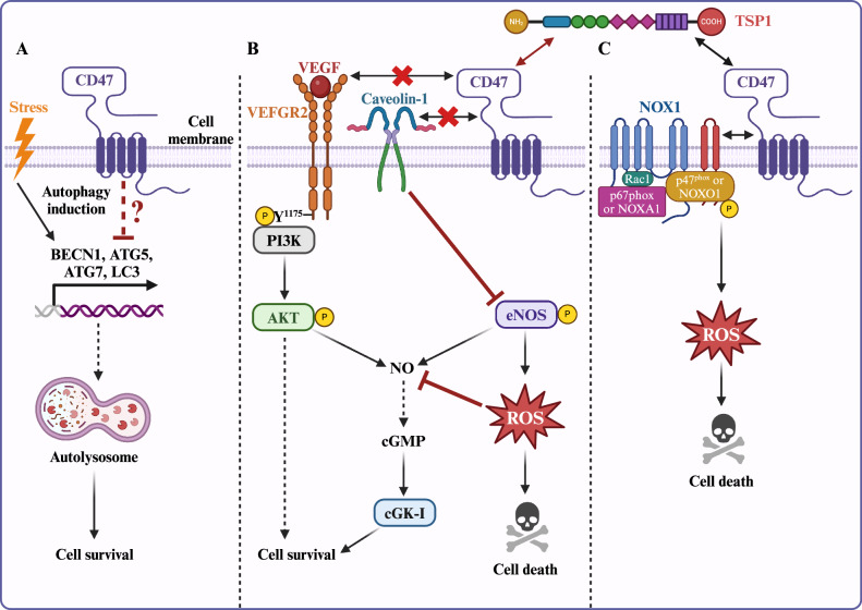
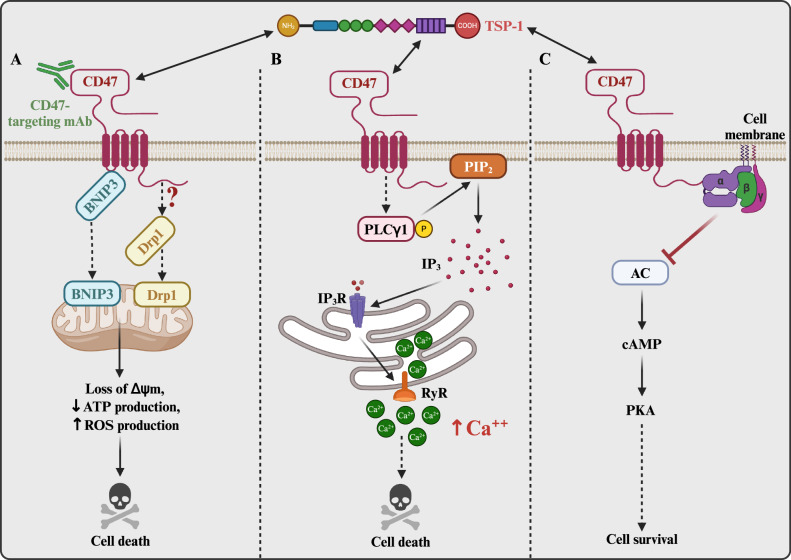
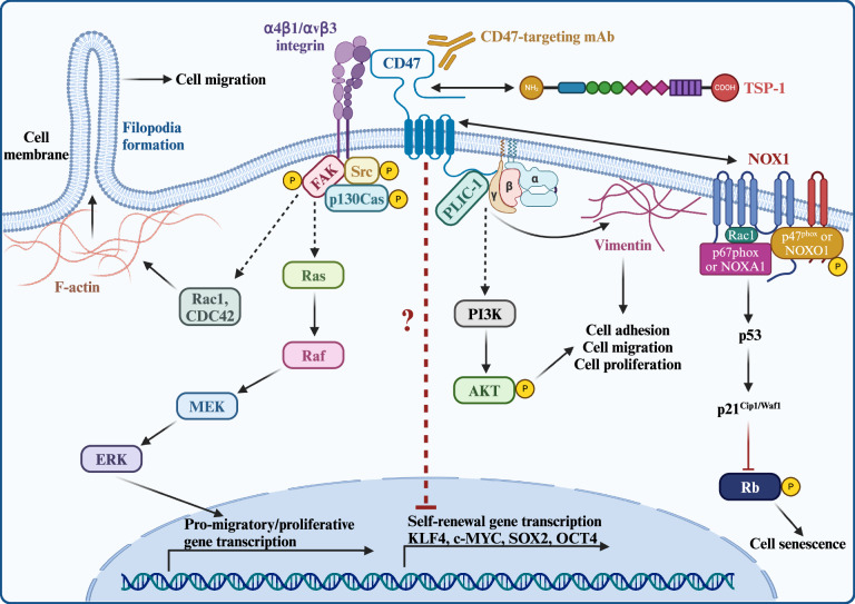
CD47 is a ubiquitously expressed cell surface receptor, which is widely known for preventing macrophage-mediated phagocytosis by interacting with signal regulatory protein α (SIRPα) on the surface of macrophages. In addition to its role in phagocytosis, emerging studies have reported numerous noncanonical functions of CD47 that include regulation of various cellular processes such as proliferation, migration, apoptosis, differentiation, stress responses, and metabolism. Despite lacking an extensive cytoplasmic signaling domain, CD47 binds to several cytoplasmic proteins, particularly upon engaging with its secreted matricellular ligand, thrombospondin 1. Indeed, the regulatory functions of CD47 are greatly influenced by its interacting partners. These interactions are often cell- and context-specific, adding a further level of complexity. This review addresses the downstream cell-intrinsic signaling pathways regulated by CD47 in various cell types and environments. Some of the key pathways modulated by this receptor include the PI3K/AKT, MAPK/ERK, and nitric oxide signaling pathways, as well as those implicated in glucose, lipid, and mitochondrial metabolism. These pathways play vital roles in maintaining tissue homeostasis, highlighting the importance of understanding the phagocytosis-independent functions of CD47. Given that CD47 expression is dysregulated in a variety of cancers, improving our understanding of the cell-intrinsic signals regulated by this molecule will help advance the development of CD47-targeted therapies.


Conflict of interest statement

The authors declare no competing interests.

Figures

Fig. 1
Fig. 1. CD47 regulates cellular stress response pathways.
A Under cellular stress, depleting CD47 triggers activation of gene expression for beclin-1 and autophagy-related genes ATG5 and ATG7. The upregulation of beclin-1, ATG5, and ATG7 enhances autophagic flux by increasing the expression of LC3, which is essential for forming the autophagosome membrane. Consequently, unwanted cellular components are targeted and degraded within autophagosomes, ultimately promoting cell survival. B TSP1 binding with CD47 disrupts constitutive association between CD47 and VEGFR2 on endothelial cells, effectively blocking VEGFR2 induced PI3K/AKT-mediated activation of eNOS and subsequent induction of NO/cGMP signaling and other signaling pathways in favor of cell survival. Additionally, TSP1 disrupts CD47 interaction with caveolin-1 on endothelial cells to enhance reactive oxygen species (ROS) production via eNOS which can also contribute to NO/cGMP signaling to enhance cell survival. C TSP1-CD47 engagement activates NOX1 through p47phox phosphorylation, resulting in ROS production and cell death. This figure was created using BioRender.com.
Fig. 2
Fig. 2. CD47-mediated regulation of cell death.
A CD47 binds to intracellular BNIP3, an interaction disrupted following CD47 ligation with TSP1 or anti-CD47 targeting antibody. Following dissociation, BNIP3 translocates to the mitochondria, inducing depolarization, ultimately triggering cell death. Concurrently, TSP1-CD47 interaction prompts DRP1 translocation to the mitochondria, inducing further mitochondrial destabilization. B TSP1 binds to CD47 leading to sustained activation of PLCγ1, which then cleaves phosphatidylinositol 4,5-bisphosphate (PIP2) into inositol 1,4,5-trisphosphate (IP3). IP3 binds to its receptors (IP3R) on the endoplasmic reticulum (ER), prompting the release of Ca2+. This release activates the ER ryanodine receptors (RyR), further triggering calcium release from the ER into the cytoplasm. Calcium overload in the cell causes mitochondrial damage, ultimately leading to cell death. C CD47 and heterotrimeric Gi protein (α, β, γ) interaction activates Gi signaling, suppressing adenyl cyclase (AC), reducing cAMP levels, and diminishing PKA activity, which results in cell death. This figure was created using BioRender.com.
Fig. 3
Fig. 3. Mechanisms underlying CD47-regulated cellular plasticity.
From left to right: CD47 associates with integrins α4β1 or ανβ3, inducing the assembly and activation of the focal adhesion complex (FAC). The FAC, composed of Src, focal adhesion kinase (FAK), and p130Cas, in turn stimulates Rac1 and CDC42 and/or MEK/ERK signaling, promoting cell migration and proliferation via increased F-actin expression and filopodia formation or the induction of gene expression, respectively. Upon CD47 activation by TSP1, CD47 associates with intracellular PLIC-1, which tethers vimentin filaments to the cell membrane, facilitating integrin-ανβ3-mediated cell spreading. PLIC-1 also forms a complex with the Gβγ dimer, stimulating PI3K/AKT signaling to promote cell motility and proliferation. TSP1-CD47 signaling further regulates cell self-renewal by downregulating SOX2, OCT4, KLF4, and c-MYC expression. TSP1 binding to CD47 also activates NOX1, inducing reactive oxygen species (ROS) generation and initiating a p53-mediated DNA damage response. This leads to p21Cip1/Waf1 upregulation and the subsequent hypophosphorylation of retinoblastoma protein (Rb), ultimately resulting in cell senescence. This figure was created using BioRender.com.
Fig. 4
Fig. 4. CD47 regulates metabolic remodeling.
From left to right: Loss of CD47 induces the expression of PGC-1α, enhancing mitochondrial biogenesis and function, consequently increasing oxidative phosphorylation (OXPHOS). Additionally, CD47 dynamically modulates the expression of PPARα and SIRT1, regulating fatty acid synthesis in response to varying dietary conditions. Through the transcriptional downregulation of CPT1b, CD47 also suppresses fatty acid oxidation. Moreover, CD47 interacts with Enolase 1 (ENO1) to inhibit its FBXW7-mediated degradation. The CD47-ENO1 interaction activates ERK signaling and promotes glycolysis in favor of tumor growth and metastasis. This figure was created using BioRender.com.
Fig. 5
Fig. 5. Overview of canonical versus noncanonical CD47 signaling.
This figure was created using BioRender.com.

References

    1. Rebres RA, Vaz LE, Green JM, Brown EJ. Normal ligand binding and signaling by CD47 (integrin-associated protein) requires a long range disulfide bond between the extracellular and membrane-spanning domains. J Biol Chem. 2001;276:34607–16. - PubMed
    1. Fenalti G, Villanueva N, Griffith M, Pagarigan B, Lakkaraju SK, Huang RYC, et al. Structure of the human marker of self 5-transmembrane receptor CD47. Nat Commun. 2021;12:5218. - PMC - PubMed
    1. Mushegian A. Refining structural and functional predictions for secretasome components by comparative sequence analysis. Proteins. 2002;47:69–74. - PubMed
    1. Reinhold MI, Lindberg FP, Plas D, Reynolds S, Peters MG, Brown EJ. In vivo expression of alternatively spliced forms of integrin-associated protein (CD47). J Cell Sci. 1995;108:3419–25. - PubMed
    1. Lee EH, Hsieh YP, Yang CL, Tsai KJ, Liu CH. Induction of integrin-associated protein (IAP) mRNA expression during memory consolidation in rat hippocampus. Eur J Neurosci. 2000;12:1105–12. - PubMed

LinkOut - more resources