. 2024 Dec;20(12):1680-1690.
doi: 10.1038/s41589-024-01685-3. Epub 2024 Jul 22.

Macrolones target bacterial ribosomes and DNA gyrase and can evade resistance mechanisms

Affiliations

Macrolones target bacterial ribosomes and DNA gyrase and can evade resistance mechanisms

Elena V Aleksandrova et al. Nat Chem Biol. 2024 Dec.

Abstract

Growing resistance toward ribosome-targeting macrolide antibiotics has limited their clinical utility and urged the search for superior compounds. Macrolones are synthetic macrolide derivatives with a quinolone side chain, structurally similar to DNA topoisomerase-targeting fluoroquinolones. While macrolones show enhanced activity, their modes of action have remained unknown. Here, we present the first structures of ribosome-bound macrolones, showing that the macrolide part occupies the macrolide-binding site in the ribosomal exit tunnel, whereas the quinolone moiety establishes new interactions with the tunnel. Macrolones efficiently inhibit both the ribosome and DNA topoisomerase in vitro. However, in the cell, they target either the ribosome or DNA gyrase or concurrently both of them. In contrast to macrolide or fluoroquinolone antibiotics alone, dual-targeting macrolones are less prone to select resistant bacteria carrying target-site mutations or to activate inducible macrolide resistance genes. Furthermore, because some macrolones engage Erm-modified ribosomes, they retain activity even against strains with constitutive erm resistance genes.


Conflict of interest statement

Competing interests: The authors declare no competing interests.

Figures

Extended Data Fig. 1
Extended Data Fig. 1. Inhibition of translation and ribosome binding of macrolones.
(a) Inhibition of the production of the green fluorescent protein (GFP) in a cell-free translation system by varying concentrations of macro lines relative to uninhibited reaction. Shown are the results of two independent experiments. (b) Competitive binding of [14C]-ERY and macrolones to the E. coli ribosome. Unlabeled ERY was used as a control (black circles). Experimental details are presented in the Online Methods section. Shown are the results of two independent experiments.
Extended Data Fig. 2
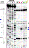
Extended Data Fig. 2. Effects of macrolones on in vitro translation.
Mapping the sites of macrolone-mediated ribosome arrests (blue arrows) at the early codons of the model ORF derived from the E. coli yrbA gene. The classic macrolide ERY is included for comparison. Due to the presence of the Thr-RS inhibitor borrelidin, the ribosomes that did not stall at the early codons are eventually trapped at the Gln12 codon when Thr13 needs to be incorporated into the growing protein (grey arrow). The AUG start codon is marked with a black arrow. The sample labeled as ‘NONE’ contained only borrelidin but no ERY or macrolones. Amino acid and nucleotide sequences of yrbA gene are shown on the left. Sequencing lanes are marked as C, U, A, G. This experiment was repeated independently twice and produced similar results.
Extended Data Fig. 3
Extended Data Fig. 3. Electron density maps of ribosome-bound MCX-66, MCX-91, and MCX-128.
(a-c) 2Fo-Fc Fourier electron density maps of MCX-66 (a, magenta), MCX-91 (b, green), and MCX-128 (c, yellow) in complex with the T. thermophilus 70S ribosome (blue mesh) shown in two mutually perpendicular views. The refined models of ribosome-bound MCX compounds are displayed in their respective electron density maps after the refinement contoured at 1.0σ. Carbon atoms are colored magenta (MCX-66), green (MCX-91), or yellow (MCX-128); nitrogen atoms are blue; oxygen atoms are red; fluorine atoms are dark green. Note that the locations of fluoroquinolone side chains can be unambiguously determined from the obtained electron density maps.
Extended Data Fig. 4
Extended Data Fig. 4. Comparison of the structures of ribosome-bound macrolones, macrolide erythromycin and tetracenomycin X.
(a, b) Superposition of the structure of ribosome-bound MCX-66 (magenta), MCX-91 (green), MCX-128 (yellow) with the previous structures of ERY (red, PDB entry 6XHX ref. 15), or TcmX (blue, PDB entry 7ZTA ref. 75). All structures were aligned based on domain V of the 23S rRNA.
Extended Data Fig. 5
Extended Data Fig. 5. Structure of ciprofloxacin (CIP) in complex with the 70S ribosome.
(a) 2Fo-Fc Fourier electron density map of ciprofloxacin (CIP, greencyan) in complex with the T. thermophilus 70S ribosome (blue mesh). The refined model of ribosome-bound CIP is displayed in its respective electron density map after the refinement contoured at 1.0σ. Carbon atoms are colored greencyan; nitrogen atoms are blue; oxygen atoms are red; fluorine atom is dark green. (b, c) Close-up views of CIP bound in the NPET of the 70S ribosome, highlighting its stacking (red arrows) interactions with the nucleotides of the 23S rRNA. (d) Superposition of the structures of ribosome-bound CIP and MCX-128 (yellow). The structures were aligned based on domain V of the 23S rRNA.
Extended Data Fig. 6
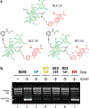
Extended Data Fig. 6. Macrolones inhibit DNA gyrase activity in vitro.
(a) Chemical structures of the macrolones used in the assay. (b) Effect of macrolones on the activity of DNA gyrase. Supercoiled or relaxed plasmid DNA bands are marked on the left. CIP and ERY were used as positive and negative controls, respectively. The sample labeled as ‘NONE’ contained no antibiotics. A control sample where gyrase was not added is marked as ‘-’. The experiment was repeated independently and produced similar results.
Extended Data Fig. 7
Extended Data Fig. 7. Electron density maps of A2058 nucleotide in Erm-modified and wild-type T. thermophilus 70S ribosome.
(a, c) Unbiased Fo-Fc (grey and green mesh) and (b, d) 2Fo-Fc (blue mesh) electron difference Fourier maps of nucleotide A2058 in the T. thermophilus 70S ribosome contoured at 3.0σ and 1.0σ, respectively. Grey mesh shows the Fo-Fc map after refinement with the entire modified nucleotide omitted. Green mesh, reflecting the presence of the two methyl groups at N6 position of the nucleobase, shows the Fo-Fc electron density map after refinement with the nucleotide A2058 built as a regular unmethylated adenine. The refined models of Erm-modified N6-dimethylated (a, b) or wild-type unmodified (c, d) nucleotide A2058 are displayed in the corresponding electron density maps. Both structures carry MCX-128 compound (not shown). Carbon atoms are colored dark blue for the Erm-modified A2058 and light blue for the unmodified A2058; nitrogens are dark blue; oxygens are red.
Figure 1 |
Figure 1 |. Macrolones inhibit DNA gyrase and protein synthesis in vivo.
(a) Chemical structures of the classic macrolide erythromycin (ERY), fluoroquinolone ciprofloxacin (CIP), and the macrolone antibiotics used in this study. (b) In vivo testing of macrolone activity using E. coli BWDK strain carrying the pDualrep2 plasmid. The expression of the red fluorescent protein (RFP, Cy3 channel) is triggered by activation of SOS response caused by inhibition of DNA toposiomerases; expression of Katushka2S protein (Cy5 channel) is activated in response to ribosome stalling. ERY and CIP, inhibiting translation and DNA topoisomerases, respectively, were used as positive controls. In the merged image, the RFP fluorescence is shown in red pseudocolor, and that of Katushka2S in green pseudocolor.
Figure 2 |
Figure 2 |. Macrolones inhibit protein synthesis and DNA gyrase in vitro.
(a) Inhibition of protein synthesis by macrolones in the cell-free transcription-translation system (see also Extended Data Fig. 1a). ERY and CIP were included as controls. All the inhibitors were present at 25 μM. The percentage of GFP fluorescence is shown relative to that obtained in a reaction devoid of antibiotics at the 3-hour timepoint. The variance from the mean of two independent experiments is too small to be represented in the plot by error bars (see source data). (b) Primer extension inhibition (toe-printing) analysis of the sites of macrolone-induced ribosome stalling during translation of the synthetic gene rst1 (blue arrows). The sites of ERY-induced arrest are indicated by red arrows. Sequencing lanes are labeled as C, U, A, G. Nucleotide sequences of the rst1 mRNA and the corresponding amino acid sequence of the encoded protein are shown on the left. (c) DNA gyrase-mediated plasmid supercoiling in the absence (NONE) or presence of macrolones. Bands corresponding to supercoiled or relaxed plasmid are indicated. A control sample where gyrase was not added is marked as “-“. CIP and ERY were used as positive and negative controls, respectively. Experiments shown in panels b and c were repeated independently and produced similar results.
Figure 3 |
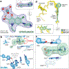
Figure 3 |. Structures of MCX-66, MCX-91, and MCX-128 in complex with the wild-type T. thermophilus 70S ribosome.
(a) Overview of the binding sites for MCX-66 (light teal), MCX-91 (green), and MCX-128 (yellow) in the 70S ribosome, viewed as a cross-cut section through the nascent peptide exit tunnel (NPET). The 30S subunit is shown in light yellow, the 50S subunit in light blue, the mRNA in blue, and the A-, P-, and E-site tRNAs are colored teal, navy blue, and orange, respectively. (b) Close-up view of MCX-128 bound in the NPET, highlighting the characteristic interactions with the two opposite walls of the NPET: one with the surface formed by nucleotides A2058 and A2059 of the 23S rRNA (E. coli numbering) and the second one with the non-Watson-Crick base pair C1782:C2586,. (c) Occlusion of the nascent peptide exit tunnel by MCX-128 viewed from the wide-open part of the tunnel onto the PTC, as indicated by the dashed arrow. Locations of the A and P sites of the PTC are labeled. (d-f) Close-up views of MCX-66/91/128 in the NPET of the 70S ribosome, highlighting their H-bond (dashed lines) and stacking (red arrows) interactions with the nucleotides of the 23S rRNA.
Figure 4 |
Figure 4 |. Identification of cellular targets of macrolones by selecting resistant mutants.
(top) Selection of the drug-resistant mutants of the antibiotic-hypersusceptible E. coli strain SQ110DTC on LB-agar plates with 3–5x MIC of antibiotics. The mutations identified in the sequenced resistant clones are shown underneath the plates. (bottom) Selection of the MCX-128 resistant mutants using the SQ110DTC strains already carrying resistance mutations either in the DNA gyrase or in the 23S rRNA genes. The primary mutations in the strains used for mutant selection and the secondary mutations found in clones that grew on the plates with 3x MIC of MCX-128 are shown.
Figure 5 |
Figure 5 |. Macrolones are poor inducers of the expression of inducible macrolide resistance genes.
(a) Schematics of inducible expression of the ermC resistant gene by macrolide-induced programmed translation arrest within the ermCL leader ORF. (b, c) Induction by macrolones of the RFP reporter gene controlled by either the regulatory ErmCL ORF (the pErmCL-RFP construct) or by a hybrid ErmAL-ErmCL ORF (pErmAL/CL-RFP). Macrolones, or the inducing macrolide ERY positive control were spotted on E. coli BWDK cells transformed with the plasmids carrying the respective constructs. (d-g) In vitro toe-printing assay mapping the site of ERY- or macrolone-induced ribosome arrest within the leader ORFs ermAL (d), ermBL (e), ermCL (f), and ermDL (g) of the respective inducible resistance genes. The arrows indicate the sites of the programmed translation arrest required for activation of expression of resistance. Samples labeled as “NONE” contained no antibiotics. Sequencing reactions are marked as C, U, A, G. Experiments shown in panels b-g were repeated independently and produced similar results.
Figure 6 |
Figure 6 |. Structure of macrolone MCX-128 bound to the Erm-methylated 70S ribosome.
(a, b) 2Fo-Fc electron density maps (blue mesh) contoured at 1.0σ of MCX-128 in complex with Erm-modified (a, teal) or wild-type (b, yellow) T. thermophilus 70S ribosomes. The N6-methyl groups of mA205826 are highlighted in orange. Carbon atoms are colored dark blue for the Erm-modified mA205826(a) and blue for the wild-type A2058 (b) nucleotides; nitrogens are dark blue; oxygens are red; sulfurs are yellow. (c, d) Superposition of MCX-128 (yellow) in complex with the WT 70S ribosome containing an unmodified residue A2058 (blue) and the structure of MCX-128 (teal) in complex with the Erm-modified 70S ribosome containing an mA205826 residue (dark blue). Hydrogen bonds are depicted with dashed lines. Note that the position of MCX-128 is almost identical in the two structures.

References

    1. Fernandes P Use of antibiotic core structures to generate new and useful macrolide antibiotics. in Antibiotics Current Innovations and Future Trends (eds. Sánchez S & Demain AL) (Caister Academic Press, Norfolk, UK, 2015).
    1. Bush NG, Diez-Santos I, Abbott LR & Maxwell A Quinolones: mechanism, lethality and their contributions to antibiotic resistance. Molecules 25(2020). - PMC - PubMed
    1. Agouridas C et al. Synthesis and antibacterial activity of ketolides (6-O-methyl-3-oxoerythromycin derivatives): a new class of antibacterials highly potent against macrolide-resistant and -susceptible respiratory pathogens. J. Med. Chem 41, 4080–4100 (1998). - PubMed
    1. Seiple IB et al. A platform for the discovery of new macrolide antibiotics. Nature 533, 338–345 (2016). - PMC - PubMed
    1. Pavlovic D, Fajdetic A & Mutak S Novel hybrids of 15-membered 8a- and 9a-azahomoerythromycin A ketolides and quinolones as potent antibacterials. Bioorg. Med. Chem 18, 8566–8582 (2010). - PubMed
REFERENCES FOR THE ONLINE METHODS
    1. Baba T et al. Construction of Escherichia coli K-12 in-frame, single-gene knockout mutants: the Keio collection. Mol. Syst. Biol 2, 2006.0008 (2006). - PMC - PubMed
    1. Datsenko KA & Wanner BL One-step inactivation of chromosomal genes in Escherichia coli K-12 using PCR products. Proc. Natl. Acad. Sci. USA 97, 6640–6645 (2000). - PMC - PubMed
    1. Bundy BC & Swartz JR Site-specific incorporation of p-propargyloxyphenylalanine in a cell-free environment for direct protein-protein click conjugation. Bioconjug. Chem 21, 255–63 (2010). - PubMed
    1. Svetlov MS, Vazquez-Laslop N & Mankin AS Kinetics of drug-ribosome interactions defines the cidality of macrolide antibiotics. Proc. Natl. Acad. Sci. USA 114, 13673–13678 (2017). - PMC - PubMed
    1. Cheng Y & Prusoff WH Relationship between the inhibition constant (K1) and the concentration of inhibitor which causes 50 per cent inhibition (I50) of an enzymatic reaction. Biochem. Pharmacol 22, 3099–3108 (1973). - PubMed

MeSH terms

LinkOut - more resources