. 2024 Aug;56(8):1644-1653.
doi: 10.1038/s41588-024-01836-1. Epub 2024 Jul 22.

Next-generation phenotyping integrated in a national framework for patients with ultrarare disorders improves genetic diagnostics and yields new molecular findings

Axel Schmidt #  1 Magdalena Danyel #  2   3 Kathrin Grundmann #  4 Theresa Brunet #  5 Hannah Klinkhammer  6   7 Tzung-Chien Hsieh  6 Hartmut Engels  1 Sophia Peters  1 Alexej Knaus  6 Shahida Moosa  8 Luisa Averdunk  9 Felix Boschann  2   3 Henrike Lisa Sczakiel  2   3 Sarina Schwartzmann  2 Martin Atta Mensah  2   3 Jean Tori Pantel  2   10 Manuel Holtgrewe  11 Annemarie Bösch  12 Claudia Weiß  12 Natalie Weinhold  12 Aude-Annick Suter  12 Corinna Stoltenburg  12 Julia Neugebauer  12 Tillmann Kallinich  12 Angela M Kaindl  13   14   15 Susanne Holzhauer  12 Christoph Bührer  12 Philip Bufler  12 Uwe Kornak  2 Claus-Eric Ott  2 Markus Schülke  2 Hoa Huu Phuc Nguyen  16 Sabine Hoffjan  16 Corinna Grasemann  17 Tobias Rothoeft  17 Folke Brinkmann  17 Nora Matar  17 Sugirthan Sivalingam  1 Claudia Perne  1 Elisabeth Mangold  1 Martina Kreiss  1 Kirsten Cremer  1 Regina C Betz  1 Martin Mücke  18 Lorenz Grigull  18 Thomas Klockgether  19 Isabel Spier  1 André Heimbach  1 Tim Bender  18 Fabian Brand  6 Christiane Stieber  18 Alexandra Marzena Morawiec  18 Pantelis Karakostas  20 Valentin S Schäfer  20 Sarah Bernsen  18 Patrick Weydt  19 Sergio Castro-Gomez  19 Ahmad Aziz  19 Marcus Grobe-Einsler  19 Okka Kimmich  19 Xenia Kobeleva  19 Demet Önder  19 Hellen Lesmann  1 Sheetal Kumar  1 Pawel Tacik  19 Meghna Ahuja Bhasin  6 Pietro Incardona  6 Min Ae Lee-Kirsch  21   22 Reinhard Berner  21   22 Catharina Schuetz  21   22 Julia Körholz  21   22 Tanita Kretschmer  21   22 Nataliya Di Donato  21   23 Evelin Schröck  21   23 André Heinen  21   22 Ulrike Reuner  21   24 Amalia-Mihaela Hanßke  21 Frank J Kaiser  25 Eva Manka  26 Martin Munteanu  25 Alma Kuechler  25 Kiewert Cordula  26 Raphael Hirtz  26 Elena Schlapakow  27 Christian Schlein  28 Jasmin Lisfeld  28 Christian Kubisch  28   29 Theresia Herget  28 Maja Hempel  28   29   30 Christina Weiler-Normann  29   31 Kurt Ullrich  29 Christoph Schramm  29   31 Cornelia Rudolph  29 Franziska Rillig  29 Maximilian Groffmann  29 Ania Muntau  32 Alexandra Tibelius  30 Eva M C Schwaibold  30 Christian P Schaaf  30 Michal Zawada  30 Lilian Kaufmann  30 Katrin Hinderhofer  30 Pamela M Okun  33 Urania Kotzaeridou  33 Georg F Hoffmann  33 Daniela Choukair  33 Markus Bettendorf  33 Malte Spielmann  34 Annekatrin Ripke  35 Martje Pauly  36   37 Alexander Münchau  35   38 Katja Lohmann  39 Irina Hüning  34 Britta Hanker  40 Tobias Bäumer  35   38 Rebecca Herzog  35   36 Yorck Hellenbroich  41 Dominik S Westphal  5 Tim Strom  5 Reka Kovacs  5 Korbinian M Riedhammer  5   42 Katharina Mayerhanser  5 Elisabeth Graf  5 Melanie Brugger  5 Julia Hoefele  5 Konrad Oexle  43 Nazanin Mirza-Schreiber  43 Riccardo Berutti  43 Ulrich Schatz  5 Martin Krenn  5   44 Christine Makowski  45 Heike Weigand  46 Sebastian Schröder  46 Meino Rohlfs  46 Katharina Vill  46 Fabian Hauck  46 Ingo Borggraefe  46 Wolfgang Müller-Felber  46 Ingo Kurth  10 Miriam Elbracht  10 Cordula Knopp  10 Matthias Begemann  10 Florian Kraft  10 Johannes R Lemke  47   48 Julia Hentschel  47 Konrad Platzer  47 Vincent Strehlow  47 Rami Abou Jamra  47 Martin Kehrer  4 German Demidov  4 Stefanie Beck-Wödl  4 Holm Graessner  49 Marc Sturm  4 Lena Zeltner  49 Ludger J Schöls  50 Janine Magg  49 Andrea Bevot  51 Christiane Kehrer  51 Nadja Kaiser  51 Ernest Turro  52 Denise Horn  2 Annette Grüters-Kieslich  53 Christoph Klein  46 Stefan Mundlos  2 Markus Nöthen  1 Olaf Riess  4 Thomas Meitinger  5 Heiko Krude  53 Peter M Krawitz  54 Tobias Haack  4 Nadja Ehmke  2   3 Matias Wagner  5   43   46
Affiliations

Next-generation phenotyping integrated in a national framework for patients with ultrarare disorders improves genetic diagnostics and yields new molecular findings

Axel Schmidt et al. Nat Genet. 2024 Aug.

Erratum in

  • Author Correction: Next-generation phenotyping integrated in a national framework for patients with ultrarare disorders improves genetic diagnostics and yields new molecular findings.
    Schmidt A, Danyel M, Grundmann K, Brunet T, Klinkhammer H, Hsieh TC, Engels H, Peters S, Knaus A, Moosa S, Averdunk L, Boschann F, Sczakiel HL, Schwartzmann S, Mensah MA, Pantel JT, Holtgrewe M, Bösch A, Weiß C, Weinhold N, Suter AA, Stoltenburg C, Neugebauer J, Kallinich T, Kaindl AM, Holzhauer S, Bührer C, Bufler P, Kornak U, Ott CE, Schülke M, Nguyen HHP, Hoffjan S, Grasemann C, Rothoeft T, Brinkmann F, Matar N, Sivalingam S, Perne C, Mangold E, Kreiss M, Cremer K, Betz RC, Mücke M, Grigull L, Klockgether T, Spier I, Heimbach A, Bender T, Brand F, Stieber C, Morawiec AM, Karakostas P, Schäfer VS, Bernsen S, Weydt P, Castro-Gomez S, Aziz A, Grobe-Einsler M, Kimmich O, Kobeleva X, Önder D, Lesmann H, Kumar S, Tacik P, Bhasin MA, Incardona P, Lee-Kirsch MA, Berner R, Schuetz C, Körholz J, Kretschmer T, Di Donato N, Schröck E, Heinen A, Reuner U, Hanßke AM, Kaiser FJ, Manka E, Munteanu M, Kuechler A, Cordula K, Hirtz R, Schlapakow E, Schlein C, Lisfeld J, Kubisch C, Herget T, Hempel M, Weiler-Normann C, Ullrich K, Schramm C, Rudolph C, Rillig F, Groffmann M, Muntau A, Tibelius A, Schwaibold EMC, Schaaf CP, Zawada M, Kaufmann L, Hinderhofer K, Okun PM, Kotzaeridou U, Hoffmann GF, Ch… See abstract for full author list ➔ Schmidt A, et al. Nat Genet. 2025 Jul;57(7):1790-1791. doi: 10.1038/s41588-025-02271-6. Nat Genet. 2025. PMID: 40555819 Free PMC article. No abstract available.

Abstract

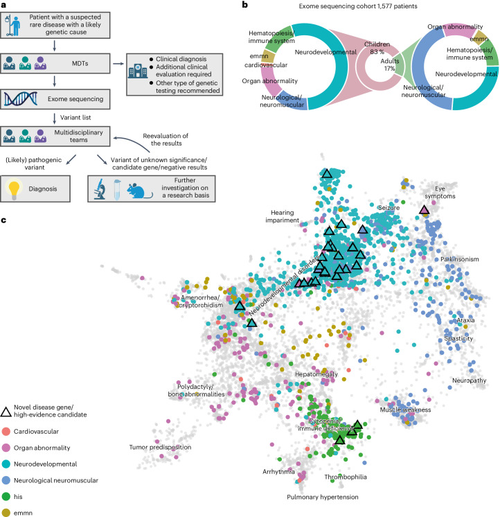
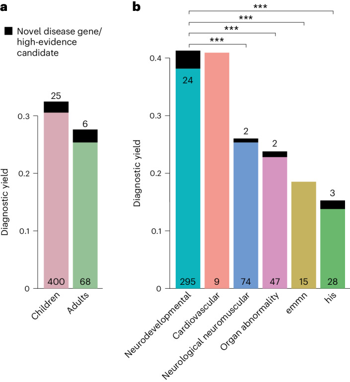
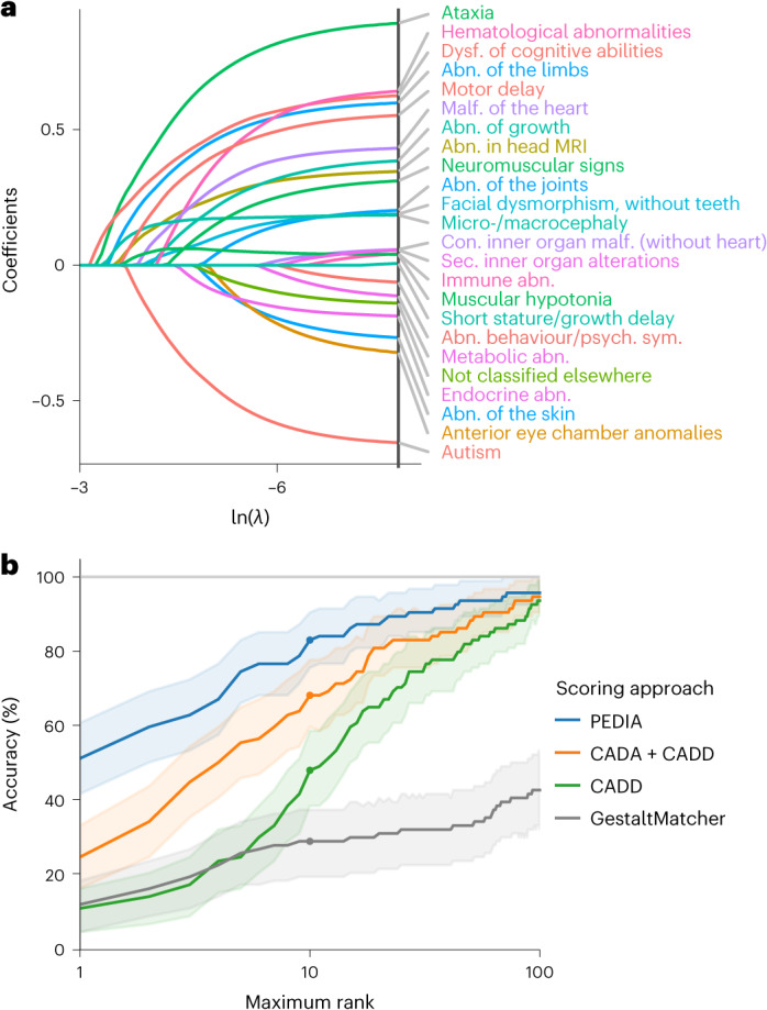
Individuals with ultrarare disorders pose a structural challenge for healthcare systems since expert clinical knowledge is required to establish diagnoses. In TRANSLATE NAMSE, a 3-year prospective study, we evaluated a novel diagnostic concept based on multidisciplinary expertise in Germany. Here we present the systematic investigation of the phenotypic and molecular genetic data of 1,577 patients who had undergone exome sequencing and were partially analyzed with next-generation phenotyping approaches. Molecular genetic diagnoses were established in 32% of the patients totaling 370 distinct molecular genetic causes, most with prevalence below 1:50,000. During the diagnostic process, 34 novel and 23 candidate genotype-phenotype associations were identified, mainly in individuals with neurodevelopmental disorders. Sequencing data of the subcohort that consented to computer-assisted analysis of their facial images with GestaltMatcher could be prioritized more efficiently compared with approaches based solely on clinical features and molecular scores. Our study demonstrates the synergy of using next-generation sequencing and phenotyping for diagnosing ultrarare diseases in routine healthcare and discovering novel etiologies by multidisciplinary teams.


Conflict of interest statement

V.S.S. has received consultant fees from Novartis, Chugai, AbbVie, Celgene, Sanofi, Lilly, Hexal, Pfizer, Amgen, BMS, Roche, Gilead, Medac, Boehringer-Ingelheim and Alexion and speaker’s bureau fees from AbbVie, Novartis, BMS, Chugai, Celgene, Medac, Sanofi, Lilly, Hexal, Pfizer, Janssen, Roche, Schire, Onkowissen, Royal College London, Boehringer-Ingelheim and UCB Fresenius. M.G.-E. has received research support from the German Ministry of Education and Research (BMBF) within the European Joint Program for Rare Diseases (EJP-RD) 2021 Transnational Call for Rare Disease Research Projects (funding number 01GM2110), from the National Ataxia Foundation (NAF) and from Ataxia UK and received consulting fees from Healthcare Manufaktur, Germany, all unrelated to this study. All other authors declare no competing interests.

Figures

Fig. 1
Fig. 1. Workflow in the TRANSLATE NAMSE project and phenotypes in which exome sequencing was performed.
a, Patients with a suspected rare disease were referred to a MDT and deeply phenotyped using HPO terminology. If a genetic etiology was considered likely, exome sequencing was performed. The MDT then evaluated the molecular findings and could order additional analyses for variants of uncertain significance or variants in potentially novel disease candidate genes (created with BioRender.com). b, Exome sequencing was performed predominantly in children. The main indications for exome sequencing in children were neurodevelopmental disorders. In adults, the main indications were neurological/neuromuscular disorders. In both children and adults, the least common disease categories were ‘cardiovascular’, ‘endocrine, metabolic, mitochondrial, nutritional’ (emmn) and ‘hematopoiesis/immune system’ (his). c, Phenotypic similarities between patients, as encoded according to their HPO terms, were visualized with UMAP. As reference, all OMIM diseases were included using their HPO annotations (gray background dots). For each patient, color coding indicates allocation to disease groups, in accordance with the leading clinical feature. An overlap is evident for patients in the neurodevelopmental and neuromuscular groups (aquamarine and blue clusters), which indicates high phenotypic similarity. This precludes the unequivocal assignment of these patients to a diagnostic group. The triangles indicate patients who contributed to the identification of a novel, high-evidence gene–phenotype association.
Fig. 2
Fig. 2. Diagnostic yield of exome sequencing depends on age and disease group.
a,b, The diagnostic yield differed according to age group (adult/child) (a) and disease category (b). For all disease categories, with the exception of cardiovascular, the diagnostic yield was increased by novel DGGs and high-evidence candidate genes (dark-colored tip of the bar). The absolute number of solved cases in which a variant was found in an established disease gene is given at the bottom of each bar, and the number of solved cases attributable to a novel DGG or high-evidence candidate gene is given at the top of each bar. The entire TRANSLATE NAMSE exome sequencing cohort was considered for a and b (n = 1,577). Diagnostic yield between disease categories were compared using two-sided Fisher’s exact test. P values were adjusted by Bonferroni correction. ***P < 0.001; exact corrected P values: neurodevelopmental (ndd) versus neurologic neuromuscular P = 5.4 × 10−5, ndd versus organ abnormality P = 5.2 × 10−5, ndd versus emmn P = 5.9 × 10−4, ndd versus his P = 1.1 × 10−11. emnn, endocrine, metabolic, mitochondrial, nutritional; his, hematopoiesis/immune system.
Fig. 3
Fig. 3. Mode of inheritance and disease burden are dependent on autozygosity.
a, Pie chart showing the distribution of modes of inheritance (MOI) for all diagnoses (n = 510). Most disease-causing variants occurred de novo and on an autosome. At least 75% of all autosomal recessive diagnoses could have been identified by expanded carrier screening (slice). b, Box plots of autozygosity for each MOI (n = 375). Individuals are indicated by gray dots. Autozygosity was substantially increased in individuals with autosomal recessive disorders due to homozygous variants. In the box plots, the center lines indicate the median values, and the bottom and top edges of the boxes are the first (25%) and the third (75%) quartiles. The whiskers extend to the minimal and maximal data points with a maximum distance of 1.5 interquartile ranges from the edges of the box. c, Bar graphs illustrating MOI in individuals with low (<2%, n = 313) and high (>2%, n = 62) autozygosity. On the right, the autosomal dominant de novo rate has been used for normalization. Individuals with high autozygosity had a higher relative burden of recessive diseases, mainly due to the presence of homozygous pathogenic variants. The box plots present the median as the center line, the upper and lower quartiles as box limits, and 1.5× the interquartile range as the whisker length (in the style of Tukey). AD, autosomal dominant inheritance, variant inherited or of unknown origin; AD (de novo), autosomal dominant inheritance with de novo variant; AR (comp het), autosomal recessive inheritance with compound heterozygous variants; AR (hom), autosomal recessive inheritance with homozygous variant; mt, mitochondrial inheritance; XL, X-linked inheritance.
Fig. 4
Fig. 4. Most variants identified in TRANSLATE NAMSE exome sequencing cohort cause ultrarare disorders that were first associated with a gene in the last decade.
a, Comparison of the number of (likely) pathogenic variants per gene in TRANSLATE NAMSE relative to the frequency of submission of (likely) pathogenic variants to ClinVar. Genes are ordered from left to right according to a decreasing frequency of ClinVar submissions. The black line corresponds to the complementary cumulative distribution (1 − CDF; cumulative distribution function) of ClinVar submissions. Diagnostic variants in TRANSLATE NAMSE (counts displayed on the right axis) were plotted as dots above their respective gene and in the color corresponding to the year in which the gene was first described as being associated with the respective disease. b, Variant counts in TRANSLATE NAMSE in genes with high (first quartile, Q1) to low (Q4) counts of submissions per gene in ClinVar. The genes in Q1–Q4 each cover approximately 1/4 of the submissions of likely or confirmed pathogenic variants to ClinVar, as shown on the x axis in a. Variants in the same gene are grouped in horizontal blocks. c, Bar graph showing the number of variants relative to the time interval in which the gene was first described as being associated with the respective disease. Note that 59 genes listed in the recommendations for reporting of secondary findings (version 2) of the ACMG were excluded from the analyses to counteract potential biases in ClinVar due to submissions of secondary findings. TNAMSE, TRANSLATE NAMSE; vars, variants.
Fig. 5
Fig. 5. Machine learning identifies features relevant to the diagnostic yield and can support variant prioritization.
a, The coefficient paths of regression analysis using the LASSO are shown. Only features that are included in the final model and are present in at least 5% of the cases that were used for training are depicted. The more to the left [lower ln(λ)] a coefficient path starts to deviate from the x axis, the more informative the corresponding feature is in terms of predicting the diagnostic yield. Features with positive coefficients increase the diagnostic yield. In contrast, features with negative coefficients render a monogenic cause less likely. For example, dysfunction of higher cognitive abilities and ataxia are associated with a higher diagnostic yield (clinical features are colored according to their higher-order HPO groups; for details, see Supplementary Note). An algorithm to predict the diagnostic yield (YieldPred) was developed on the basis of these data and can be found online (https://translate-namse.de). b, The performances of variant prioritization approaches were compared. All disease-associated genes were ranked using the respective variant prioritization method. Subsequently, the proportion of cases detected with the correct disease-associated gene (sensitivity) was shown as a function of the number of disease-associated genes considered, beginning at the top score. The following four approaches for variant prioritization were tested in solved cases from the PEDIA cohort (n = 94): (1) only a molecular pathogenicity score (CADD) with top-10 accuracy of 48%; (2) feature-based score (CADA) in addition to CADD with top-10 accuracy of 68%; and (3 and 4) a gestalt score from facial image analysis (GestaltMatcher) alone or in addition to both CADD and CADA referred to as PEDIA score with top-10 accuracy of 82%. Note that the bold lines indicate the observed top-k accuracy and bootstrapped 95% CIs are indicated by the lighter shading around the lines. MRI, magnetic resonance imaging; abn., abnormality; con., congenital; dysf., dysfunction; psych., psychiatric; sym., symptoms; sec, secondary.

References

    1. Nguengang Wakap, S. et al. Estimating cumulative point prevalence of rare diseases: analysis of the Orphanet database. Eur. J. Hum. Genet.28, 165–173 (2020). - PMC - PubMed
    1. Blöß, S. et al. Diagnostic needs for rare diseases and shared prediagnostic phenomena: results of a German-wide expert Delphi survey. PLoS ONE12, e0172532 (2017). - PMC - PubMed
    1. Boycott, K. M. et al. International cooperation to enable the diagnosis of all rare genetic diseases. Am. J. Hum. Genet.100, 695–705 (2017). - PMC - PubMed
    1. Austin, C. P. et al. Future of rare diseases eesearch 2017–2027: an IRDiRC Perspective. Clin. Transl. Sci.11, 21–27 (2018). - PMC - PubMed
    1. Hochstenbach, R. et al. Array analysis and karyotyping: workflow consequences based on a retrospective study of 36,325 patients with idiopathic developmental delay in the Netherlands. Eur. J. Med. Genet.52, 161–169 (2009). - PubMed

LinkOut - more resources