. 2024 Jun 28:47:207-222.
doi: 10.1016/j.jot.2024.06.001. eCollection 2024 Jul.

TBK1 pharmacological inhibition mitigates osteoarthritis through attenuating inflammation and cellular senescence in chondrocytes

Affiliations

TBK1 pharmacological inhibition mitigates osteoarthritis through attenuating inflammation and cellular senescence in chondrocytes

Rui Lu et al. J Orthop Translat. .

Abstract

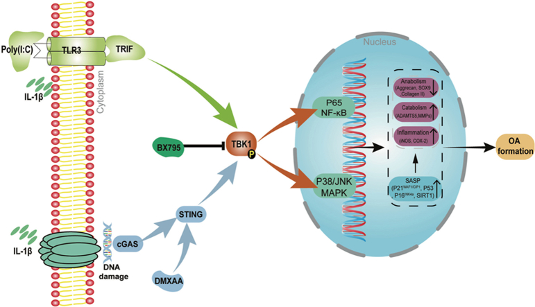
Objectives: TANK-binding kinase 1 (TBK1) is pivotal in autoimmune and inflammatory diseases, yet its role in osteoarthritis (OA) remains elusive. This study sought to elucidate the effect of the TBK1 inhibitor BX795 on OA and to delineate the underlying mechanism by which it mitigates OA.

Methods: Interleukin-1 Beta (IL-1β) was utilized to simulate inflammatory responses and extracellular matrix degradation in vitro. In vivo, OA was induced in 8-week-old mice through destabilization of the medial meniscus surgery. The impact of BX795 on OA was evaluated using histological analysis, X-ray, micro-CT, and the von Frey test. Additionally, Western blot, RT-qPCR, and immunofluorescence assays were conducted to investigate the underlying mechanisms of BX795.

Results: Phosphorylated TBK1 (P-TBK1) levels were found to be elevated in OA knee cartilage of both human and mice. Furthermore, intra-articular injection of BX795 ameliorated cartilage degeneration and alleviated OA-associated pain. BX795 also counteracted the suppression of anabolic processes and the augmentation of catabolic activity, inflammation, and senescence observed in the OA mice. In vitro studies revealed that BX795 reduced P-TBK1 levels and reversed the effects of anabolism inhibition, catabolism promotion, and senescence induction triggered by IL-1β. Mechanistically, BX795 inhibited the IL-1β-induced activation of the cGAS-STING and TLR3-TRIF signaling pathways in chondrocytes.

Conclusions: Pharmacological inhibition of TBK1 with BX795 protects articular cartilage by inhibiting the activation of the cGAS-STING and TLR3-TRIF signaling pathways. This action attenuates inflammatory responses and cellular senescence, positioning BX795 as a promising therapeutic candidate for OA treatment.

The translational potential of this article: This study furnishes experimental evidence and offers a potential mechanistic explanation supporting the efficacy of BX795 as a promising candidate for OA treatment.

Keywords: BX795; Chondrocyte; Inflammation; Osteoarthritis; Senescence; TBK1.


Figures

Image 1
Graphical abstract
Figure 1
Figure 1
Expression levels of P-TBK1 and TBK1 in human and mouse knee articular cartilage. Immunohistochemical and quantitative analysis of positive chondrocytes were performed to determine the expression of P-TBK1 and TBK1 in human knee cartilage (A–D, n = 20) and mice knee cartilage (E–H, n = 3). (I, J) Expression levels of P-TBK1 in chondrocytes following stimulation with IL-1β at different concentrations (0, 1, 2, 5, and 10 ng/mL IL-1β for 24 h) and durations (5 ng/mL IL-1β for 0, 1, 6, 12, 24, and 48 h) (n = 3) as determined using western blot analysis. (K–N) Expression levels of P-TBK1 in chondrocytes following stimulation with IL-1β at different concentrations (0, 1, 2, 5, and 10 ng/mL IL-1β for 24 h) and durations (5 ng/mL IL-1β for 0, 1, 6, 12, 24, and 48 h) as determined using immunofluorescence staining (n = 3). Data are presented as means ± SD. *p < 0.05, **p < 0.01, ***p < 0.001. ns, no significance.
Figure 2
Figure 2
Effects of BX795 on IL-1β-induced chondrocyte anabolism, catabolism, and inflammatory response. (A) Western blot and (B) quantitative analyses of proteins (P-TBK1 and TBK1) in chondrocytes treated with IL-1β along with or without 5, 10, or 20 μM BX795. Expression of anabolic proteins (C, D), catabolic proteins (E, F), and inflammatory proteins (H, I) was evaluated using western blot and quantitative analysis. (G) RT-PCR was performed to determine the expression levels of Aggrecan, Collagen II, SOX9, ADAMTS5, and MMP13. Immunofluorescence staining of Aggrecan (J, K) and Collagen II (L, M) and quantitative analysis of their expression levels. Data are presented as means ± SD (n = 3). ##p < 0.01; ###p < 0.001 compared with control group. ns, no significance; *p < 0.05; **p < 0.01; ***p < 0.001 compared with IL-1β group.
Figure 3
Figure 3
Effects of BX795 on IL-1β-induced chondrocyte senescence (A, C) Western blot data and (B, D) quantitative analysis of the expression levels of senescence-associated phenotypic proteins (SIRT1, P53, P21WAF1/CIP1, and P16INK4a) in chondrocytes treated with different concentrations of IL-1β (0, 1, 2, 5, and 10 ng/mL IL-1β for 24 h) and different stimulation times (5 ng/mL IL-1β for 0, 1, 6, 12, 24, and 48 h). (E) Western blot data and (F) quantitative analysis of the expression levels of senescence-associated phenotypic proteins (SIRT1, P53, P21WAF1/CIP1, and P16INK4a) in chondrocytes treated with 5 ng/mL IL-1β along with or without 5, 10, and 20 μM BX795 for 24 h. (G) SA-β-Gal staining and (H) quantitative analysis of senescence-positive chondrocytes (SA-β-Gal-positive cells) in chondrocytes treated with IL-1β along with or without 20 μM BX795. Data are presented as means ± SD (n = 3). ##p < 0.01; ###p < 0.001 compared with control group. ns, no significance; *p < 0.05; **p < 0.01; ***p < 0.001 compared with IL-1β group.
Figure 4
Figure 4
Protection of BX795 against DMXAA-induced cGAS–STING–TBK1 signaling pathway-dependent chondrocyte senescence, anabolism, and catabolism. (A, B) Protein expression levels of cGAS and STING evaluated by western blot analysis. (C, D) Expression of proteins (STING, P-TBK1, and TBK1) in the cGAS–STING–TBK1 signaling pathway was evaluated using western blot analysis. Expression of senescence-associated phenotypic proteins (E, F) and anabolic and catabolic proteins (G, H) in chondrocytes were determined using western blot analysis. (I, J) Immunofluorescence staining and quantitative analysis of the expression levels of P16INK4a. Data are presented as means ± SD (n = 3). ns, no significance, *p < 0.05; **p < 0.01; ***p < 0.001.
Figure 5
Figure 5
Protection of BX795 against Poly(I:C)-induced TLR3–TRIF–TBK1 signaling pathway-dependent chondrocyte senescence, anabolism, and catabolism. (A, B) Protein expression levels of TRIF evaluated by western blot analysis. (C, D) Expression of proteins (TRIF, P-TBK1, and TBK1) in the TLR3–TRIF–TBK1 signaling pathway was evaluated using western blot analysis. Expression of chondrocyte senescence-associated phenotypic proteins (E, F) and anabolic and catabolic proteins (G, H) were determined using western blot analysis. (I, J) Immunofluorescence staining and quantitative analysis of the expression levels of P16INK4a. Data are presented as means ± SD (n = 3). ns, no significance, *p < 0.05; **p < 0.01; ***p < 0.001.
Figure 6
Figure 6
Effects of BX795 on MAPK and NF-κB signaling pathways. Western blot and quantitative analysis showed the activation of MAPK (A, B) and NF-κB (C, D) signaling pathways in chondrocytes treated with 5 ng/mL IL-1β at different time points (0, 15, 30, 60, and 120 min). Western blot and quantitative analyses showed that BX795 affected MAPK (E, F) and NF-κB(G, H) signal-related proteins. (I) Immunofluorescence staining and (J) quantitative analysis reflected the nuclear accumulation of P–P65 in chondrocytes treated with IL-1β alone or together with 20 μM BX795. Data are presented as means ± SD (n = 3). #p < 0.05; ###p < 0.001 compared with control group. ns, no significance, *p < 0.05; **p < 0.01; ***p < 0.001 compared with IL-1β group.
Figure 7
Figure 7
BX795 attenuated the progression of OA in DMM mice. (A, B) Images of X-Ray, Micro-CT-3D, and Coronal Plane about the knee joints exhibited a general view of knee cartilage changes in the SHAM, SHAM + BX795, DMM, DMM + BX795 groups. (C) Pain behavioral test assessed by von Frey test. Paw withdrawal threshold in response to von Frey filament stimulation at several time points. Results are presented as mean ± SD of 6 mice per group. # represents differences in values between the DMM and SHAM groups; * represents differences in values between the DMM and DMM + BX795 groups; (D, E) Quantitative analysis of subchondral bone remodeling parameters (osteophyte volume, BV, BV/TV, Tb.Sp, Tb.N, and Tb.Th) of tibial plateau in knee joints among the SHAM, SHAM + BX795, DMM, DMM + BX795 groups (n = 6). *p < 0.05; **p < 0.01; ***p < 0.001; ##p < 0.01.
Figure 8
Figure 8
BX795 alleviated cartilage degradation and chondrocyte senescence in DMM mice. (A) HE, toluidine blue, and safranin O/fast green staining reflect the articular cartilage surfaces. (B) Quantitative analysis of the OARSI scores (n = 8). (C) Immunohistological staining and (D) quantitative analysis of anabolic (Aggrecan, Collagen II), catabolic (MMP13), inflammation (iNOS), and senescence-related (P16INK4a) phenotypic proteins (n = 3). *p < 0.05; **p < 0.01; ***p < 0.001.
figs1
figs1
figs2
figs2

References

    1. Hunter D.J., Bierma-Zeinstra S. Osteoarthritis. Lancet. 2019;393(10182):1745–1759. - PubMed
    1. Dieppe P.A., Lohmander L.S. Pathogenesis and management of pain in osteoarthritis. Lancet. 2005;365(9463):965–973. - PubMed
    1. Hiligsmann M., Cooper C., Arden N., Boers M., Branco J.C., Luisa Brandi M., et al. Health economics in the field of osteoarthritis: an expert's consensus paper from the European Society for Clinical and Economic Aspects of Osteoporosis and Osteoarthritis (ESCEO) Semin Arthritis Rheum. 2013;43(3):303–313. - PubMed
    1. Bolduc J.A., Collins J.A., Loeser R.F. Reactive oxygen species, aging and articular cartilage homeostasis. Free Radic Biol Med. 2019;132:73–82. - PMC - PubMed
    1. Rim Y.A., Nam Y., Ju J.H. The role of chondrocyte Hypertrophy and senescence in osteoarthritis initiation and progression. Int J Mol Sci. 2020;21(7) - PMC - PubMed

LinkOut - more resources