Induction of DR/IA antigens in human liver allografts. An immunocytochemical and clinicopathologic analysis of twenty failed grafts
- PMID: 3904089
- PMCID: PMC3035980
- DOI: 10.1097/00007890-198511000-00007
Induction of DR/IA antigens in human liver allografts. An immunocytochemical and clinicopathologic analysis of twenty failed grafts
Abstract
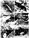
Twenty failed human liver allograft specimens obtained at the time of retransplantation procedures were studied using a panel of monoclonal antibodies (T11, T4, T8, NK, B1, OKM1, OKM5, Ia, DR). A clinicopathologic analysis was used to distinguish between graft failures secondary to rejection (n = 10) and those due, at least in part, to other causes (n = 10). T lymphocytes constituted the major infiltrating cellular population in the liver in rejection cases, but significant numbers of B cells and monocytes/macrophages were present also. Following transplantation, but not before, the bile duct epithelium, as well as portal and central vein and hepatic artery endothelium expressed DR/Ia antigens. These structures are preferential targets of the rejection reaction. The selective destruction of bile ducts in livers undergoing rejection was manifested in these patients by striking elevations of serum gamma glutamyl transpeptidase (GGTP) activity, a marker of biliary epithelial damage. The induced expression of DR/Ia antigens on structures targeted for immune destruction may be an important event in the pathogenesis of liver allograft rejection.
Figures

References
-
- Porter KA. Pathology of liver transplantation. Transplant Rev. 1969;2:129. - PubMed
-
- McLean IW, Nakane PK. Periodate-lysine-paraformaldehyde fixative: a new fixative for immunoelectron microscopy. J Histochem Cytochem. 1974;22:1077. - PubMed
-
- Hayry P. Intragraft events in allograft destruction. Transplantation. 1984;38:1. - PubMed
MeSH terms
Substances
Grants and funding
LinkOut - more resources
Full Text Sources
Medical
