

Changes in the ocular surface microbiome of patients with coronavirus disease 2019 (COVID-19)
- PMID: 39040901
- PMCID: PMC11262004
- DOI: 10.3389/fmicb.2024.1389139
Changes in the ocular surface microbiome of patients with coronavirus disease 2019 (COVID-19)
Abstract
Purpose: To elucidate the reasons behind the increased incidence of ocular disease in patients with coronavirus disease 2019 (COVID-19), this study delved deeper into the specific effects of COVID-19 on patients' ocular surface microbiome (OSM) and investigated its relationship with the increased incidence of ocular disease.
Methods: In this study, conjunctival sac swabs were collected from 43 participants for 16S rRNA amplicon sequencing. The participants were categorized into three groups based on their COVID-19 status: the control group (C group) consisted of 15 participants who showed no evidence of COVID-19, the experimental group (E group) included 15 participants who tested positive for COVID-19, and the COVID-19 recovery period group (R group) comprised 13 participants.
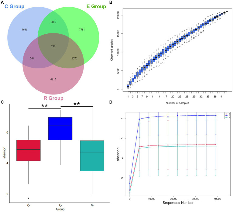
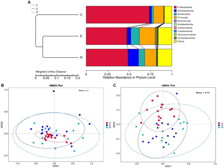
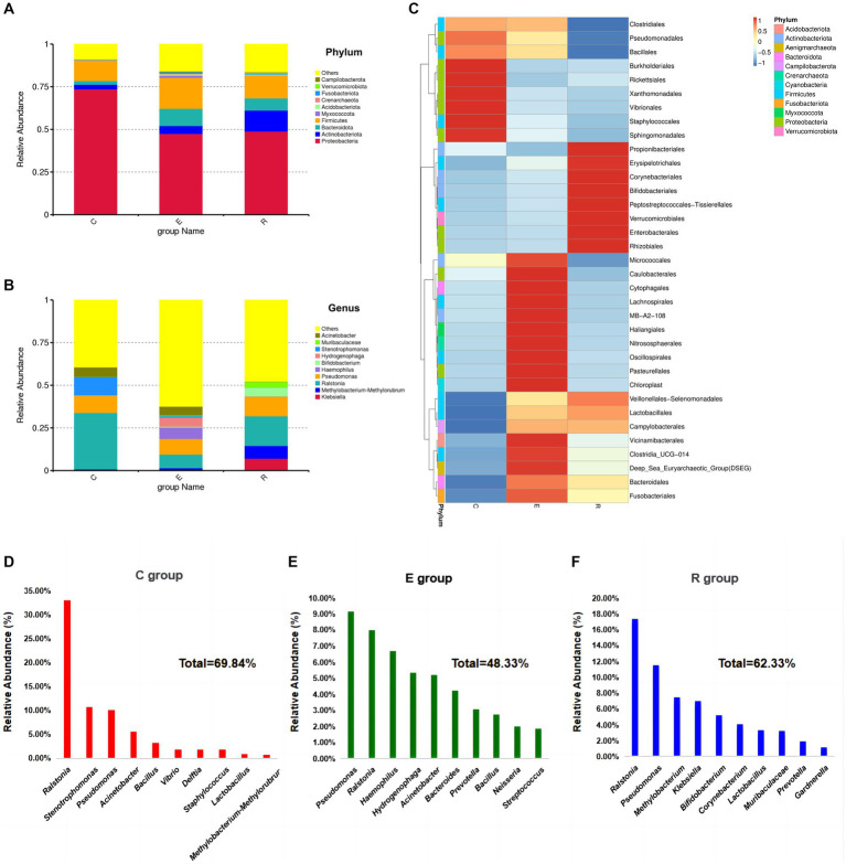
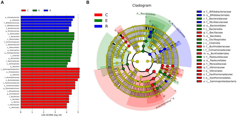
Results: In the comparison of alpha diversity, group E had a higher Shannon, Chao1 and Goods coverage index. When comparing beta diversity, groups E and R were more similar to each other. At the phylum level, although the OSM of the three groups was dominated by Proteobacteria, Actinobacteriota, Bacteroidota and Firmicutes, the compositional proportions were significantly different. At the genus level, the dominant species in the three OSM groups were significantly different, with Pseudomonas becoming the dominant genus in groups E and R compared to group C, and the abundance of Ralstonia decreasing significantly.
Conclusion: This study provides additional evidence supporting the association between the OSM and COVID-19, which contributes to our understanding of the potential mechanisms underlying ocular symptoms and complications associated with COVID-19 in the future.
Keywords: 16S rRNA amplicon sequencing; COVID-19; COVID-19 recovery period; ocular surface; ocular surface microbiome.
Copyright © 2024 Lin, Wang, Feng, Zhu, Guo, Dong, Zhang and Jin.
Conflict of interest statement
The authors declare that the research was conducted in the absence of any commercial or financial relationships that could be construed as a potential conflict of interest.
Figures
Similar articles
-
Characterization of Conjunctival Sac Microbiome from Patients with Allergic Conjunctivitis.J Clin Med. 2022 Feb 21;11(4):1130. doi: 10.3390/jcm11041130. J Clin Med. 2022. PMID: 35207407 Free PMC article.
-
Analysis of Conjunctival Sac Microbiome in Dry Eye Patients With and Without Sjögren's Syndrome.Front Med (Lausanne). 2022 Mar 8;9:841112. doi: 10.3389/fmed.2022.841112. eCollection 2022. Front Med (Lausanne). 2022. PMID: 35350577 Free PMC article.
-
The ocular surface microbiome of rhesus macaques.Res Sq [Preprint]. 2025 Mar 14:rs.3.rs-6205866. doi: 10.21203/rs.3.rs-6205866/v1. Res Sq. 2025. PMID: 40162216 Free PMC article. Preprint.
-
Alterations to the bovine bacterial ocular surface microbiome in the context of infectious bovine keratoconjunctivitis.Anim Microbiome. 2023 Nov 23;5(1):60. doi: 10.1186/s42523-023-00282-4. Anim Microbiome. 2023. PMID: 37996960 Free PMC article.
-
Investigating the Ocular Surface Microbiome: What Can It Tell Us?Clin Ophthalmol. 2023 Jan 19;17:259-271. doi: 10.2147/OPTH.S359304. eCollection 2023. Clin Ophthalmol. 2023. PMID: 36698849 Free PMC article. Review.
References
LinkOut - more resources
Full Text Sources