. 2024;56(5):579-591.
doi: 10.1080/00222895.2024.2364657. Epub 2024 Jul 23.

Grasp Posture Variability Leads to Greater Ipsilateral Sensorimotor Beta Activation During Simulated Prosthesis Use

Affiliations

Grasp Posture Variability Leads to Greater Ipsilateral Sensorimotor Beta Activation During Simulated Prosthesis Use

Bennett L Alterman et al. J Mot Behav. 2024.

Abstract

Motor behaviour using upper-extremity prostheses of different levels is greatly variable, leading to challenges interpreting ideal rehabilitation strategies. Elucidating the underlying neural control mechanisms driving variability benefits our understanding of adaptation after limb loss. In this follow-up study, non-amputated participants completed simple and complex reach-to-grasp motor tasks using a body-powered transradial or partial-hand prosthesis simulator. We hypothesised that under complex task constraints, individuals employing variable grasp postures will show greater sensorimotor beta activation compared to individuals relying on uniform grasping, and activation will occur later in variable compared to uniform graspers. In the simple task, partial-hand variable and transradial users showed increased neural activation from the early to late phase of the reach, predominantly in the hemisphere ipsilateral to device use. In the complex task, only partial-hand variable graspers showed a significant increase in neural activation of the sensorimotor cortex from the early to the late phase of the reach. These results suggest that grasp variability may be a crucial component in the mechanism of neural adaptation to prosthesis use, and may be mediated by device level and task complexity, with implications for rehabilitation after amputation.

Keywords: amputation; electrophysiology; rehabilitation; upper limb.


Conflict of interest statement

Declaration of interests

The authors report no conflict of interest. This work was supported by the National Institutes of Health under Grant 1R03NS103006-01.

Figures

Figure 1
Figure 1
(A) The TrPS is an orthosis that encompasses the hand, wrist, and forearm. It features a body-powered, voluntary opening of a split-hook effector via a figure-of-nine harness to the contralateral shoulder through glenohumeral flexion and scapular abduction. The palm was fit with padding to reduce sensory feedback and control by the hand. (B) The PhPS is an orthosis than mimics an amputation of the first 3 digits of the hand. The thumb is constrained at a right angle along the palm, and the fore- and middle fingers are secured to a roof plate, limiting sensory feedback and control by the fingers. The PhPS operates via voluntary opening and closing through wrist flexion and extension, respectively. TrPS: transradial prosthesis simulator, PhPS: partial-hand prosthesis simulator.
Figure 2
Figure 2
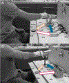
(A) Participants are seated at the experimental setup wearing the prosthesis simulator, completing the Translation task. This task consists of a reach-to-grasp (outlined arrow) from a starting position to pick up a small metal disk. The disk is then translated and placed onto a target location (solid arrow). After object placement, the participants return to the starting position (striped arrow) in preparation for the subsequent movement. (B) Participants are completing the Rotation task. This follows the same structure as the Translation task but requires the additional action of rotating the object (a marker) during translation (solid arrow) before placing it vertically on its end at the target location.
Figure 3
Figure 3
Top: The top row portrays scalp maps from early phase of the reach (-100 to 200 ms from movement onset) for the PhPS Uniform, PhPS Variable, and TrPS, from left to right. The PhPS Variable group shows left hemisphere laterality, while the PhPS Uniform group shows bilateral activation, closely resembling the TrPS group. Bottom: The bottom row portrays scalps maps of the late phase (200 to 400 ms from movement onset). There is a general increase in ERD, particularly in the right hemisphere. Frequency range of 20–26 Hz. Regions of interest are outlined in black.
Figure 4
Figure 4
Line graph of EEG power in the Translation task over the left frontal ROI. (B) Line graph of EEG power in the Translation task over the right frontal ROI. (C) Line graph of EEG power in the Translation task over the left parietal ROI. (D) Line graph of EEG power in the Translation task over the right parietal ROI. Significance at p < .05. EEG: electroencephalography, ROI: region of interest.
Figure 5
Figure 5
Top: The top row portrays scalp maps from early phase of the reach (-100 to 200 ms from movement onset) for the PhPS Uniform, PhPS Variable, and TrPS, from left to right. The PhPS Variable group shows hemispheric ERD, while the PhPS Uniform group resembles the TrPS group, showing right hemispheric laterality. Bottom: The bottom row portrays scalps maps of the late phase (200 to 400 ms from movement onset). There is a general increase in sensorimotor beta activation in the right hemisphere of the PhPS Variable group. Frequency range of 20–26 Hz. Regions of interest are outlined in black.
Figure 6
Figure 6
(A) Line graph of EEG power in the Rotation task over the left frontal ROI. (B) Line graph of EEG power in the Translation task over the right frontal ROI. (C) Line graph of EEG power in the Translation task over the left parietal ROI. (D) Line graph of EEG power in the Translation task over the right parietal ROI. Significance at p < .05. EEG: electroencephalography, ROI: region of interest.

Similar articles

References

    1. Acharya JN, Hani A, Cheek J, Thirumala P, & Tsuchida TN (2016). American clinical neurophysiology society guideline 2: Guidelines for standard electrode position nomenclature. Journal of clinical neurophysiology, 33(4), 308–311. 10.1097/WNP.0000000000000316 - DOI - PubMed
    1. Alterman BL, Keeton E, Ali S, Binkley K, Hendrix W, Lee PJ, Wang S, Kling J, Johnson JT, & Wheaton LA (2022). Partial-hand prosthesis users show improved reach-to-grasp behaviour compared to transradial prosthesis users with increased task complexity. Journal of motor behavior, 54(6), 706–718. 10.1080/00222895.2022.2070122 - DOI - PMC - PubMed
    1. Aoh Y, Hsiao H-J, Lu M-K, Macerollo A, Hunag H-C, Hamada M, Tsai C-H, & Chen J-C (2019). Event-related desynchronization/synchronization in spinocerebellar ataxia type 3. Frontiers in neurology, 10, 822. 10.3389/fneur.2019.00822 - DOI - PMC - PubMed
    1. Badre D, & D’Esposito M (2009). Is the rostro-caudal axis of the frontal lobe hierarchical? Nature reviews neuroscience, 10(9), 659–669. 10.1038/nrn2667 - DOI - PMC - PubMed
    1. Bayani KY, Lawson RR, Levinson L, Mitchell S, Atawala N, Otwell M, Rickerson B, & Wheaton LA (2019). Implicit development of gaze strategies support motor improvements during action encoding training of prosthesis use. Neuropsychologia, 127, 75–83. 10.1016/j.neuropsychologia.2019.02.015 - DOI - PMC - PubMed