. 2024 Dec;71(6):646-658.
doi: 10.1165/rcmb.2024-0003OC.

Enzymatic Modulation of the Pulmonary Glycocalyx Enhances Susceptibility to Streptococcus pneumoniae

Affiliations

Enzymatic Modulation of the Pulmonary Glycocalyx Enhances Susceptibility to Streptococcus pneumoniae

Cengiz Goekeri et al. Am J Respir Cell Mol Biol. 2024 Dec.

Abstract

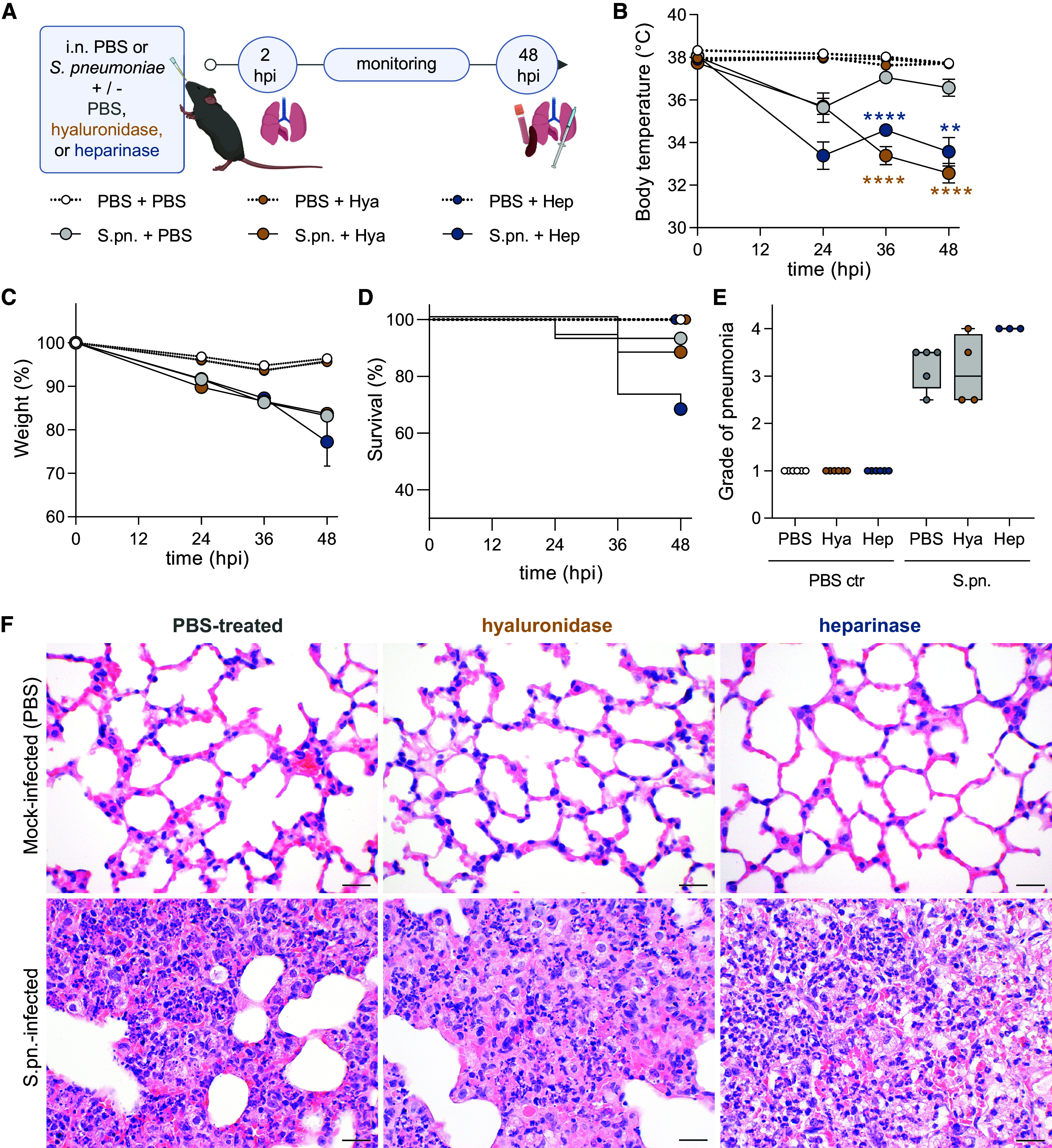
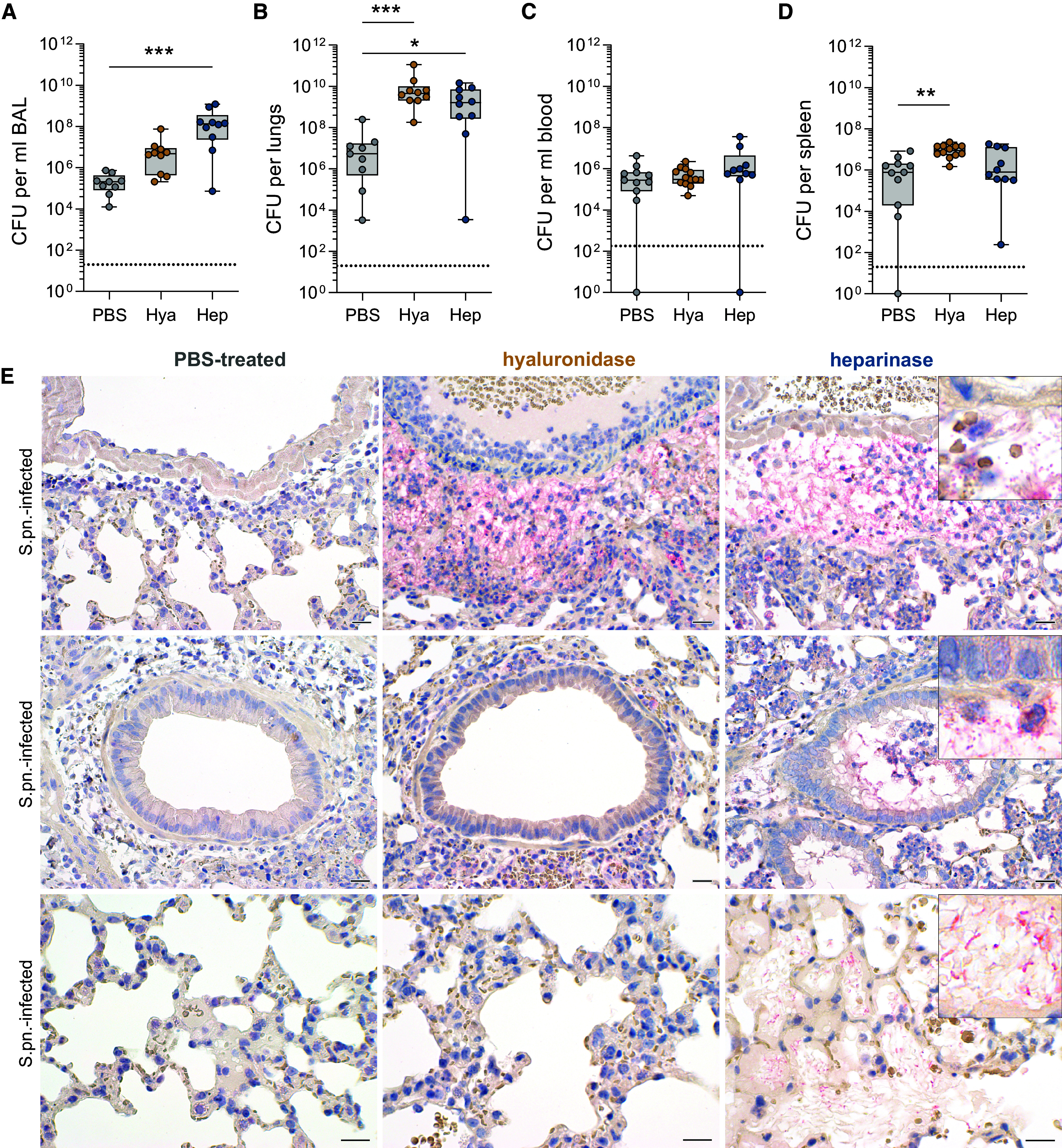
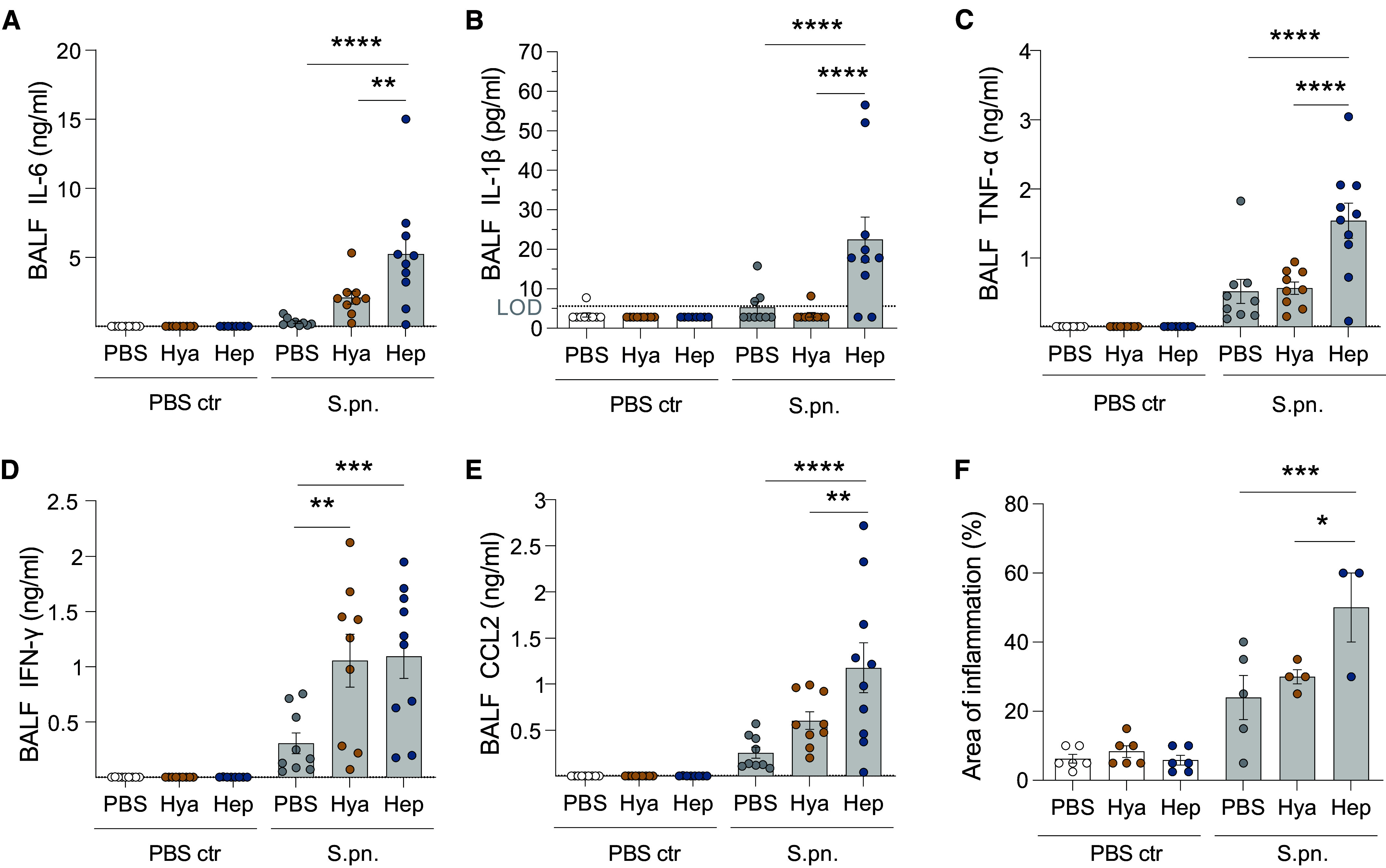
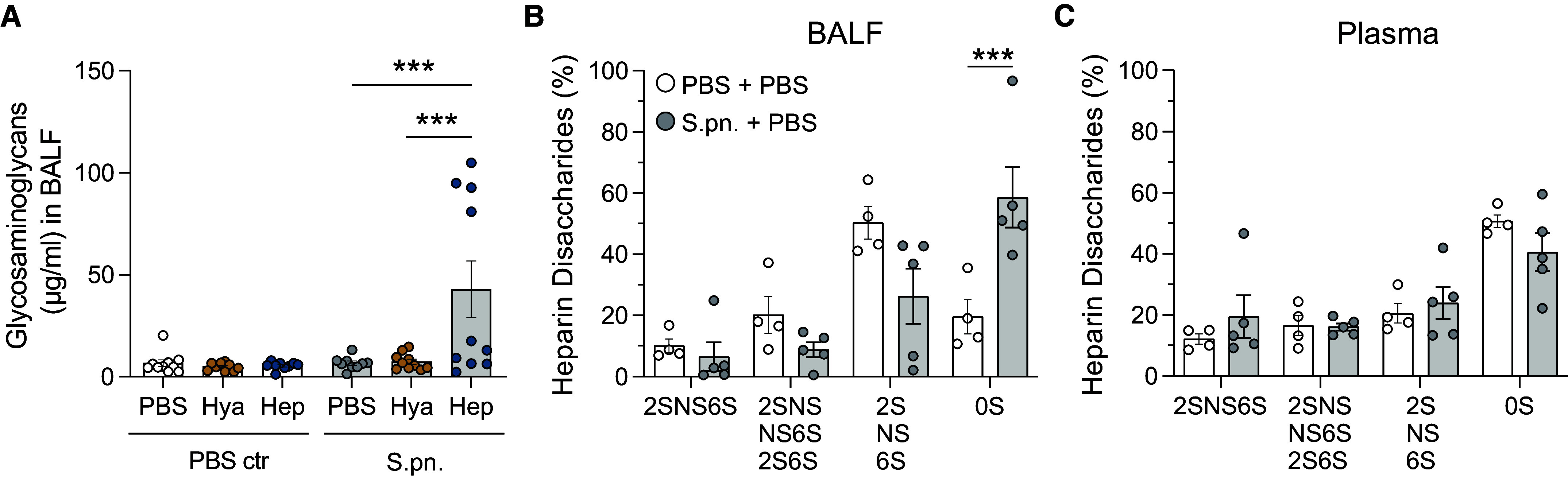
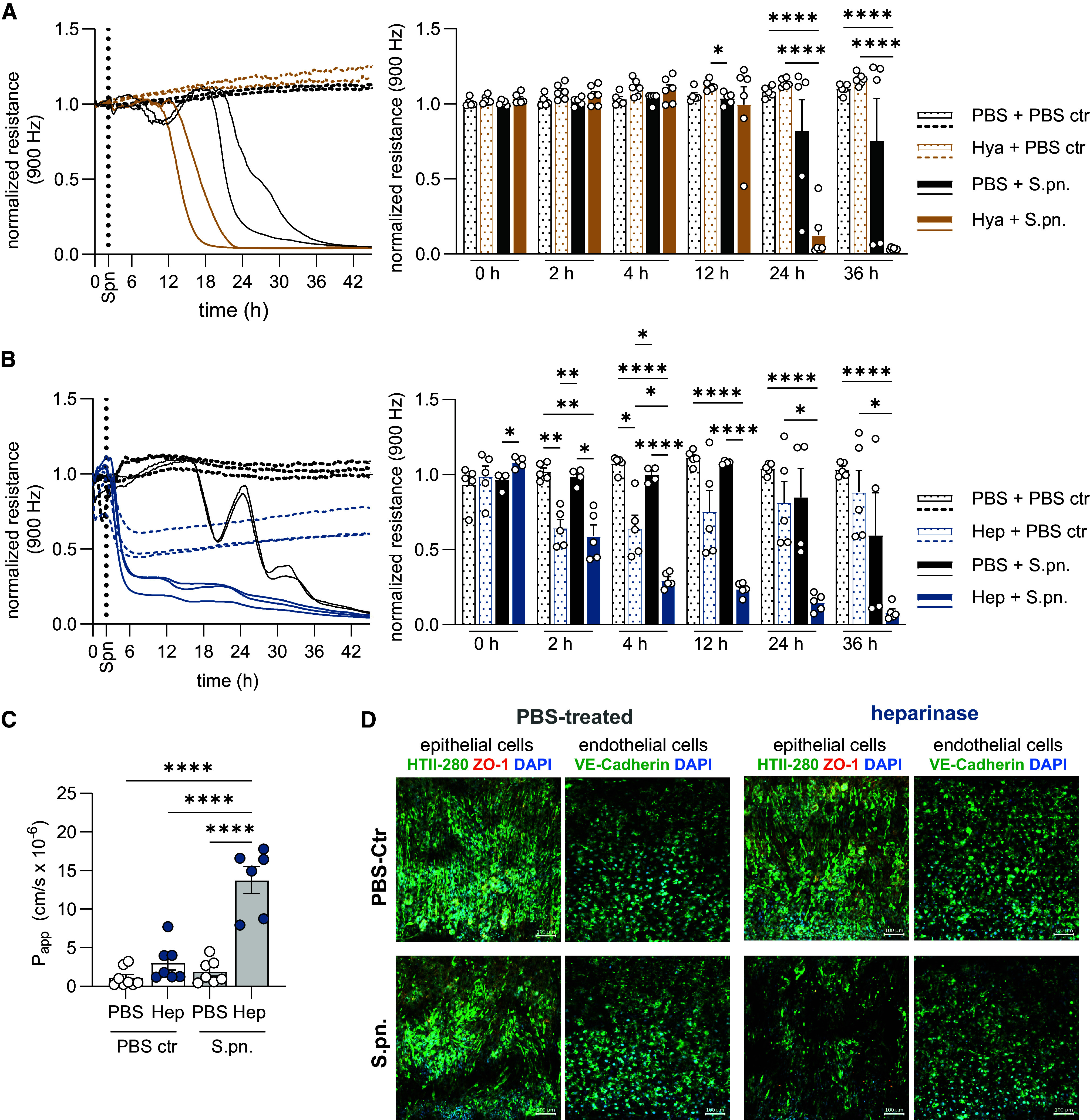
The pulmonary epithelial glycocalyx is rich in glycosaminoglycans such as hyaluronan and heparan sulfate. Despite their presence, the importance of these glycosaminoglycans in bacterial lung infections remains elusive. To address this, we intranasally inoculated mice with Streptococcus pneumoniae in the presence or absence of enzymes targeting pulmonary hyaluronan and heparan sulfate, followed by characterization of subsequent disease pathology, pulmonary inflammation, and lung barrier dysfunction. Enzymatic degradation of hyaluronan and heparan sulfate exacerbated pneumonia in mice, as evidenced by increased disease scores and alveolar neutrophil recruitment. However, targeting epithelial hyaluronan in combination with S. pneumoniae infection further exacerbated systemic disease, indicated by elevated splenic bacterial load and plasma concentrations of proinflammatory cytokines. In contrast, enzymatic cleavage of heparan sulfate resulted in increased bronchoalveolar bacterial burden, lung damage, and pulmonary inflammation in mice infected with S. pneumoniae. Accordingly, heparinase-treated mice also exhibited disrupted lung barrier integrity as evidenced by higher alveolar edema scores and vascular protein leakage into the airways. This finding was corroborated in a human alveolus-on-a-chip platform, confirming that heparinase treatment also disrupts the human lung barrier during S. pneumoniae infection. Notably, enzymatic pretreatment with either hyaluronidase or heparinase also rendered human epithelial cells more sensitive to pneumococci-induced barrier disruption, as determined by transepithelial electrical resistance measurements, consistent with our findings in murine pneumonia. Taken together, these findings demonstrate the importance of intact hyaluronan and heparan sulfate in limiting pneumococci-induced damage, pulmonary inflammation, and epithelial barrier function and integrity.

Keywords: acute lung injury; community-acquired pneumonia; glycosaminoglycans; heparan sulfate; hyaluronan.


Figures

Figure 1.
Figure 1.
Enzymatically targeting pulmonary glycosaminoglycans (GAGs) worsens symptoms and severity of pneumococcal pneumonia. Mice were infected intranasally (i.n.) with Streptococcus pneumoniae (S.pn.) or PBS (PBS ctr) with addition of hyaluronidase (Hya) or heparinase (Hep) or PBS as a control and killed at 48 hours postinfection (hpi). (A) Experimental layout. Graphs display (B) body temperature (°C), (C) weight change (%), and (D) survival of experimental animals over the infection course. (B, C) Mixed-effect analysis, Dunnett’s multiple comparisons test, tested against S.pn. + PBS. Test results displayed for S.pn. groups; n = 14–19 mice per group. **P ≤ 0.01 and ****P ≤ 0.0001. (D) Log-rank (Mantel-Cox) test; no significant differences between infected groups. (BD) Data derived from 17 independent experiments. (E) Histopathological degree of pneumonia ranging from (0) absent to (4) severe at 48 hpi. n = 3–6 mice per group; data derived from six independent experiments. Kruskal-Wallis and Dunn’s multiple comparisons tests. Significance was tested between treatments (PBS and enzymes) for sham (PBS ctr) and S.pn.-infected groups. Data are displayed as box plots. Middle line displays median, box indicates first and third quartiles, and whiskers indicate minimum to maximum. (F) Hematoxylin and eosin (H&E) staining of murine lungs. Although mock infection resulted in almost no lesions across all groups (upper panel), all infected groups developed diffuse alveolar damage with highest severity, density, and damage in heparinase-treated mice (H&E stain). Scale bars, 20 μm.
Figure 2.
Figure 2.
Enzymatic targeting of epithelial glycocalyx alters susceptibility to streptococcal pneumonia. Mice were infected i.n. with S.pn. with addition of Hya or Hep or PBS as a control and killed at 48 hpi. (AD) Colony-forming units (CFU) for (A) BAL, (B) lungs, (C) blood, and (D) spleen. Kruskal-Wallis and Dunn’s multiple comparison tests; n = 9–13, data derived from 11 independent experiments, significant differences were tested between treatments (PBS and enzymes) for S.pn.-infected groups. Data are displayed as box plots. Middle line displays median, box indicates first and third quartiles, and whiskers indicate minimum to maximum. Dotted lines indicate the limit of detection (LOD); BAL, 20 CFU/ml; lungs and spleen, 20 CFU/organ; blood, 200 CFU/ml. Samples below LOD were set to 1 or 1/ml. *P ≤ 0.05, **P ≤ 0.01, and ***P ≤ 0.001. (E) Immunohistochemical visualization of S.pn. (New fuchsin, red) in lungs from S.pn-infected and PBS-treated (left panel), hyaluronidase-treated (middle panel), or heparinase-treated (right panel) mice with hematoxylin (blue) as a counterstain. Upper row: perivascular region; middle row: intrabronchiolar region; bottom row, intraalveolar region. n = 3–6 mice per group; data derived from six independent experiments. Scale bars, 20 μm.
Figure 3.
Figure 3.
Inflammatory cell recruitment is enhanced after targeting of epithelial glycocalyx in pneumonia. Mice were infected i.n. with S.pn. or PBS (PBS ctr) with addition of Hya or Hep or PBS as a control and killed at 48 hpi. (AG) Flow cytometry–based analysis of immune cells. Total numbers of (A) CD45+ leukocytes, (B) polymorphonuclear neutrophils (PMNs), and (C) Ly6Chi inflammatory monocyte–derived macrophages (Ly6Chi iM) in BAL were determined by use of counting beads, whereas cell frequencies (DG) were determined as a percentage of CD45+. (E, G) Dot plots representing cellular frequencies among CD45+ leukocytes. (HJ) Protein concentrations of IL-17A (LOD, 4.42 pg/ml), CXCL5, and CXCL1 in BAL fluid (BALF) as quantified by ELISA. Dotted lines indicate LOD. Values below LOD were set to half LOD for statistical analysis. Two-way ANOVA and Tukey’s multiple comparisons test. n = 8–10; data derived from 11 independent experiments. Significant differences were tested between treatments (PBS and enzymes) for sham (PBS ctr) and S.pn.-infected groups. *P ≤ 0.05, **P ≤ 0.01, ***P ≤ 0.001, and ****P ≤ 0.0001.
Figure 4.
Figure 4.
Targeting of epithelial glycocalyx enhances release of proinflammatory mediators. Mice were infected i.n. with S.pn. or PBS (PBS ctr) with addition of Hya or Hep or PBS as a control and killed at 48 hpi. The concentrations of proinflammatory cytokines and chemokine CCL2 in BALF were measured by multiplex ELISA: (A) IL-6 (LOD, 1.72 pg/ml), (B) IL-1β (LOD, 5.66 pg/ml), (C) TNF-α (LOD, 13.49 pg/ml), (D) IFN-γ (LOD, 1.06 pg/ml), and (E) CCL2 (LOD, 5.54 pg/ml). Dotted lines indicate LOD. Values below LOD were set to half LOD for statistical analysis. (F) Area of inflammation detected during histopathological examination of H&E-stained lung tissue. (AF) Two-way ANOVA, Tukey’s multiple comparisons test, control group versus treated groups. (AE) n = 8–10; data derived from 11 independent experiments. (F) n = 3–6; data derived from six independent experiments. (AF) Significant differences were tested between treatments (PBS and enzymes) for sham (PBS ctr) and S.pn.-infected groups. *P ≤ 0.05, **P ≤ 0.01, ***P ≤ 0.001, and ****P ≤ 0.0001.
Figure 5.
Figure 5.
Heparin disaccharides are shed and remodeled during pneumococcal infection and after heparinase treatment in mice. Mice were infected i.n. with S.pn. or PBS (PBS ctr) with addition of Hya or Hep or PBS as a control and killed at 48 hpi. (A) GAG shedding into BALF was determined by dimethylmethylene blue (DMMB) assay. Two-way ANOVA, Tukey’s multiple comparisons test; n = 9–10, data derived from 11 independent experiments. Significant differences were tested between treatments (PBS and enzymes) for sham (PBS ctr) and S.pn.-infected groups. High-performance liquid chromatography was performed on pooled (B) BALF and (C) plasma samples to analyze sulfation of heparin disaccharides. Two-way ANOVA, Tukey’s multiple comparisons test; n = 4–5 (pooled from two or three independent samples derived from 11 independent experiments). Significant differences were tested between sham (PBS + PBS) and S.pn.-infected (S.pn. + PBS) groups. HexA-GlcNAc = 0S, HexA(2S)-GlcNAc = 2S, HexA-GlcNS = NS, HexA-GlcNAc(6S) = 6S, HexA(2S)-GlcNAc(6S) = 2S6S, HexA(2S)-GlcNS = 2SNS, HexA-GlcNS(6S) = NS6S, HexA(2S)-GlcNS(6S) = 2SNS6S. ***P ≤ 0.001.
Figure 6.
Figure 6.
Lung barrier function is impaired by enzymatic glycocalyx modulation during pneumococcal pneumonia. Mice were infected i.n. with S.pn. or PBS (PBS ctr) with addition of Hya or Hep or PBS as a control and killed at 48 hpi. (A) Protein content in BALF was determined by colorimetric assay. Two-way ANOVA, Tukey’s multiple comparisons test. n = 9–10; data derived from 11 independent experiments. (B, C) Graph displaying results of histopathological scoring of (B) perivascular edema and (C) alveolar edema in H&E-stained lung sections. Scoring 0 = none, 1 = scattered, 2 = low grade, 3 = medium grade, 4 = severe, 5 = massive. Kruskal-Wallis, Dunn’s multiple comparisons test. Data are displayed as box plots; middle line displays median, box indicates first and third quartiles, and whiskers indicate minimum to maximum. n = 3–6; data derived from six independent experiments. (D) Representative H&E staining of murine lungs. Upper panels display representative perivascular edema, ovals highlight perivascular hemorrhage, and # highlights alveolar hemorrhage only found in heparinase-treated groups. Lower panels display representative alveolar edema in corresponding groups indicated by dotted circles. Scale bars, 20 μm. (AC) Significance tested between treatments (PBS ctr and enzymes) for sham (PBS ctr) and S.pn.-infected groups and between sham (PBS ctr) and S.pn-infected per treatment. *P ≤ 0.05, **P ≤ 0.01, ***P ≤ 0.001, and ****P ≤ 0.0001.
Figure 7.
Figure 7.
Human epithelial barrier function is impaired by enzymatic glycocalyx disruption in vitro. (A, B) Transepithelial electrical resistance (TEER) measurement by electric cell substrate impedance sensing to assess barrier function of human primary alveolar epithelial cells (HPAECs) after (A) Hya or (B) Hep or PBS treatment with subsequent S.pn. infection or mock infection (PBS ctr). Left: Continuous TEER measurements over time of two or three replicates of one representative experiment; n = 2–3. Right: Individual datapoints of TEER measurements across all experimental groups at designated time points during baseline and postinfection; summary data derived from two or three independent experiments. Two-way ANOVA, Tukey’s multiple comparisons test. Significant differences were tested between all groups. (C) Determination of alveolus-on-a-chip barrier integrity upon 24 hours of heparinase treatment and S.pn. infection by apparent permeability (Papp) measurement of tracer molecules. Experiment was performed two to four times with two or three different cell donors per group. Graph shows a summary of experiments and replicates. Two-way ANOVA, Tukey’s multiple comparisons test. Significant differences were tested between all groups. (D) Immunofluorescence of HPAECs (HTII-280, green; zona occludens [ZO]-1, orange [pseudocolored]; DAPI, blue) on the alveolar side and human pulmonary microvascular endothelial cells (HPMECs, VE-cadherin, green; DAPI, blue) on the vascular side. Nuclei are stained with DAPI (blue). Scale bars, 100 μm. *P ≤ 0.05, **P ≤ 0.01, and ****P ≤ 0.0001.

Comment in

References

    1. Welte T, Torres A, Nathwani D. Clinical and economic burden of community-acquired pneumonia among adults in Europe. Thorax . 2012;67:71–79. - PubMed
    1. Ghia CJ, Dhar R, Koul PA, Rambhad G, Fletcher MA. Streptococcus pneumoniae as a cause of community-acquired pneumonia in Indian adolescents and adults: a systematic review and meta- analysis. Clin Med Insights Circ Respir Pulm Med . 2019;13:1179548419862790. - PMC - PubMed
    1. Howard LS, Sillis M, Pasteur MC, Kamath AV, Harrison BD. Microbiological profile of community-acquired pneumonia in adults over the last 20 years. J Infect . 2005;50:107–113. - PubMed
    1. Ferrer M, Travierso C, Cilloniz C, Gabarrus A, Ranzani OT, Polverino E, et al. Severe community-acquired pneumonia: characteristics and prognostic factors in ventilated and non-ventilated patients. PLoS One . 2018;13:e0191721. - PMC - PubMed
    1. Ewig S, Woodhead M, Torres A. Towards a sensible comprehension of severe community-acquired pneumonia. Intensive Care Med . 2011;37:214–223. - PubMed

MeSH terms

LinkOut - more resources