. 2024 Aug 5;63(31):14673-14690.
doi: 10.1021/acs.inorgchem.4c02235. Epub 2024 Jul 23.

Minimal Functionalization of Ruthenium Compounds with Enhanced Photoreactivity against Hard-to-Treat Cancer Cells and Resistant Bacteria

Affiliations

Minimal Functionalization of Ruthenium Compounds with Enhanced Photoreactivity against Hard-to-Treat Cancer Cells and Resistant Bacteria

Geângela de Fátima Sousa Oliveira et al. Inorg Chem. .

Abstract

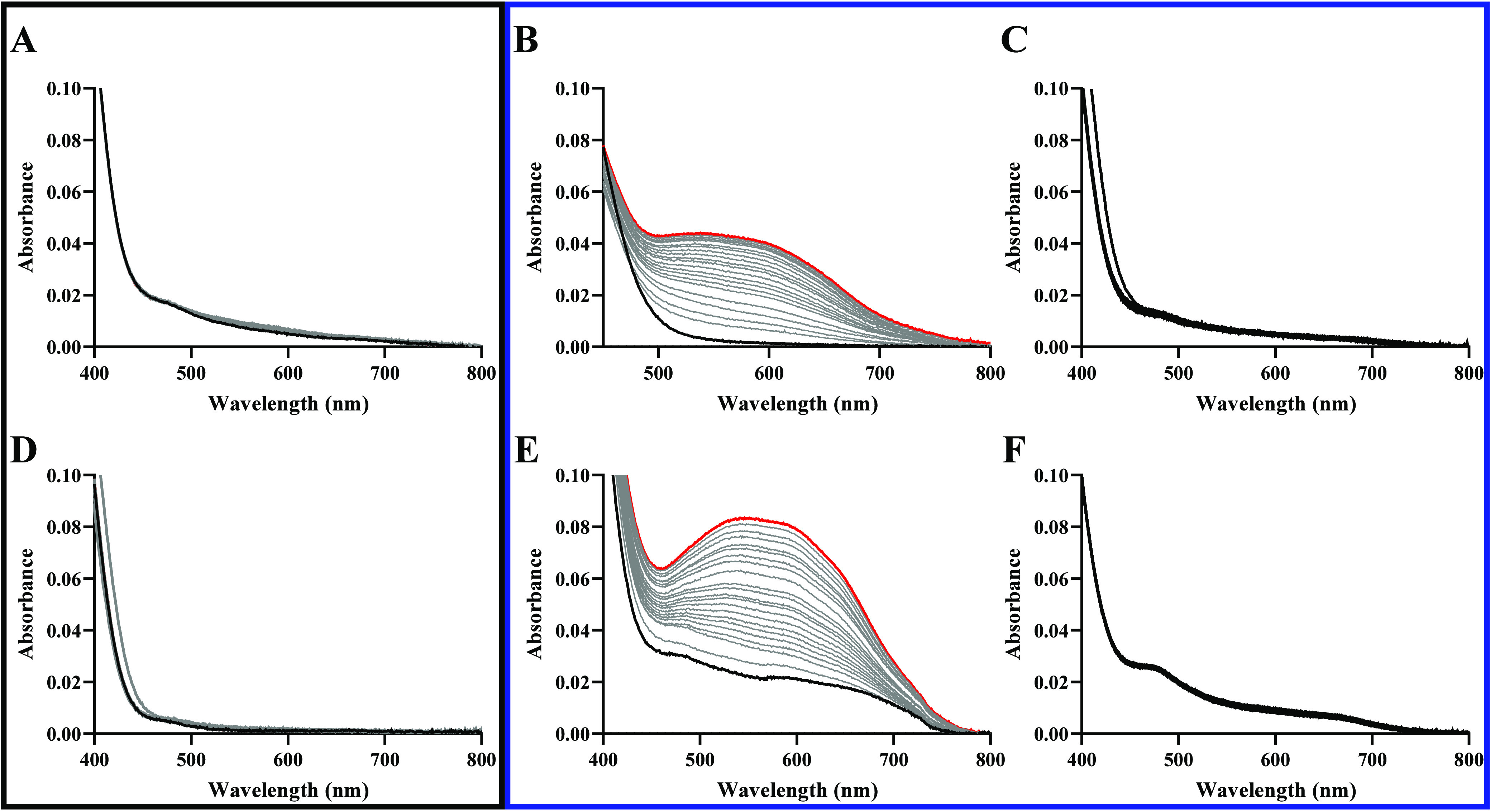
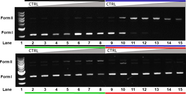
Metallocompounds have emerged as promising new anticancer agents, which can also exhibit properties to be used in photodynamic therapy. Here, we prepared two ruthenium-based compounds with a 2,2'-bipyridine ligand conjugated to an anthracenyl moiety. These compounds coded GRBA and GRPA contain 2,2'-bipyridine or 1,10-phenathroline as auxiliary ligands, respectively, which provide quite a distinct behavior. Notably, compound GRPA exhibited remarkably high photoproduction of singlet oxygen even in water (ϕΔ = 0.96), almost twice that of GRBAΔ = 0.52). On the other hand, this latter produced twice more superoxide and hydroxyl radical species than GRPA, which may be due to the modulation of their excited state. Interestingly, GRPA exhibited a modest binding to DNA (Kb = 4.51 × 10⁴), while GRBA did not show a measurable interaction only noticed by circular dichroism measurements. Studies with bacteria showed a great antimicrobial effect, including a synergistic effect in combination with commercial antibiotics. Besides that, GRBA showed very low or no cytotoxicity against four mammalian cells, including a hard-to-treat MDA-MB-231, triple-negative human breast cancer. Potent activities were measured for GRBA upon blue light irradiation, where IC50 of 43 and 13 nmol L-1 were seen against hard-to-treat triple-negative human breast cancer (MDA-MB-231) and ovarian cancer cells (A2780), respectively. These promising results are an interesting case of a simple modification with expressive enhancement of biological activity that deserves further biological studies.


Conflict of interest statement

The authors declare no competing financial interest.

Figures

Figure 1
Figure 1
Structures of GRPA (A) and GRBA (B).
Figure 2
Figure 2
Electronic absorption (A) and emission (B) spectra of GRPA in methanol (2 × 10⁻⁵ mol L–1) at 25 °C (λexc at 446 nm).
Figure 3
Figure 3
Schematic of the energy levels for GRPA (black line) and GRBA (blue line).
Figure 4
Figure 4
Measurement of the production of singlet oxygen upon blue light irradiation using SOSG probe (1 μmol L–1) in ultrapure water (λexc at 490 nm). Panel A shows a linear change of fluorescence during light irradiation in water for SOSG (alone, black circle), GRPA (green inverted triangle), GRBA (red triangle) and [Ru(bpy)3]2+ (blue square). Panel B and C show the raw emission spectra for SOSG with GRPA (10 μmol L–1) and GRBA (10 μmol L–1) during blue light irradiation.
Figure 5
Figure 5
Measurement of the production of hydroxyl radical upon blue light irradiation using APF probe (5 μmol L–1) in 100 mmol L–1 phosphate buffer pH 7.4 (λexc at 463 nm). Panels A and B show emission spectra for APF with GRBA (10 μmol L–1) and GRPA (10 μmol L–1) during blue light irradiation. Panels C and D show a linear change of fluorescence during light irradiation in phosphate buffer for APF with GRPA and GRBA, and also with the addition of mannitol (10 mmol L–1) or sodium azide (10 mmol L–1), respectively.
Figure 6
Figure 6
Superoxide detection using NBT (50 μmol L–1), GSH (1.5 mmol L–1) and GRPA (5 μmol L–1), monitored for 100 min in the dark (A), with blue light irradiation for 100 min (B) and in the presence of SOD with blue light irradiation (C); similarly, for GRBA (5 μmol L–1) in the dark (D), with blue light irradiation (E) and in the presence of SOD with blue light irradiation (F). All reaction were carried out at 25 °C.
Figure 7
Figure 7
DNA binding measurements. Panels show the titration of GRPA with calf thymus DNA monitored by UV–vis absorption electronic spectra (A), plot of εa – εfb – εf vs [DNA] (B), luminescence with excitation at 450 nm (C) and plot of (IaIf)/(IbIf) vs [DNA]) (D) in 0.1 mol L–1 Tris-HCl pH 7.4 at 25 °C.
Figure 8
Figure 8
Interaction of the metal complexes with DNA investigated by circular dichroism. Panel A shows the CD spectra of calf thymus DNA (black line, at 10 μmol L–1 in base pairs) and GRPA complex (gray line, at 20 μmol L–1) and mixtures of DNA and metal complex at a concentration of 2, 5, 7, 10, 15, 20, and 25 μmol L–1. Panel B shows CD spectra of calf thymus DNA (black line) and GRBA metal complex (gray line) and mixtures of DNA and this metal complex as specified above.
Figure 9
Figure 9
Photocleavage assay of pBR322 DNA (20 μmol L–1, in base pair) in the presence of GRPA, in the dark and after 1 h of irradiation with blue, green and red LEDs. In all experiments, lane 1 contains only linear DNA ladder (1 kb) and lane 2 only pBR322 DNA, while lanes 3–8 and 10–15 contained the following concentrations of 0.5, 1.0, 3.0, 5.0, 7.0, and 10 μmol L–1 of GRPA. Dark, blue, green and red lines indicate either the experiment was carried out in the dark or with blue, green or red-light irradiation.
Figure 10
Figure 10
Suppression of ROS in a photocleavage assay using pBR322 DNA (20 μmol L–1) in the presence of GRPA (5 μmol L–1) after 1 h of blue LED irradiation, and with radical scavengers. Lane 1: pBR322 DNA only with blue light irradiation. Lane 2: DNA + complexes in the dark. Lane 3: DNA + complexes with blue light irradiation. Other lanes 4–7: pBR322 DNA + GRPA + suppressor: pyruvate (4), histidine (5), D-mannitol (6), and tiron (7), respectively.

References

    1. Zeng L.; Gupta P.; Chen Y.; Wang E.; Ji L.; Chao H.; Chen Z.-S. The Development of Anticancer Ruthenium(ii) Complexes: From Single Molecule Compounds to Nanomaterials. Chem. Soc. Rev. 2017, 46 (19), 5771–5804. 10.1039/C7CS00195A. - DOI - PMC - PubMed
    1. Dixon I. M.; Lebon E.; Sutra P.; Igau A. Luminescent Ruthenium–Polypyridine Complexes & Phosphorus Ligands: Anything but a Simple Story. Chem. Soc. Rev. 2009, 38 (6), 1621.10.1039/b804763b. - DOI - PubMed
    1. Monro S.; Colón K. L.; Yin H.; Roque J.; Konda P.; Gujar S.; Thummel R. P.; Lilge L.; Cameron C. G.; McFarland S. A. Transition Metal Complexes and Photodynamic Therapy from a Tumor-Centered Approach: Challenges, Opportunities, and Highlights from the Development of TLD1433. Chem. Rev. 2019, 119 (2), 797–828. 10.1021/acs.chemrev.8b00211. - DOI - PMC - PubMed
    1. Abid M.; Shamsi F.; Azam A. Ruthenium Complexes: An Emerging Ground to the Development of Metallopharmaceuticals for Cancer Therapy. Mini-Rev. Med. Chem. 2016, 16 (10), 772–786. 10.2174/1389557515666151001142012. - DOI - PubMed
    1. Frei A.; Verderosa A. D.; Elliott A. G.; Zuegg J.; Blaskovich M. A. T. Metals to Combat Antimicrobial Resistance. Nat. Rev. Chem. 2023, 7 (3), 202–224. 10.1038/s41570-023-00463-4. - DOI - PMC - PubMed

MeSH terms

LinkOut - more resources