

Evaluating stereotactic accuracy with patient-specific MRI distortion corrections for frame-based radiosurgery
- PMID: 39042450
- PMCID: PMC11492306
- DOI: 10.1002/acm2.14472
Evaluating stereotactic accuracy with patient-specific MRI distortion corrections for frame-based radiosurgery
Abstract
Purpose: This study examines how MRI distortions affect frame-based SRS treatments and assesses the need for clinical distortion corrections.
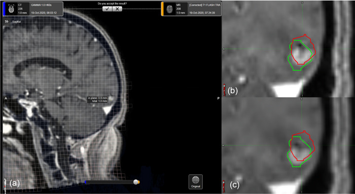
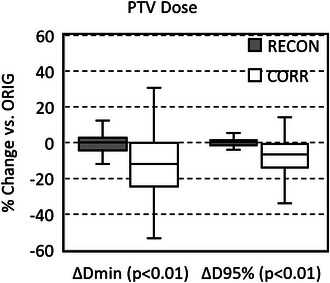
Methods: The study included 18 patients with 80 total brain targets treated using frame-based radiosurgery. Distortion within patients' MRIs were corrected using Cranial Distortion Correction (CDC) software, which utilizes the patient's CT to alter planning MRIs to reduce inherent intra-cranial distortion. Distortion was evaluated by comparing the original planning target volumes (PTVORIG) to targets contoured on corrected MRIs (PTVCORR). To provide an internal control, targets were also re-contoured on uncorrected (PTVRECON) MRIs. Additional analysis was done to assess if 1 mm expansions to PTVORIG targets would compensate for patient-specific distortions. Changes in target volumes, DICE and JACCARD similarity coefficients, minimum PTV dose (Dmin), dose to 95% of the PTV (D95%), and normal tissue receiving 12 Gy (V12Gy), 10 Gy (V10Gy), and 5 Gy (V5Gy) were calculated and evaluated. Student's t-tests were used to determine if changes in PTVCORR were significantly different than intra-contouring variability quantified by PTVRECON.
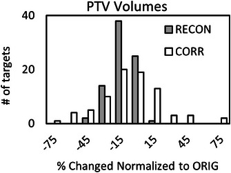
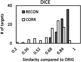
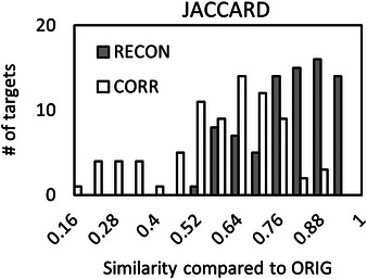
Results: PTVRECON and PTVCORR relative changes in volume were 6.19% ± 10.95% and 1.48% ± 32.92%. PTVRECON and PTVCORR similarity coefficients were 0.90 ± 0.08 and 0.73 ± 0.16 for DICE and 0.82 ± 0.12 and 0.60 ± 0.18 for JACCARD. PTVRECON and PTVCORR changes in Dmin were -0.88% ± 8.77% and -12.9 ± 17.3%. PTVRECON and PTVCORR changes in D95% were -0.34% ± 5.89 and -8.68% ± 13.21%. The 1 mm expanded PTVORIG targets did not entirely cover 14 of the 80 PTVCORR targets. Normal tissue changes (V12Gy, V10Gy, V5Gy) calculated with PTVRECON were (-0.09% ± 7.39%, -0.38% ± 5.67%, -0.08% ± 2.04%) and PTVCORR were (-2.14% ± 7.34%, -1.42% ± 5.45%, -0.61% ± 1.93%). Except for V10Gy, all PTVCORR changes were significantly different (p < 0.05) than PTVRECON.
Conclusion: MRIs used for SRS target delineation exhibit notable geometric distortions that may compromise optimal dosimetric accuracy. A uniform 1 mm expansion may result in geometric misses; however, the CDC algorithm provides a feasible solution for rectifying distortions, thereby enhancing treatment precision.
Keywords: MRI; SRS; distortions.
© 2024 The Author(s). Journal of Applied Clinical Medical Physics published by Wiley Periodicals LLC on behalf of American Association of Physicists in Medicine.
Conflict of interest statement
The authors declare no conflicts of interest.
Figures

References
-
- Wang D, Doddrell DM. Geometric distortion in structural magnetic resonance imaging. Current Medical Imaging. 2005;1(1):49‐60.
Publication types
MeSH terms
LinkOut - more resources
Full Text Sources
Medical
