. 2024 Jul 23;9(17):e179755.
doi: 10.1172/jci.insight.179755.

Enteric neural stem cell transplant restores gut motility in mice with Hirschsprung disease

Affiliations

Enteric neural stem cell transplant restores gut motility in mice with Hirschsprung disease

Ahmed A Rahman et al. JCI Insight. .

Abstract

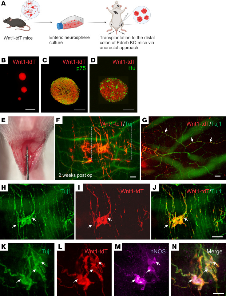
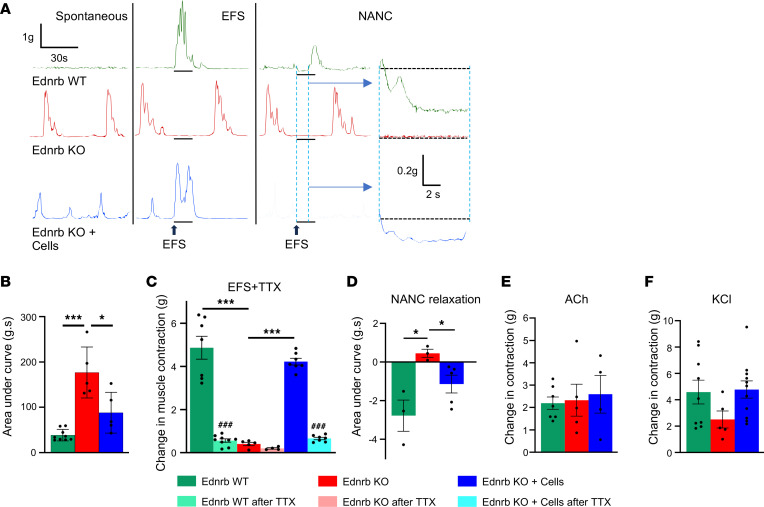
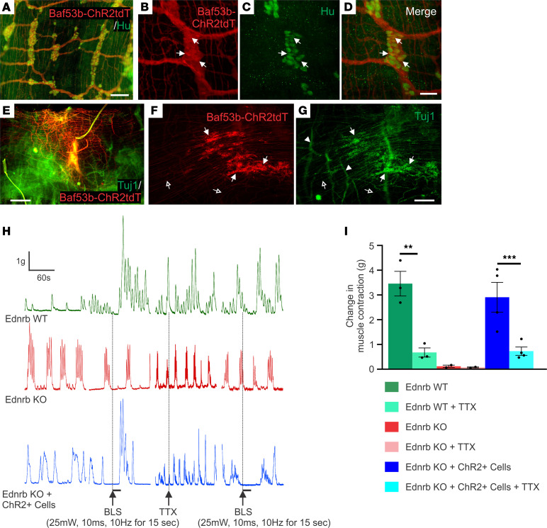
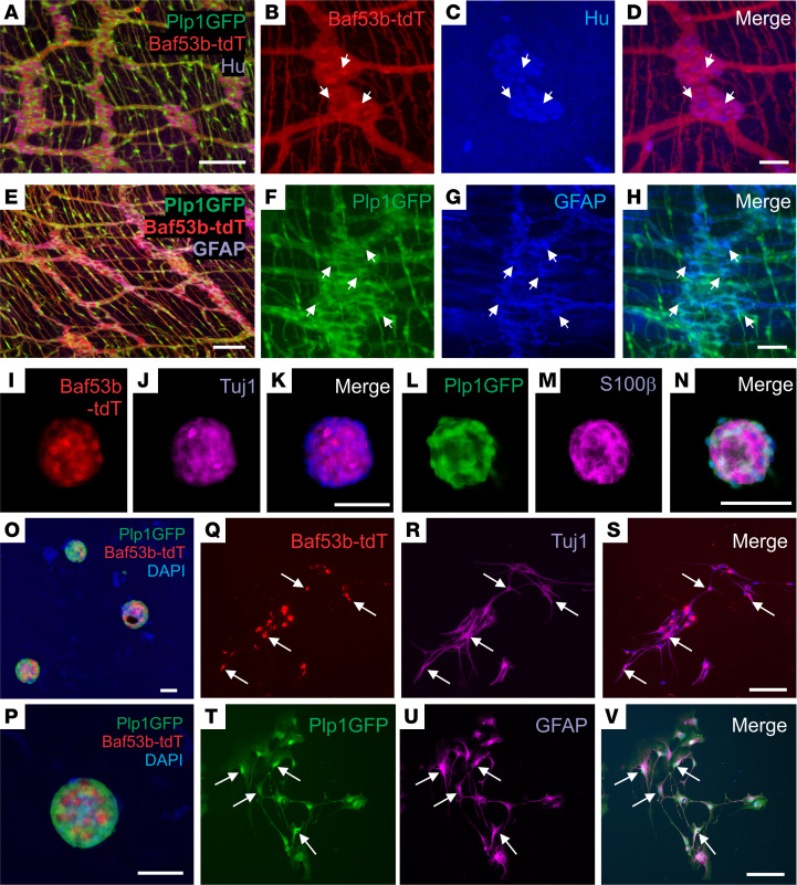
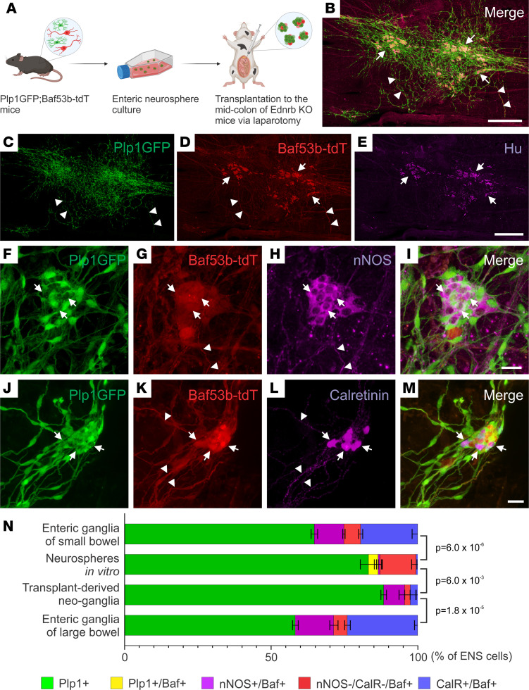
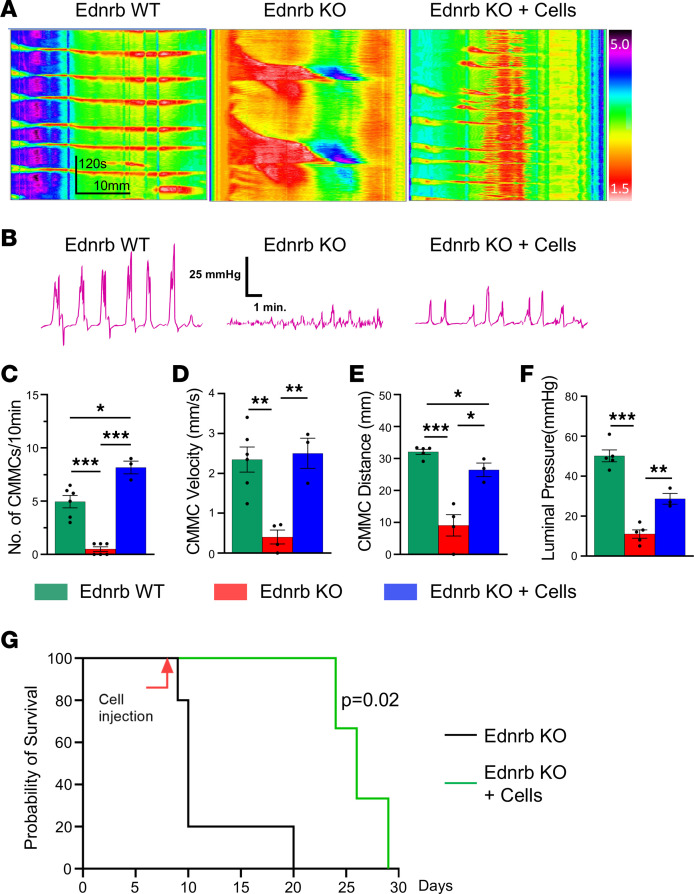
The goal of this study was to determine if transplantation of enteric neural stem cells (ENSCs) can rescue the enteric nervous system, restore gut motility, reduce colonic inflammation, and improve survival in the Ednrb-KO mouse model of Hirschsprung disease (HSCR). ENSCs were isolated from mouse intestine, expanded to form neurospheres, and microinjected into the colons of recipient Ednrb-KO mice. Transplanted ENSCs were identified in recipient colons as cell clusters in "neo-ganglia." Immunohistochemical evaluation demonstrated extensive cell migration away from the sites of cell delivery and across the muscle layers. Electrical field stimulation and optogenetics showed significantly enhanced contractile activity of aganglionic colonic smooth muscle following ENSC transplantation and confirmed functional neuromuscular integration of the transplanted ENSC-derived neurons. ENSC injection also partially restored the colonic migrating motor complex. Histological examination revealed a significant reduction in inflammation in ENSC-transplanted aganglionic recipient colon compared with that of sham-operated mice. Interestingly, mice that received cell transplant also had prolonged survival compared with controls. This study demonstrates that ENSC transplantation can improve outcomes in HSCR by restoring gut motility and reducing the severity of Hirschsprung-associated enterocolitis, the leading cause of death in human HSCR.

Keywords: Cell biology; Gastroenterology; Neurodevelopment; Neuronal stem cells; Stem cell transplantation.


Figures

Figure 1
Figure 1. Transplant of Wnt1-tdT ENSCs to Ednrb-KO mice.
Schematic of experimental overview (A), including isolation of ENSCs from the gastrointestinal tract of Wnt1-tdT mice, their expansion as neurospheres (B), and subsequent transplantation into the aganglionic distal colons of Ednrb-KO mice via anorectal needle injection (A and E). Enteric neurospheres contain p75+ neural crest cells (C) and Hu+ neurons (D). Transplanted cells were observed 2 weeks following surgery (F), projecting fibers along host-derived Tuj1+ extrinsic nerves (G, arrows) and forming neo-ganglia (HJ, arrows) that contain donor-derived nNOS immunoreactive neurons (KN, arrows). Scale bars: 50 μm (C, D, and KN), 100 μm (FJ), and 200 μm (B).
Figure 2
Figure 2. EFS demonstrates functional recovery of smooth muscle contractility in Ednrb-KO mice after cell transplantation.
Representative traces of smooth muscle contractions during the spontaneous, after EFS, and under NANC conditions (A). Quantifications of spontaneous muscle contractility (B), EFS-induced contractility (C), and under NANC conditions (D) are shown. The amplitude of EFS contractions reflects maximal contractility as an absolute change from baseline and is markedly reduced in the presence of TTX (C). Effects of ACh (E) and KCl (F) on muscle activity. All the values represent the mean of 2–4 animals for each group, repeated 2–3 times. Data are shown as the mean ± SEM. Statistical significance was determined by the 1-way ANOVA with a post hoc Tukey’s test; *P < 0.05, ***P < 0.001, and ###P < 0.001 are statistically significant. EFS, electrical field stimulation; TTX, tetrodotoxin.
Figure 3
Figure 3. Optogenetics demonstrates neuromuscular connectivity between ENSCs and recipient aganglionic colon.
Immunohistochemical evaluation of ENS in the Baf53b-ChR2tdT mice confirmed that Hu+ enteric neurons express ChR2tdT (AD, arrows). Two weeks after surgery, transplanted cells were visualized (E). High-power images show that transplanted cells form neuronal cell clusters (F and G, arrows) with projecting fibers (F and G, open arrows), and hypertrophic nerve bundles (F and G, arrowheads) within the aganglionic colon. Traces depict spontaneous contractions and smooth muscle responses to BLS (H). While Ednrb-KO and WT colon show no response to BLS, transplantation of ChR2-expressing ENSCs leads to robust smooth muscle contraction (I), which is significantly reduced by the addition of TTX (I). Scale bars: 50 μm (BD), 100 μm (A), 200 μm (F and G), and 500 μm (E). All the values represent the mean of 2–4 animals for each group, repeated 2–3 times. Data are shown as the mean ± SEM. Statistical significance was determined by the 1-way ANOVA with a post hoc Tukey’s test. **P < 0.01 and ***P < 0.001 are statistically significant. BLS, blue light stimulation; ChR2, channelrhodopsin-2; TTX, tetrodotoxin.
Figure 4
Figure 4. Isolation, expansion, and differentiation of ENSCs from Plp1GFP;Baf53b-tdT mice.
Plp1GFP;Baf53b-tdT mice, in which Baf53b/Hu+ neurons express tdT (AD, arrows) and PLP1/GFAP+ glial cells express GFP (EH, arrows) were used to isolate ENSCs and generate enteric neurospheres (IP), which express markers for neurons (Tuj1;J) and glia (S100β;M). Upon dissociation and culturing on fibronectin, neurospheres give rise to neurons (QS, Tuj1, arrows) and glial cells (TV, GFAP, arrows). Scale bars: 50 μm (BD, IK, and LN), and 100 μm (A, EH, and OV).
Figure 5
Figure 5. ENSCs transplanted into Ednrb-KO mice via laparotomy formed neo-ganglia that contain enteric neuron subtypes.
The experimental design involves isolation of ENSCs from Plp1GFP;Baf53b-tdT mice, expansion as enteric neurospheres, and transplantation into the midcolon of recipient HSCR mice by multiple injections via laparotomy (A). Two weeks following surgery, transplanted cells are present in the aganglionic recipient colon (B). Many cell clusters contain neurons (CE, arrows), and extensive fiber projections are seen (C and D, arrowheads). Transplanted ENSC-derived neo-ganglia contain nNOS-immunoreactive (FI, arrows) and calretinin-immunoreactive (JM, arrows) neurons with fibers (G, H, K, and L, arrowheads). Cell compositions in “Neurospheres in vitro” and “Transplant-derived neo-ganglia” were compared with those in the enteric ganglia of small or large bowel of 1- to 2-month-old WT mice (N). Statistical significance was determined by Fishers’ exact test. Scale bars: 25 μm (FM) and 500 μm (BE).
Figure 6
Figure 6. ENSC transplantation restores colonic motility in mice with HSCR and prolongs their survival.
Representative spatiotemporal map kymographs generated from video recordings of colonic motility from Ednrb WT (n = 6), Ednrb-KO (n = 5), and Ednrb-KO + cells (n = 3) mice 2 weeks after cell transplant, depicting colonic contraction (red) and relaxation (yellow) along the length of the colon over time. The propagating CMMCs observed in WT mice are absent in KO mice but are partially restored following cell transplantation (Ednrb-KO + Cells) (A). Simultaneous intraluminal pressure recordings show effective colorectal contractility in WT mice, minimal pressure generation in Ednrb-KO mice, and significant restoration after cell transplant (B and F). CMMC frequency (C), velocity (D), and distance propagated (E) are all markedly increased in the Ednrb-KO + Cells group compared with the Ednrb-KO group. (G) Survival curve of Ednrb-KO mice that underwent ENSC transplantation (n = 3) or no treatment (n = 5). Statistical significance was determined by log-rank (Mantel-Cox) test (G). All the values represent the mean of 2–4 animals for each group, repeated 2–3 times. Data are shown as the mean ± SEM. Statistical significance was determined by the 1-way ANOVA with a post hoc Tukey’s test (C–F). *P < 0.05, **P < 0.01, and ***P < 0.001 are statistically significant.

References

    1. Goldstein AM, et al. Building a brain in the gut: development of the enteric nervous system. Clin Genet. 2013;83(4):307–316. doi: 10.1111/cge.12054. - DOI - PMC - PubMed
    1. Heuckeroth RO. Hirschsprung disease - integrating basic science and clinical medicine to improve outcomes. Nat Rev Gastroenterol Hepatol. 2018;15(3):152–167. doi: 10.1038/nrgastro.2017.149. - DOI - PubMed
    1. Montalva L, et al. Hirschsprung disease. Nat Rev Dis Primers. 2023;9(1):54. doi: 10.1038/s41572-023-00465-y. - DOI - PubMed
    1. Collins L, et al. Quality of life outcomes in children with Hirschsprung disease. J Pediatr Surg. 2017;52(12):2006–2010. doi: 10.1016/j.jpedsurg.2017.08.043. - DOI - PubMed
    1. Wester T, Granstrom AL. Hirschsprung disease-bowel function beyond childhood. Semin Pediatr Surg. 2017;26(5):322–327. doi: 10.1053/j.sempedsurg.2017.09.008. - DOI - PubMed

Substances