. 2024 Sep 10;13(9):898-911.
doi: 10.1093/stcltm/szae047.

GMP-compliant iPS cell lines show widespread plasticity in a new set of differentiation workflows for cell replacement and cancer immunotherapy

Affiliations

GMP-compliant iPS cell lines show widespread plasticity in a new set of differentiation workflows for cell replacement and cancer immunotherapy

Daniel Terheyden-Keighley et al. Stem Cells Transl Med. .

Abstract

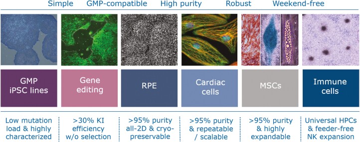
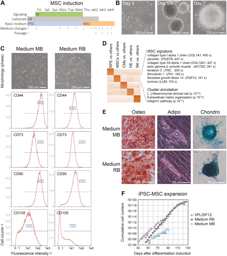
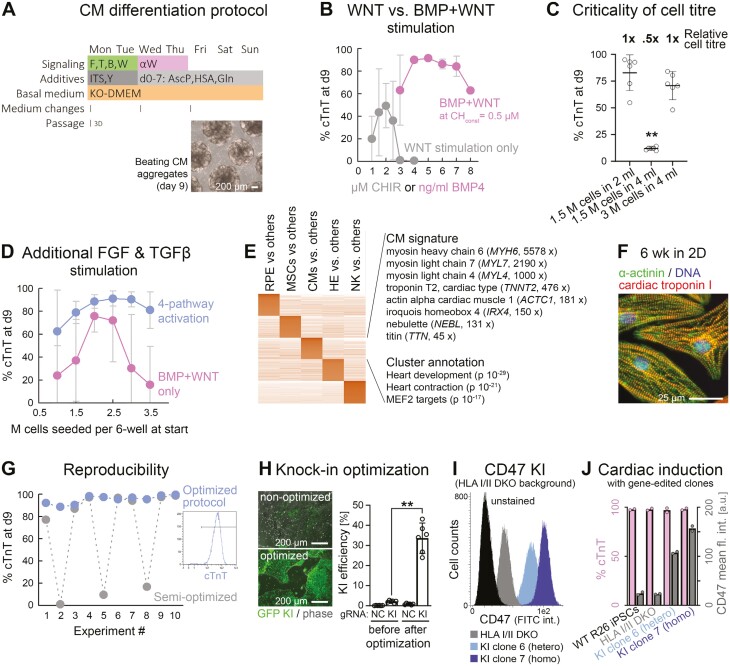
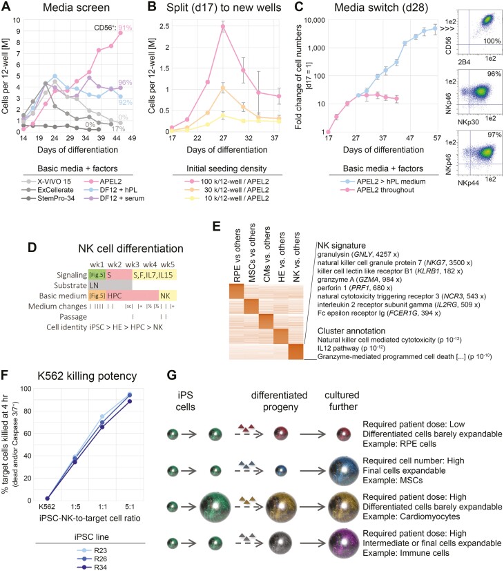
Cell therapeutic applications based on induced pluripotent stem cells (iPSCs) appear highly promising and challenging at the same time. Good manufacturing practice (GMP) regulations impose necessary yet demanding requirements for quality and consistency when manufacturing iPSCs and their differentiated progeny. Given the scarcity of accessible GMP iPSC lines, we have established a corresponding production workflow to generate the first set of compliant cell banks. Hence, these lines met a comprehensive set of release specifications and, for instance, displayed a low overall mutation load reflecting their neonatal origin, cord blood. Based on these iPSC lines, we have furthermore developed a set of GMP-compatible workflows enabling improved gene targeting at strongly enhanced efficiencies and directed differentiation into critical cell types: A new protocol for the generation of retinal pigment epithelium (RPE) features a high degree of simplicity and efficiency. Mesenchymal stromal cells (MSCs) derived from iPSCs displayed outstanding expansion capacity. A fully optimized cardiomyocyte differentiation protocol was characterized by a particularly high batch-to-batch consistency at purities above 95%. Finally, we introduce a universal immune cell induction platform that converts iPSCs into multipotent precursor cells. These hematopoietic precursors could selectively be stimulated to become macrophages, T cells, or natural killer (NK) cells. A switch in culture conditions upon NK-cell differentiation induced a several thousand-fold expansion, which opens up perspectives for upscaling this key cell type in a feeder cell-independent approach. Taken together, these cell lines and improved manipulation platforms will have broad utility in cell therapy as well as in basic research.


Conflict of interest statement

Catalent is a commercial entity that licenses iPSC lines described in this study and offers services and IP licensing based on the disclosed methodology. Several aspects of it form the basis of ongoing patent applications or provisional filings. All of the other authors declared no potential conflicts of interest.

Figures

Graphical Abstract
Graphical Abstract
Figure 5.
Figure 5.
Universal immune cell differentiation platform based on the induction of an HPC intermediate state. (A) 2D protocol for the induction of HPCs by days 14-17. (B) W = BMP and WNT signaling stimulation; V = VEGFA; S, X = target cell-dependent factors such as SCF (see Methods). After 1 week, the cells resembled hemogenic endothelium-like precursors (HE). (B) Expression profiling of the day 7 cells indicates a hybrid signature of endothelial and hematopoietic precursor cells. (C) Flow cytometry data of day 7 cells suggesting near-homogeneous differentiation into HE-like cells (n = 3). (D) Cellular morphology in experimental 12-well format and illustration of EHT process. (E) Representative time course data (flow cytometry) highlighting a homogeneous transition into hematopoietic suspension cells after day 7 (CD34+/CD45+), at the expense of endothelial commitment marked by VE-cadherin. (F) Methylcellulose assays using independent iPSC lines indicate robust formation of hematopoietic colonies from days 14 to 17 cells, comparable to primary HSCs. (G) Expression signature of iPSC-derived HPCs depends on basic medium used, with defined conditions giving rise to naïve CD34+/CD43+/CD117+/CD38 cells. (H) Proof-of-concept experiment demonstrating directed differentiation competence of day 14 cells into monocytes (myeloid lineage). (I) Proof-of-concept experiment demonstrating directed differentiation competence of iPSC-derived HPCs into immature T cells marked by CD4 and 8a. A small fraction also showed mature alpha beta T-cell receptor expression (bottom right panel).
Figure 1.
Figure 1.
Reprogramming process and iPSC characterization. (A) Flowchart of iPSC manufacturing process with QC. (B) Top 7 most frequent Caucasian HLA haplotypes accessible for reprogramming from homozygous donors. R25 and R26 are iPSC lines derived from distinct individuals. (C) Probabilities of identifying matching patients with a given set of HLA-homozygous iPSC lines in the Caucasian population. Open circles in the bottom chart: reprogrammed iPSC lines with pending QC. Filled circles in same chart: released iPSC lines. Two released lines share the same (most frequent) HLA haplotype. (D) Exemplary characterization data of 4 reprogrammed iPSC lines showing the indicated features and markers.
Figure 2.
Figure 2.
Directed all-2D RPE induction. (A) RT-qPCR analysis following RPE induction with 2 protocols (line R26, n = 2-6 per data point). PD: PD0325901 (αFGF), SB: SB431542 (αTGFβ), DM: Dorsomorphin (αBMP). (B) RT-qPCR data showing importance of Activin A supplementation in optimized induction cocktail (line R26, n = 5 for + ActA). (C) Analysis of individual contributions by 3 small molecules in the RPE induction cocktail (RT-qPCR data at 4 weeks). (D) Comparison of media supplements used in the new differentiation protocol. Low RLPB1 expression was consistently observed with the B27 supplement. (E) Optimized RPE differentiation protocol. The FGF, TGFβ, and BMP pathways were (moderately) inhibited during the first week. Overlapping ActA treatment was productive for RPE induction. (F) Light and immunofluorescence analysis of maturated iPSC-RPE cells. A minority of (likely more immature) cells was MITFhigh/CRALBP-negative. (G) RNA-seq analysis of iPSC-RPE cells against other derivatives. Selected markers with ratios against other cell types as well as annotation terms of the RPE cluster are highlighted. (H) Scanning EM pictures at different magnifications reveal pronounced microvilli on all RPE cells. (I) Transmission EM analysis reveals apical-basal polarity with biased localization of melanosomes and nuclei (left) as well as tight junctions (right, marked with arrow heads). (J) Freeze-thawing preserves RPE marker expression (RT-qPCR analysis, n = 2). K, iPSC-RPE cells can efficiently be recovered from cryopreserved stocks by 6 but not 7 weeks of differentiation. All data in this figure are based on iPSC line R26.
Figure 3.
Figure 3.
Single molecule-based induction of highly proliferative iPSC-MSCs. (A) Outline of protocol. W = stimulation of canonical WNT signaling. (B) Cellular morphology at different stages. Cells growing out from re-attached spheres had a fibroblastoid morphology from the beginning. (C) Morphology and surface marker expression of iPSC-MSCs derived from line R26 in 2 commercial MSC media. (D) RNA-seq analysis of R26 iPSC-MSCs against other iPSC-derived cell types showing a pronounced ECM signature. (E) Representative pictures/stainings indicating tri-lineage differentiation competence of R26 iPSC-MSCs. (F) Expansion potential of iPSC-MSCs in different media (n = 2-4 per data series).
Figure 4.
Figure 4.
High-efficiency cardiac induction hallmarked by improved robustness. (A) Outline of optimized protocol based on initial co-stimulation of the FGF, TGFβ, BMP, and WNT pathways. Differentiation commenced upon—not after—EB formation. (B) BMP and WNT co-stimulation (without FGF/ TGFβ) enables superior CM differentiation as compared to WNT stimulation alone, in this protocol (n = 2-10 per data point). (C) Critical dependency of BMP + WNT protocol on cell titer (flow cytometry data). (D) Additional FGF and TGFβ pathway stimulation enhances protocol robustness with regard to cell titer dependency (n = 6-8—dropouts were scored as 0%). (E) Markers and annotation terms of cardiac differentiation cluster (RNA-seq analysis). (F) Immunostaining of iPSC-CMs maturated under adherent conditions. (G) Fully optimized procedure overcomes inter-experimental variation. Data shows flow cytometry data of 10 independent experiments conducted in a row. (H) Optimization of CRISPR-mediated knockin efficiencies in iPSCs using a GFP vector targeting the AAVS1 locus. NC: Negative control using an irrelevant gRNA. (I) Representative flow cytometry plot showing negative controls as well as isolated hetero and homozygous knockin clones of a CD47 transgene targeted to AAVS1 on an HLA I/II deficient background. Note the correlation between signal and gene dosage. (J) Analysis of these iPSC lines after directed cardiac differentiation according to panel A. Data imply consistent differentiation after KO/KI editing and preserved gene dosage-dependent transgene expression.
Figure 6.
Figure 6.
Basic media switch enables significant cell expansion upon NK-cell differentiation. (A) Screening of indicated basic media to support NK-cell differentiation and proliferation from iPSC-HPCs. All media were supplemented with SCF, FLT3L, IL-7, and IL-15. Cells remained in the original differentiation wells throughout (media switch on day 14). (B) Defined APEL2 medium with factors does not support longer-term cell expansion after transfer of HPCs to independent culture wells, regardless of cell density (n = 3 per data point). (C) Media switch at transition point between precursors and NK cells enables cell expansion upon differentiation (n = 6 similar conditions). Note the logarithmic scale. Right panel: Flow cytometric analysis at end of time course. (D) Resulting protocol with indicated media changes and added signaling molecules. S = SCF, F = FLT3L. Refer to Figure 5A for treatments over the first 7 days. (E) RNA-seq analysis highlighting NK-cell-specific cluster with selected marker genes and enrichment terms (also see Supplementary Table S2). (F) Killing assays using NK cells derived from 3 iPSC lines. (G) Illustration linking differentiation paradigms of the present study to context-dependent strategies for upscaling. Ball sizes are to reflect cell numbers.

References

    1. Bertolino GM, Maumus M, Jorgensen C, Noël D.. Recent advances in extracellular vesicle-based therapies using induced pluripotent stem cell-derived mesenchymal stromal cells. Biomedicines. 2022;10(9):2281. 10.3390/biomedicines10092281 - DOI - PMC - PubMed
    1. Zhou Y, Li M, Zhou K, et al.. Engineering induced pluripotent stem cells for cancer immunotherapy. Cancers (Basel). 2022;14(9):2266. 10.3390/cancers14092266 - DOI - PMC - PubMed
    1. Kobold S, Guhr A, Mah N, et al.. A manually curated database on clinical studies involving cell products derived from human pluripotent stem cells. Stem Cell Rep. 2020;15(2):546-555. 10.1016/j.stemcr.2020.06.014 - DOI - PMC - PubMed
    1. Sharma R, Bose D, Montford J, Ortolan D, Bharti K.. Triphasic developmentally guided protocol to generate retinal pigment epithelium from induced pluripotent stem cells. STAR Protoc. 2022;3(3):101582. 10.1016/j.xpro.2022.101582 - DOI - PMC - PubMed
    1. Petrus-Reurer S, Winblad N, Kumar P, et al.. Generation of retinal pigment epithelial cells derived from human embryonic stem cells lacking human leukocyte antigen class I and II. Stem Cell Rep. 2020;14(4):648-662. 10.1016/j.stemcr.2020.02.006 - DOI - PMC - PubMed

MeSH terms