. 2024 Jul 23;17(846):eadp8569.
doi: 10.1126/scisignal.adp8569. Epub 2024 Jul 23.

Generation of antitumor chimeric antigen receptors incorporating T cell signaling motifs

Affiliations

Generation of antitumor chimeric antigen receptors incorporating T cell signaling motifs

Lakshmi Balagopalan et al. Sci Signal. .

Abstract

Chimeric antigen receptor (CAR) T cells have been used to successfully treat various blood cancers, but adverse effects have limited their potential. Here, we developed chimeric adaptor proteins (CAPs) and CAR tyrosine kinases (CAR-TKs) in which the intracellular ζ T cell receptor (TCRζ) chain was replaced with intracellular protein domains to stimulate signaling downstream of the TCRζ chain. CAPs contain adaptor domains and the kinase domain of ZAP70, whereas CAR-TKs contain only ZAP70 domains. We hypothesized that CAPs and CAR-TKs would be more potent than CARs because they would bypass both the steps that define the signaling threshold of TCRζ and the inhibitory regulation of upstream molecules. CAPs were too potent and exhibited high tonic signaling in vitro. In contrast, CAR-TKs exhibited high antitumor efficacy and significantly enhanced long-term tumor clearance in leukemia-bearing NSG mice as compared with the conventional CD19-28ζ-CAR-T cells. CAR-TKs were activated in a manner independent of the kinase Lck and displayed slower phosphorylation kinetics and prolonged signaling compared with the 28ζ-CAR. Lck inhibition attenuated CAR-TK cell exhaustion and improved long-term function. The distinct signaling properties of CAR-TKs may therefore be harnessed to improve the in vivo efficacy of T cells engineered to express an antitumor chimeric receptor.


Conflict of interest statement

Competing interests: L.B., J.Y., K.M., and L.E.S. are co-applicants for a provisional patent on CAPs and CAR-TKs. The other authors declare that they have no competing interests.

Figures

Fig. 1.
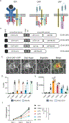
Fig. 1.. Screening of CD19-CAP constructs containing LAT, SLP-76, and ZAP-70 domains.
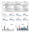
(A) Schematic of TCR signaling, CAR signaling, and CAP signaling. Whereas the TCR and CARs must cross the signaling threshold (indicated as the yellow highlighted bar) for T cell activation, CAPs bypass the upstream steps. (B) Schematic of CD19-CAP constructs. (C) Image of a Jurkat cell expressing the indicated fluorescent proteins forming an immunological synapse with a Raji cell. Scale bar, 2 μm. (D) The percentage lysis of target cells by T cells incubated at a 5:1 effector cell–to–target cell (E:T) ratio. (E) IFN-γ production by T cells incubated with the indicated target cells at a 1:1 E:T ratio. (F) Proliferation of T cells. Data in (D) and (E) were analyzed by one-way ANOVA with Tukey’s post hoc test for antigen-negative samples [NALM6 KO in (D) and K562 in (E)] and antigen-positive samples [NALM6 in (D) and K562 CD19 in (E)] separately. P values in black refer to comparisons of CAPs with the 4–1BBζ control. Data in (F) were analyzed by two-way ANOVA with repeated measure selection with Tukey’s post hoc test, and P values shown refer to comparisons between CAPs and the 4–1BBζ control on day 10. Data are means ± SEM and are representative of three independent experiments from three different donors. **P ≤ 0.01, ****P ≤ 0.0001; ns, not significant.
Fig. 2.
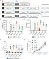
Fig. 2.. Screening of CD19-CAR-TK constructs that contain ZAP-70 domains.
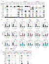
(A) Schematic of the CD19-CAR-TK constructs. (B and C) IL-2 and IFN-γ production by T cells incubated with the indicated target cells at a 1:1 E:T ratio. (D) The percentage lysis of target cells by T cells incubated with the indicated target cells at a 5:1 E:T ratio. (E) Proliferation of T cells. Data in (B) to (D) were analyzed by one-way ANOVA with Tukey’s post hoc test for antigen-negative samples [K562 in (B and C) and NALM6 KO in (D)] and antigen-positive samples [K562 CD19 in (B and C) and NALM6 in (D)] separately. In (B), P values in red refer to comparisons with CAR-TK2, whereas in (C), P values in black refer to comparisons with 4–1BBζ. Data in (E) were analyzed by two-way ANOVA with repeated measure selection with Tukey’s post hoc test. Data are means ± SEM and are representative of three independent experiments from three different donors. *P ≤ 0.05, **P ≤ 0.01, ****P ≤ 0.0001.
Fig. 3.
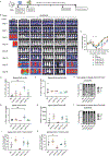
Fig. 3.. CD19-CAR-TK constructs show robust efficacy in an in vivo NSG leukemia model.
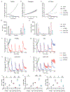
(A) Schematic of the NSG mouse model of leukemia. (B) Leukemia growth was evaluated at the indicated times by bioluminescent imaging (BLI), and IVIS images are shown. (C) Quantification of the BLI radiance data. Data are means ± SEM. Statistical differences were assessed with a Mann Whitney U-test to compare CAR-TK2 (P values in red) or CAR-TK3 (P values in blue) with the 28ζ-CAR. *P ≤ 0.05, **P ≤ 0.01. (D to F) Flow cytometric analysis of peripheral blood on day 30 (D30) assessing CD3+ cells (D), CAR+ cells (E), and T cell subsets as follows: Tn (CD62L+CD45RA+), Tcm (CD62L+, CD45RA), Tem (CD62LCD45RA), and Temra (CD62LCD45RA) (F). (G to I) Flow cytometric analysis of splenocytes on day 44 (D44) assessing CD3+ T cells (G), CAR+ T cells (H), and T cell subsets (I). (J and K) Flow cytometric analysis of splenocytes and bone marrow (BM) cells measuring CD3+CAR+ cells that were PD1+Tim3+LAG3+. Statistical differences were assessed by one-way ANOVA with Tukey’s post hoc test (for D, E, G, H, J, and K) or by two-way ANOVA with Tukey’s post hoc test (F and I). Data are means ±SEM. *P ≤ 0.05, **P ≤ 0.01; ns, not significant.
Fig. 4.
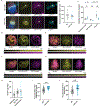
Fig. 4.. CAR-TK cells display reduced recruitment of activated proteins and delayed and prolonged kinetics of protein recruitment compared to CAR-T cells.
(A) Fixed-cell image of a 28ζ-CAR-GFP cell and a CAR-TK3-GFP cell (GFP is pseudocolored in yellow) interacting with a Raji cell (blue). Cells were also transfected with a ZAP70-Apple constructed (magenta) and immunostained for pSLP-76 (turquoise). (B) Volume of the immunological synapse formed by 28ζ-CAR or CAR-TK3 cells. (C) Fluorescence intensities of the indicated proteins at the IS. (D to G) Top: TIRF images of 28ζ-CAR-GFP (D and E) and CAR-TK3-GFP (F and G), as well as of Zap70-Apple (D and F) and Grb2-Apple (E and G). Bottom: A time-lapse montage showing a single microcluster at 3 s/frame over the course of 24 frames (72 s). Scale bar, 2 μm. (H) Kinetic lags between indicated proteins. (I and J) Accumulation time of CAR or CAR-TK3 (I) and Grb2 (J). Data were analyzed by unpaired t test with Welch’s correction. Dot plots show means ± SEM. Data are representative of three independent experiments. **P ≤ 0.01, ****P ≤ 0.0001; ns, not significant.
Fig. 5.
Fig. 5.. Phosphorylation kinetics of CARs and CAR-TKs.
(A to D) Jurkat cells stably expressing 28ζ-CAR or the indicated CAR-TK constructs were mixed with antigen-presenting cells (APCs) that were either antigen-negative (parental K562 cells) or antigen-positive (K562 CD19). Cell mixtures were lysed at the indicated times and analyzed by Western blotting to detect the phosphorylated forms of the indicated proteins. (A) Representative Western blots of 28ζ, CAR-TK2 and CAR-TK3 and signaling protein phosphorylation at the indicated times. (B) Averaged graphs of phosphorylation curves for the indicated markers of CAR and CAR-TK activation and signaling. (C and D) Phosphorylation and dephosphorylation rates for the indicated signaling proteins in each construct. Rates were determined by fitting an expression modeling exponential rise and decay. Paired Student’s t-tests were performed to compare the phosphorylation and dephosphorylation rates in 28ζ vs. CAR-TK2 and 28ζ vs. CAR-TK3 samples. Data are means ± SEM and are representative of three independent experiments. *P ≤ 0.05, **P ≤ 0.01; ns, not significant.
Fig. 6.
Fig. 6.. Dependency of CARs and CAR-TKs on receptor-proximal signaling proteins.
(A to C) CAR or CAR-TK constructs were used to stably transduce Jurkat E6.1 cells that had wild-type, CRISPR Lck KO, P116 ZAP KO, or CRSIPR TCRβ KO genetic backgrounds. Those Cells were mixed with antigen-positive or antigen-negative APCs, incubated at 37°C for 8 min, lysed, and analyzed by Western blotting for the phosphorylated forms of the indicated signaling proteins. (A) Representative Western blots of the components of 28ζ, CAR-TK2, and CAR-TK3 signaling in WT, Lck KO, ZAP70 KO, and TCRβ KO cells. (B) Averaged graphs of the band intensities of the phosphorylated forms of the indicated components of CAR and CAR-TK activation and signaling. Paired Student’s t-tests were performed to compare the abundances of the indicated phosphorylated signaling proteins in stimulated WT vs. Lck KO or ZAP70 KO cells. (C) Representative Western blot of 28ζ and CAR-TK3 signaling components in WT and Lck KO cells after preincubation with DMSO or the SFK inhibitor PP1. Data are means ± SEM and are representative of three independent experiments. *P ≤ 0.05, **P ≤ 0.01, ***P ≤ 0.001, ****P ≤ 0.0001; ns, not significant.
Fig. 7.
Fig. 7.. CAR-TKs function in the absence of Lck activity.
(A to I) CAR and CAR-TK constructs PBMCs from healthy donors transduced to express the indicated CAR and CAR-TK constructs were treated with DMSO, Dasatinib, or A770041 (A77) and then incubated with NALM6-GFP+ cells in the Incucyte ZOOM Live-Cell analysis system for 48 hours (A to C) or over 10 days with repeated stimulations (F to I). (A to C) Representative graphs of total integrated GFP intensity to assess the amount of tumor cell killing that occurred over 48 hours. (D) Measurement of IFN-γ production at 48 hours. (E) Analysis of CAR surface expression at 48 hours. (F to I) CAR-T and CAR-TK T cells were treated as described for (A) with DMSO, Dasatinib, or A77 and subjected to repeat NALM6-GFP+ stimulations (indicated by arrows) over 10 days. The tumor cell killing indices for a representative donor are shown. (J to L) Analysis of the expression of exhaustion markers on CAR-T and CAR-TK cells treated with DMSO or A77 after repeated restimulations at day 10. Data are means ± SEM and are representative of three independent experiments from three different donors. Statistical differences between DMSO- and A77-treated samples were determined by paired Student’s t test. *P ≤ 0.05, **P ≤ 0.01, ***P ≤ 0.001; ns, not significant.

References

    1. June CH, Sadelain M, Chimeric Antigen Receptor Therapy. N Engl J Med 379, 64–73 (2018); published online EpubJul 5 (10.1056/NEJMra1706169). - DOI - PMC - PubMed
    1. Kochenderfer JN, Dudley ME, Feldman SA, Wilson WH, Spaner DE, Maric I, Stetler-Stevenson M, Phan GQ, Hughes MS, Sherry RM, Yang JC, Kammula US, Devillier L, Carpenter R, Nathan DA, Morgan RA, Laurencot C, Rosenberg SA, B-cell depletion and remissions of malignancy along with cytokine-associated toxicity in a clinical trial of anti-CD19 chimeric-antigen-receptor-transduced T cells. Blood 119, 2709–2720 (2012); published online EpubMar 22 (10.1182/blood-2011-10-384388). - DOI - PMC - PubMed
    1. Kochenderfer JN, Wilson WH, Janik JE, Dudley ME, Stetler-Stevenson M, Feldman SA, Maric I, Raffeld M, Nathan DA, Lanier BJ, Morgan RA, Rosenberg SA, Eradication of B-lineage cells and regression of lymphoma in a patient treated with autologous T cells genetically engineered to recognize CD19. Blood 116, 4099–4102 (2010); published online EpubNov 18 (10.1182/blood-2010-04-281931). - DOI - PMC - PubMed
    1. Porter DL, Levine BL, Kalos M, Bagg A, June CH, Chimeric antigen receptor-modified T cells in chronic lymphoid leukemia. N Engl J Med 365, 725–733 (2011); published online EpubAug 25 (10.1056/NEJMoa1103849). - DOI - PMC - PubMed
    1. Majzner RG, Mackall CL, Clinical lessons learned from the first leg of the CAR T cell journey. Nat Med 25, 1341–1355 (2019); published online EpubSep (10.1038/s41591-019-0564-6). - DOI - PubMed

MeSH terms

Substances

LinkOut - more resources