. 2024 Jul 23;12(7):e009221.
doi: 10.1136/jitc-2024-009221.

Directing B7-H3 chimeric antigen receptor T cell homing through IL-8 induces potent antitumor activity against pediatric sarcoma

Affiliations

Directing B7-H3 chimeric antigen receptor T cell homing through IL-8 induces potent antitumor activity against pediatric sarcoma

Jessica A Lake et al. J Immunother Cancer. .

Abstract

Background: Advances in pediatric oncology have occurred for some cancers; however, new therapies for sarcoma have been inadequate. Cellular immunotherapy using chimeric antigen receptor (CAR) T cells has shown dramatic benefits in leukemia, lymphoma, and multiple myeloma but has been far less successful in pediatric solid tumors such as rhabdomyosarcoma (RMS) and osteosarcoma (OS). Balancing issues of "on-target, off-tumor toxicity", investigators have identified B7-H3 as a broadly expressed tumor antigen with otherwise restricted expression on normal tissues. We hypothesized that rapid homing via a chemokine receptor and CAR engagement through B7-H3 would enhance CAR T cell efficacy in solid tumors.

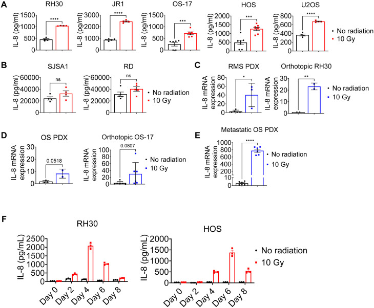
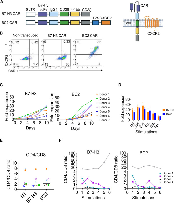
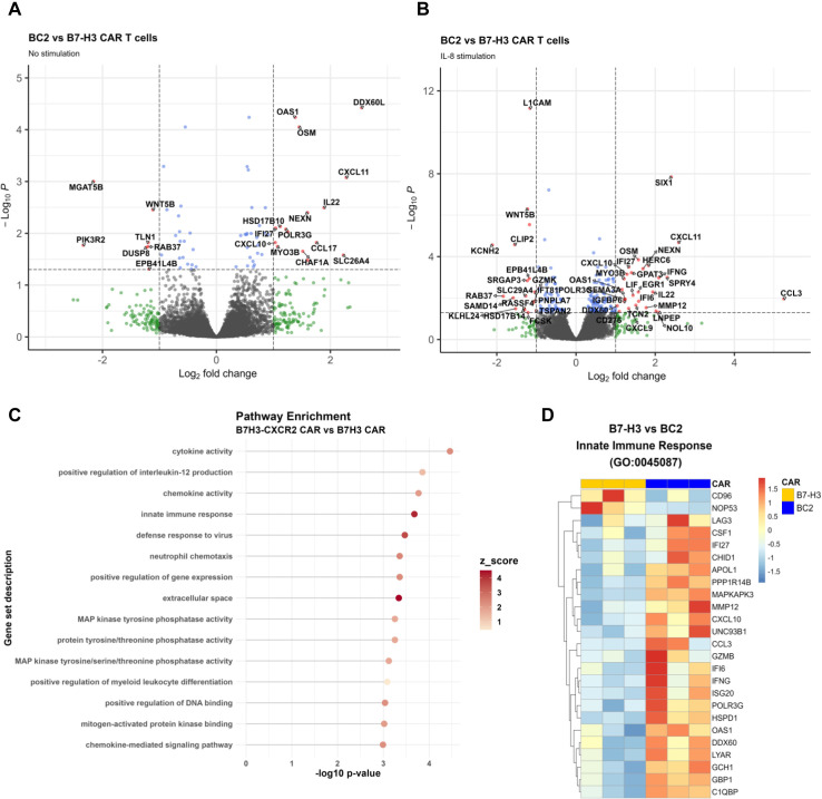
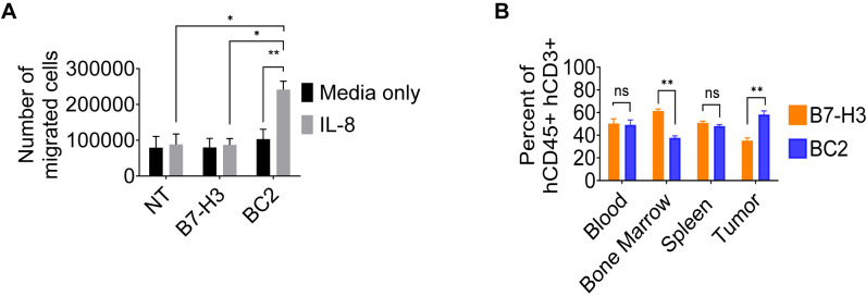
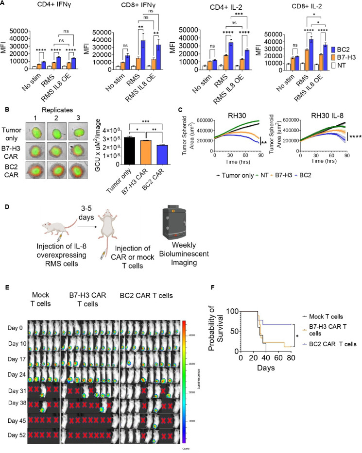
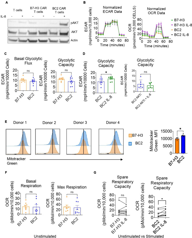
Methods: We generated B7-H3 CAR T cells that also express the Interleukin-8 (IL-8) receptor, CXCR2. Cytokine production, flow cytometry, Seahorse assays and RNA sequencing were used to compare the B7-H3 CXCR2 (BC2) CAR T cells with B7-H3 CAR T cells. We developed an IL-8 overexpressing human RMS mouse model to test homing and cytotoxicity in vivo.

Results: We demonstrate that IL-8 is expressed by RMS and OS and expression significantly increases after radiation. Overexpression of an IL-8 receptor, CXCR2, on B7-H3 CAR T cells enhances homing into IL-8 expressing tumors, augments T cell metabolism and leads to significant tumor regression.

Conclusion: These findings warrant further investigation into the use of BC2 CAR T cells as a treatment for patients with RMS, OS and other B7-H3-expressing, IL-8 producing solid tumors.

Keywords: Adoptive cell therapy - ACT; Chimeric antigen receptor - CAR; Immunotherapy; Solid tumor.


Conflict of interest statement

Competing interests: US Patent (University of Colorado): W02022221592: Compositions and methods for producing and using cell-based immunotherapies to target tumors (inventors: MRV and JAL).

Figures

Figure 1
Figure 1. Radiation induces IL-8 expression by pediatric OS and RMS cell lines and PDX models. (A, B) IL-8 protein expression by OS and RMS cell lines was measured by ELISA, without irradiation or 4 days after 10 Gy irradiation (n=4–7). (C–E) Cells were engrafted orthotopically into NSG mice until tumors were palpable (near 1 cm). Tumors were irradiated (or not irradiated) and then all tumors were removed 4 days later. IL-8 gene expression by qPCR (n=2–6 mice). Each data point represents a replicate experiment. Comparisons were made using unpaired t tests. (F) IL-8 protein expression by RH30 and HOS cell lines as measured by ELISA, without irradiation and at different time points after 10 Gy irradiation (n=3). *p<0.05, **p<0.01, ***p<0.001, ****p<0.0001. OS, osteosarcoma; RMS, rhabdomyosarcoma; PDX, patient-derived xenograft.
Figure 2
Figure 2. Engineering and characterization of B7-H3 and BC2 CAR T cells. (A) Schematic of the B7-H3 and BC2 CAR construct modular designs and membrane location of the proteins once the construct is expressed and the T2A linker is cleaved. (B) Representative retroviral transduction efficiency of human T cells with the B7-H3 and BC2 CAR by flow cytometry. (C) Fold expansion in the first 10 days after transduction of B7-H3 and BC2 CAR T cells, in the presence of IL-2 (100 µ/mL). Comparisons made by unpaired t-test at day 10 (p=ns; n=7 donors). (D) RH30 IL-8 OE cells were plated at 50,000 cells/well and CAR T cells (either B7-H3 or BC2) were added at equal numbers. Every week for 6 weeks, 50,000 CAR T cells were removed from the prior week’s culture and replated with equal numbers of fresh tumor cells. Fold expansion of B7-H3 and BC2 CAR T cells was measured over 6 weeks of chronic IL-8 overexpressing tumor stimulation. Comparisons made by unpaired t-test (n=4; p=ns). (E) CD4/CD8 ratio of non-transduced, B7-H3 CAR and BC2 CAR T cells at day 10 of culture. Comparison made by Kruskal-Wallis test between T cell groups (p=ns; n=7). (F) CD4/CD8 ratio of B7-H3 and BC2 CAR T cells after chronic tumor (IL-8) stimulation (n=4).
Figure 3
Figure 3. Transcriptional signatures of BC2 CAR T cells favor increased chemotaxis and innate immunity pathways. (A, B) Volcano plots showing the top differentially expressed genes (DEG) between BC2 and B7-H3 CAR T cells with either (A) no IL-8 stimulation (n=3 donors) or (B) after IL-8 stimulation (50 ng/mL for 24 hours; n=3). (C) Top enriched pathways in BC2 vs B7-H3 CAR T cells after IL-8 stimulation. (D) Heatmap showing top DEGs in the innate immune response pathway between BC2 and B7-H3 CAR T cells after IL-8 stimulation. Gene names are listed on the right side and each CAR is marked at the top.
Figure 4
Figure 4. Coexpression of CXCR2 with a B7H3 CAR improves homing of CAR T cells. (A) In vitro Boyden chamber transwell migration assay of non-transduced, B7-H3 CAR and BC2 CAR T cells with either media alone (black bars) or media+IL-8 at 50 ng/mL (gray bars) in the bottom chamber. Migration was assessed after 6 hours. Comparisons made by unpaired t-tests (n=4) (B) RH30 IL-8 OE cells were engrafted orthotopically into NSG mice until tumors were palpable (near 1 cm). B7-H3 and BC2 CAR T cells were injected 1:1 (B7-H3 and BC2 labeled with either Celltrace far-red or Celltrace violet) via tail vein. Four days later, mice were bled and euthanized to collect organs and tumors for processing and analysis by flow cytometry. The percentage of each CAR construct gated on human CD45+ and human CD3+ cells from mouse blood, bone marrow, spleen and IL-8 OE human RMS tumor were analyzed by flow cytometry. Comparisons made by unpaired t-test between treatment groups (n=6). *p<0.05, **p<0.01. RMS, rhabdomyosarcoma.
Figure 5
Figure 5. B7H3 CAR+CXCR2 combination increases antitumor efficacy of CAR T cells and eradicates tumor in an NSG mouse model. (A) Mean fluorescent intensity (MFI) by flow cytometry of Interferon-γ and IL-2 (by intracellular staining) expression in non-transduced (mock), B7-H3 and BC2 CAR T cells, 24 hours after tumor stimulation. Cells were either unstimulated or stimulated with RH30 or RH30 IL8 OE cells at 1:1 E:T ratio. Analysis done by two-way ANOVA with multiple comparisons (n=4). (B) Incucyte live cell images of GFP-expressing RMS IL-8 OE tumor spheroids, alone and with B7-H3 CAR or BC2 CAR T cells at 24 hours. Apoptotic activity was detected using a red Annexin V conjugate. Bar graph shows integrated intensity and area of the tumor (green) (GCU×µm2/image) of each group and analyzed with one-way ANOVA with multiple comparisons. (C) Incucyte cytotoxicity assay measuring tumor spheroid area over time with different T/CAR T cell treatments. Analysis by one-way ANOVA with multiple comparisons. (D) Schematic of mouse experiment, created with BioRender.com. (E) IVIS imaging of luciferase-expressing RMS IL-8 OE mice receiving non-transduced (mock; n=5), B7-H3 CAR (n=9) or BC2 CAR T cells (n=9). (F) Survival curve to 80 days. Survival analysis by the Mantel-Cox test. *p<0.05, **p<0.01, ***p<0.001, ****p<0.0001. ANOVA, analysis of variance.
Figure 6
Figure 6. BC2 CAR T cells have increased metabolic potential. (A) Western blot analysis for phospho-(Serine 473) and total AKT in lysates from T cells, B7H3 CAR T or BC2 CAR T cells with and without IL-8 stimulation (50 ng/mL) for 10 min. Blots are representative of three different experiments. (B) Representative Seahorse traces (normalized) for B7-H3 and BC2 CAR T cells without and after IL-8 stimulation. (C) Seahorse assay measuring basal glycolytic flux and glycolytic capacity of B7H3 and BC2 CAR T cells (n=9). (D) Glycolytic capacity of BC2 CAR T cells with and without IL-8 stimulation alone and after treatment by AKT inhibitor VIII (4 uM; n=6–9). (E) Quantification of mitochondria using Mitotracker green dye and flow cytometry of B7H3 CAR and BC2 CAR T cells. Individual histograms shown by donor (n=4). (F) Mitochondrial stress test showing basal and maximum respiration of both CAR T cell groups (n=8). (G) Mitochondrial respiration of each CAR T cell group with and without a 24-hour IL-8 stimulation (n=8). Comparisons were made by unpaired (C, E, F) and paired (D, G) t-tests. *p<0.05.

Similar articles

Cited by

References

    1. Maude SL, Laetsch TW, Buechner J, et al. Tisagenlecleucel in children and young adults with B-cell Lymphoblastic leukemia. N Engl J Med. 2018;378:439–48. doi: 10.1056/NEJMoa1709866. - DOI - PMC - PubMed
    1. Ahmed N, Brawley VS, Hegde M, et al. Human epidermal growth factor receptor 2 (HER2)-specific chimeric antigen receptor-modified T cells for the immunotherapy of HER2-positive sarcoma. J Clin Oncol. 2015;33:1688–96. doi: 10.1200/JCO.2014.58.0225. - DOI - PMC - PubMed
    1. Haas AR, Tanyi JL, O’Hara MH, et al. Phase I study of lentiviral-transduced chimeric antigen receptor-modified T cells recognizing mesothelin in advanced solid cancers. Mol Ther. 2019;27:1919–29. doi: 10.1016/j.ymthe.2019.07.015. - DOI - PMC - PubMed
    1. June CH, O’Connor RS, Kawalekar OU, et al. CAR T cell immunotherapy for human cancer. Science. 2018;359:1361–5. doi: 10.1126/science.aar6711. - DOI - PubMed
    1. O’Rourke DM, Nasrallah MP, Desai A, et al. A single dose of peripherally infused EGFRvIII-directed CAR T cells mediates antigen loss and induces adaptive resistance in patients with recurrent glioblastoma. Sci Transl Med. 2017;9:eaaa0984. doi: 10.1126/scitranslmed.aaa0984. - DOI - PMC - PubMed

LinkOut - more resources