. 2024 Jul 23;15(1):6218.
doi: 10.1038/s41467-024-50650-0.

Dissemination of pathogenic bacteria is reinforced by a MARTX toxin effector duet

Affiliations

Dissemination of pathogenic bacteria is reinforced by a MARTX toxin effector duet

Sanghyeon Choi et al. Nat Commun. .

Abstract

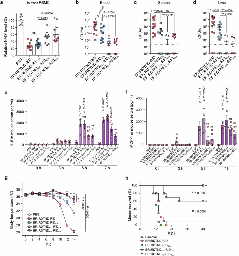
Multiple bacterial genera take advantage of the multifunctional autoprocessing repeats-in-toxin (MARTX) toxin to invade host cells. Secretion of the MARTX toxin by Vibrio vulnificus, a deadly opportunistic pathogen that causes primary septicemia, the precursor of sepsis, is a major driver of infection; however, the molecular mechanism via which the toxin contributes to septicemia remains unclear. Here, we report the crystal and cryo-electron microscopy (EM) structures of a toxin effector duet comprising the domain of unknown function in the first position (DUF1)/Rho inactivation domain (RID) complexed with human targets. These structures reveal how the duet is used by bacteria as a potent weapon. The data show that DUF1 acts as a RID-dependent transforming NADase domain (RDTND) that disrupts NAD+ homeostasis by hijacking calmodulin. The cryo-EM structure of the RDTND-RID duet complexed with calmodulin and Rac1, together with immunological analyses in vitro and in mice, provide mechanistic insight into how V. vulnificus uses the duet to suppress ROS generation by depleting NAD(P)+ and modifying Rac1 in a mutually-reinforcing manner that ultimately paralyzes first line immune responses, promotes dissemination of invaders, and induces sepsis. These data may allow development of tools or strategies to combat MARTX toxin-related human diseases.


Conflict of interest statement

The authors declare no competing interests.

Figures

Fig. 1
Fig. 1. Pathogenic bacteria use the DUF1-RID effector duet within the MARTX toxin to hijack calcium-free calmodulin during infection.
a Schematic diagram showing the domains of the DUF1-RID module within the MARTX toxin secreted by V. vulnificus MO6-24/O strain. DUF1 domain of unknown function in the first position, RID Rho inactivation domain, ND N-terminal domain (RIDND), MLD_C membrane localization domain-containing domain (RIDMLD_C), CD catalytic domain (RIDCD). b Analysis of the DUF1-RID module, and its variants containing RID, within the MARTX toxin from different bacterial genera. c Characteristics of effectors within the MARTX toxin from clinical V. cholerae and V. vulnificus strains. Not aligned, not aligned with the DUF1-RID module. d Cytopathogenicity of the indicated proteins. GFP or GFP-fused proteins were expressed in HEK293T cells and analyzed by confocal imaging. Note that the expression level of DUF1-RID, DUF1-RIDC/A, and RID was much lower than that of RIDC/A due to their inherent cytotoxicity. e In vitro pull-down assay showing the interactions between DUF1-RID and its variants with calmodulin (CaM). The data shown in d and e are representative of three independent experiments, each with similar results. Source data are provided as a Source Data file.
Fig. 2
Fig. 2. DUF1 is a NAD(P)+ hydrolase that is transformed via RID-dependent CaM-binding.
Overall structures of DUF1-RIDCBD (a) and DUF1-RIDCBD complexed with CaM (b). The structures are shown as cartoon diagrams. c Superimposition of the DUF1-RIDCBD structure (gray) onto the CaM-binding DUF1-RIDCBD structure. The arrows indicate significant conformational changes induced in DUF1-RIDCBD upon interaction with CaM. d Structural comparison between DUF1 within DUF1-RIDCBD complexed with CaM, human CD38 (PDB ID 1YH3), and A. californica ADP-ribosyl cyclase (PDB ID: 1R12). The catalytic residues in each protein are represented by sticks. e Switching of non-functional catalytic residues (gray) within DUF1 to functional residues (yellow) within DUF1-RIDCBD complexed with CaM. f Superimposition of the functional catalytic site of DUF1 onto that of human CD38 or A. californica ADP-ribosyl cyclase. Catalytic residues are displayed. g In vitro NAD+-hydrolyzing assay for DUF1-RID and its truncates, with or without CaM. Levels of NAD+ relative to PBS are represented (n = 3 per group). In vitro NAD+- or NADP+-hydrolyzing assay for DUF1-RID and its mutants complexed with CaM or Ca2+-bound CaM. NAD+ (h) or NADP+ (i) levels relative to PBS are represented (n = 3 per group). Data shown in gi are representative of three independent experiments, each with similar results (mean ± SEM). P-values were calculated using one-way ANOVA with multiple comparisons. ns, not significant. Source data are provided as a Source Data file.
Fig. 3
Fig. 3. Detailed molecular structure of RDTND-RIDCBD/CaM/Ca2+ complexed with NAD+.
a Overall structure of RDTND-RIDCBD/CaM complexed with NAD+, and the binding mode of NAD+ at the catalytic site (magnified panel). The mFo-DFc omit electron density map of NAD+ contoured at 3.0 σ is shown as a mesh (red). b Superimposition of the NAD+-bound RDTND-RIDCBD/CaM structure onto the RDTND-RIDCBD/CaM structure (gray). Significant conformational changes in RDTNDLid are shown (magnified panel). c In vitro NAD+-hydrolyzing assay for CaM-bound RDTND-RIDC/A and its RDTND mutants. The NAD+ levels relative to the PBS control are presented (n = 3 per group). Data are representative of three independent experiments, each with similar results (mean ± SEM). P-values were calculated using one-way ANOVA with multiple comparisons. ns, not significant. d Structural comparison of active NAD+-bound RDTND with that of NAD+-bound human CD38 (PDB ID 2I65) and NAD+-bound A. californica ADP-ribosyl cyclase (PDB ID: 3ZWM). Structures are shown as cartoon (upper panel) or surface diagrams (lower panel). Source data are provided as a Source Data file.
Fig. 4
Fig. 4. Cryo-EM structure of RDTND-RIDC/A complexed with CaM and Rac1Q61L.
Reconstructed 3D cryo-EM map (a) and the fitted model (cartoon representation) of RDTND-RIDC/A complexed with CaM and Rac1Q61L (b). Catalytic residue C2838 in RID, and the C-terminal polybasic (PBR, red dashed line) and Switch 1 regions in Rac1Q61L, are indicated. c Surface charge distribution of the RDTND-RIDC/A/CaM/Rac1Q61L complex. The negative and positive charges are shown as red and blue, respectively. The positively charged surface of RIDMLD_C and RIDCD is highlighted by a dashed line. d Subcellular localization of the RDTND-RID module and its truncates. The GFP, GFP-fused RDTNDE/Q-RIDC/A, or GFP-fused RDTND-RID truncates (RDTNDE/Q-RIDCBD/MLD_C, RDTNDE/Q-RIDCBD/MLD, and RDTND) were expressed in HeLa cells and analyzed. The HeLa cell plasma membrane and nucleus were stained with CF® 594-conjugated wheat germ agglutinin (WGA) and Hoechst 33342, respectively. Data are representative of three independent experiments, each with similar results. e Cartoon summarizing the re-localization of RDTND-RID to the membrane after CPD-mediated release from the MARTX toxin, as well as the concerted invasion strategy used by the effector duet at the membrane and near the membrane.
Fig. 5
Fig. 5. The RDTND-RID duet silences the ROS/ERK/JNK/NF-κB axis, which is critical for first line immune responses.
a Schematic diagram showing MARTX toxins from engineered V. vulnificus mutant strains. Substituted residues in RDTND-RID within the MARTX toxin from each mutant strain are indicated. Intracellular NAD+ (b) and NADP+ (c) levels in BMDMs infected for 2 h with the indicated V. vulnificus strains (MOI = 5, 5 × 10⁶ CFU). NAD(P)+ levels relative to PBS are presented (n = 3 per group). ROS level in RAW 264.7 cells infected for 2 h with the indicated V. vulnificus strains (MOI = 5, 5 × 10⁶ CFU). ROS production was measured by flow cytometry (d), and fold changes in mean fluorescence intensity (MFI) compared with the PBS control are shown (n = 3 per group) (e). f Immunoprecipitation assay. Lysates of BMDMs infected for 2 h with the indicated V. vulnificus strains were immunoprecipitated by an anti-gp91phox antibody, followed by immunoblot analysis with anti-Rac1, anti-p47phox, anti-p67phox, and anti-gp91phox antibodies. g Immunoblot analysis of MAPK signaling molecules and transcription factors in BMDMs infected for 2 h with the indicated V. vulnificus strains. EF, effector-free; WT-WT, EF::RDTND-RID; WT-C/A, EF::RDTND-RIDC/A; E/Q-C/A, EF::RDTNDE/Q-RIDC/A; 4mt-C/A, EF::RDTND4mt-RIDC/A. ELISA of pro-inflammatory cytokines IL-6 (h), TNF-α (i), and MCP-1 (j) in supernatants from BMDMs infected for 2 h with the indicated V. vulnificus strains (n = 6 per group). Data shown are representative of three independent experiments, each with similar results (mean ± SEM). P-values were calculated using one-way ANOVA with multiple comparisons. ns, not significant. Source data are provided as a Source Data file.
Fig. 6
Fig. 6. The RDTND-RID duet promotes bacterial dissemination and sepsis in mice.
a Intracellular NAD+ levels in PBMCs isolated from 5-week-old female ICR mice 7 h post-infection with the indicated V. vulnificus strains. NAD+ levels relative to PBS are presented (n = 8 for PBS; n = 12 for EF::RDTND-RID, EF::RDTND-RIDC/A, EF::RDTNDE/Q-RIDC/A, and EF::RDTND4mt-RIDC/A). Single bacterial colonies in blood (b), spleen (c), and liver (d) collected from 5-week-old female ICR mice at 10 h post-infection with the indicated V. vulnificus strains; normalized data are shown (n = 14 per group). ELISA of pro-inflammatory cytokines of IL-6 (e) and MCP-1 (f) in serum collected from 5-week-old female ICR mice infected with the indicated V. vulnificus strains at the designated time (n = 2 for 0 h, and n = 6 for 3 h, 5 h, and 7 h post-infection per group). P-values were calculated versus the PBS control at each time point, and are indicated above each graph. ICR mice were challenged with indicated V. vulnificus strains (n = 10, 5 × 10⁶ CFU per group), and the ventral surface temperature (g) and survival rates (h) of the infected mice are shown. h.p.i., hours post-infection. The ventral surface temperature at 14 h was analyzed, and P-values compared with those from PBS-treated mice. Data shown in ad are pooled from three independent experiments (mean ± SEM). Data shown in e and f are representative of three independent experiments, each with similar results (mean ± SEM). Data shown in g and h are pooled from n = 5 per group from n = 2 independent experiments. P-values were calculated using one-way ANOVA with multiple comparisons (a, g), a non-parametric Mann-Whitney U-test (two-tailed) (bd), or a two-way ANOVA with multiple comparisons (e, f) or log-rank test (h). ns, not significant. Source data are provided as a Source Data file.

References

    1. Lee, Y. et al. Makes caterpillars floppy-like effector-containing MARTX toxins require host ADP-ribosylation factor (ARF) proteins for systemic pathogenicity. Proc. Natl Acad. Sci. USA116, 18031–18040 (2019). 10.1073/pnas.1905095116 - DOI - PMC - PubMed
    1. Choi, S., Kim, B. S., Hwang, J. & Kim, M. H. Reduced virulence of the MARTX toxin increases the persistence of outbreak-associated Vibrio vulnificus in host reservoirs. J. Biol. Chem.296, 100777 (2021). 10.1016/j.jbc.2021.100777 - DOI - PMC - PubMed
    1. Kim, B. S. et al. MARTX Toxin-Stimulated Interplay between Human Cells and Vibrio vulnificus. mSphere5, 10.1128/mSphere.00659-20 (2020). - PMC - PubMed
    1. Gavin, H. E. & Satchell, K. J. F. RRSP and RID Effector Domains Dominate the Virulence Impact of Vibrio vulnificus MARTX Toxin. J. Infect. Dis.219, 889–897 (2019). 10.1093/infdis/jiy590 - DOI - PMC - PubMed
    1. Gavin, H. E. & Satchell, K. J. MARTX toxins as effector delivery platforms. Pathog. Dis.73, ftv092 (2015). 10.1093/femspd/ftv092 - DOI - PMC - PubMed

LinkOut - more resources