. 2024 Jul 23;14(1):16970.
doi: 10.1038/s41598-024-67912-y.

B cells secrete functional antigen-specific IgG antibodies on extracellular vesicles

Affiliations

B cells secrete functional antigen-specific IgG antibodies on extracellular vesicles

Claudia Rival et al. Sci Rep. .

Abstract

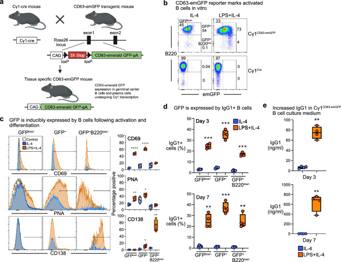
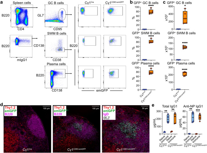
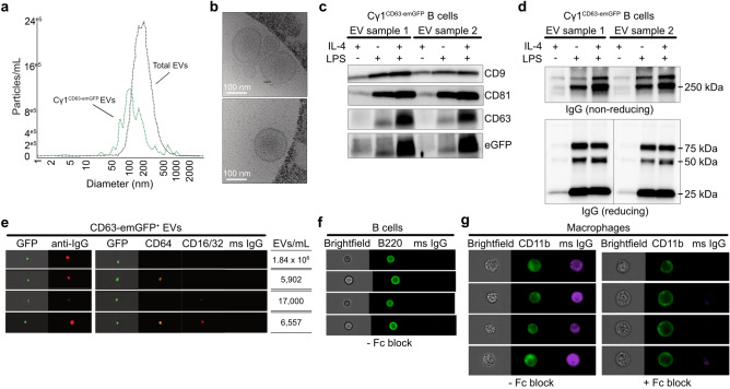
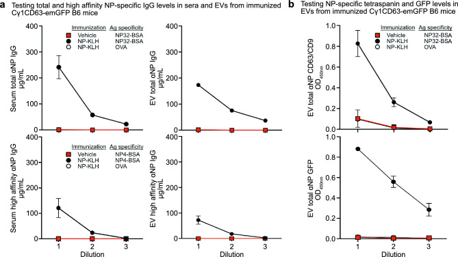
B cells and the antibodies they produce are critical in host defense against pathogens and contribute to various immune-mediated diseases. B cells responding to activating signals in vitro release extracellular vesicles (EV) that carry surface antibodies, yet B cell production of EVs that express antibodies and their function in vivo is incompletely understood. Using transgenic mice expressing the Cre recombinase in B cells switching to IgG1 to induce expression of fusion proteins between emerald green fluorescent protein (emGFP) and the EV tetraspanin CD63 as a model, we identify emGFP expression in B cells responding to foreign antigen in vivo and characterize the emGFP+ EVs they release. Our data suggests that emGFP+ germinal center B cells undergoing immunoglobulin class switching to express IgG and their progeny memory B cells and plasma cells, also emGFP+, are sources of circulating antigen-specific IgG+ EVs. Furthermore, using a mouse model of influenza virus infection, we find that IgG+ EVs specific for the influenza hemagglutinin antigen protect against virus infection. In addition, crossing the B cell Cre driver EV reporter mice onto the Nba2 lupus-prone strain revealed increased circulating emGFP+ EVs that expressed surface IgG against nuclear antigens linked to autoimmunity. These data identify EVs loaded with antibodies as a novel route for antibody secretion in B cells that contribute to adaptive immune responses, with important implications for different functions of IgG+ EVs in infection and autoimmunity.

Keywords: Adaptive immunity; Antibody production; B cells; Extracellular vesicles.


Conflict of interest statement

The authors declare no competing interests.

Figures

Figure 1
Figure 1
Activated Cγ1CD63-emGFP B cells undergoing Cre-mediated recombination in vitro express emGFP. (a) Schematic for Cγ1CD63-emGFP reporter strain design. Transgenic mice expressing Cre recombinase driven by transcription of the Ig γ1 constant region gene segment (Cγ1) was crossed with a silenced reporter mouse, resulting in CD63-emerald GFP expression driven by the CAG promoter. (b) Percentage of emGFP+ Cγ1CD63-emGFP and Cγ1Cre control B cells following stimulation with IL-4 or LPS + IL-4 for 3 days. Gates indicate GFPlow/- B cells, GFP+ B cells and GFP+B220low/− B cells. (c) Percentage of cell surface expression of CD69, PNA and CD138 in GFPlow/−, GFP+ and GFP+B220low/− B cells at day 3, for the Cγ1CD63-emGFP reporter mice in panel (b). Gates were set on negative control samples (dotted black histograms). (d) Frequencies of IgG1+ B cells in B cells gated on GFP expression at days 3 and 7, for the reporter mice in (b). (e) Concentration of IgG1 in the culture medium of B cells from the reporter mice in (b). All data are expressed as mean ± SEM. Results shown are representative of at least three independent experiments. P = **0.01 and ***0.001, with unpaired, two-tailed t-test.
Figure 2
Figure 2
In vivo generation of Cγ1CD63-emGFP GC B cells and progeny IgG1+ memory B cells and plasma cells express emGFP after immunization with NP-KLH. (a) Flow cytometry gating strategy of GC B cells, IgG1+ SWM B cells, and plasma cells that express emGFP in the spleens of Cγ1Cre and Cγ1CD63-emGFP mice immunized with NP-KLH for 14 days. (b) Percentages of emGFP+ cells within each gated B cell subset are shown. (c) Numbers of emGFP+ B cells in spleens for the reporter mice shown in panel (b). (d) Immunofluorescence confocal microscopy of emGFP+ B cells in the lymph nodes of mice immunized with NP-KLH for 14 days. (e) Serum total and NP-specific IgG1 antibody levels in naïve Cγ1Cre and Cγ1CD63-emGFP mice and after immunization with NP-KLH for 14 days. Data are expressed as mean ± SEM. Results shown in panels (ac) and (e) are representative of three independent experiments and in panel d from > 20 images of two independent experiments. P = *0.05, **0.01, ***0.001 and ****0.0001, with unpaired, two-tailed t-test.
Figure 3
Figure 3
Activated Cγ1CD63-emGFP B cells undergoing Cre-mediated recombination in vitro produce EVs that express emGFP, tetraspanins, and IgG. (a) Nanoparticle tracking analysis of total and GFP+ EVs isolated from cell culture medium from Cγ1CD63-emGFP B cells stimulated with LPS + IL-4 for 7 days. (b) EVs were visualized by cryoelectron microscopy. Images are representative of three independent EV preparations. (c, d) Lysates from EVs isolated from cell culture medium from Cγ1CD63-emGFP B cells stimulated with IL-4, LPS, and LPS + IL-4 for 7 days were probed by Western blot for the presence of CD9, CD81, CD63, GFP, and IgG under non-reducing conditions, and IgG under reducing conditions (10 μg/lane). Under reducing conditions, IgG bands from EV samples 1 and 2 are juxtaposed from the same blot as indicated by vertical line, from full length blots shown in Supplementary Fig. S6b. Results show two EV preparations and are representative of at least six independent experiments. (e) ImageStream analysis of individual emGFP+ EVs isolated from the cell culture medium of Cγ1CD63-emGFP B cells stimulated with LPS + IL-4 and stained for surface IgG or CD16/32 (Alexa Fluor 647), CD64 (PE), and binding of exogenous mouse IgG (DyLight 405). (f) ImageStream analysis of peritoneal B cells stained for surface B220 (FITC) and binding of exogenous mouse IgG. (g) ImageStream analysis of peritoneal macrophages stained for surface CD11b (FITC) and binding of exogenous mouse IgG in the absence (left panel) or presence of 2.4G2 Fc block (right panel). The results shown are representative of three independent experiments.
Figure 4
Figure 4
Circulating EVs produced from Cγ1CD63-emGFP B cells bind specific antigen and express GFP and EV tetraspanin markers after immunization with NP-KLH. (a) ELISA measurements of NP-specific total (NP32-BSA) and high-affinity (NP4-BSA) IgG antibody levels in whole serum (left two panels) and EVs isolated from serum (right two panels) from Cγ1CD63-emGFP mice unimmunized or immunized with NP-KLH for 14 days. Sample dilutions shown are 1:1000–1:30,000 for serum and 3.3 × 107–3.7 × 106 particles/ml for EVs. (b) ELISA measurements for the presence of both CD63 and CD9 and GFP from NP-specific EVs, for the reporter mice in panel (a). Data are expressed as mean ± SEM. The results shown are representative of three independent experiments.
Figure 5
Figure 5
HA-specific EVs neutralize influenza infection. (a) Experimental strategy for testing HA-specific EVs in influenza infection in vivo. (b) Four groups of mice (n = 6 mice per group) were intranasally infected with PR8 virus only (Vehicle), PR8 virus mixed with high dose of EVs isolated from serum from Cγ1CD63-emGFP mice immunized with influenza A hemagglutinin (HA) protein for 21 days (HA-high), PR8 virus mixed with low dose of EVs from the same mice immunized with HA (HA-low), or PR8 virus mixed with high dose of EVs isolated from serum from Cγ1CD63-emGFP mice immunized with NP-KLH for 21 days. Host survival and body weight change following PR8 challenge were monitored. Data are expressed as mean ± SEM. The results shown are representative of two independent experiments. P = **0.001 between the HA-high and HA-low body weight change, calculated by unpaired, two-tailed t-test. P = * < 0.05 for survival in HA-high and HA-low groups as compared with NP-high and Vehicle groups, calculated by log-rank test. (c) Analysis of EV samples from serum from Cγ1CD63-emGFP mice immunized with HA protein by size exclusion chromatography collected in 0.5 ml fractions. The number of particles per fraction was measured by nanoparticle tracking analysis and the amount of soluble IgG per fraction was measured by ELISA. Results shown are representative of two independent experiments.
Figure 6
Figure 6
EVs derived from spontaneous germinal center B cell responses in Cγ1CD63-emGFP Nba2 lupus-prone mice express surface IgG with self-reactivity. (a) Schematic for generating the Cγ1CD63-emGFP reporter strain on the Nba2 lupus-prone congenic background. (b) Flow cytometry gating strategy of GC B cells, IgG1+ SWM B cells, and plasma cells that express emGFP in the spleens of 7-mo-old Cγ1Cre Nba2 and Cγ1CD63-emGFP Nba2 mice. (c) Percentages of emGFP+ cells within each gated B cell subset are shown (n = 9 mice per group). (d) Numbers of emGFP+ B cells in spleens for the reporter mice shown in panel (c). (e) Immunofluorescence confocal microscopy of emGFP+ B cells in the spleens of 7-mo-old Cγ1CD63-emGFP Nba2 mice. (f) ELISA measurements of antinuclear IgG antibody levels in whole serum and EVs isolated from serum from Cγ1CD63-emGFP Nba2 mice and Cγ1CD63-emGFP B6 controls, for the reporter mice in panel (c). Sample dilutions shown are 1:500–1:3000 for serum and 6.6 × 108–7.4 × 107 particles/ml for EVs. (g, h) ELISA measurements for the presence of antinuclear binding EVs that express either CD9 and CD81, CD63 and CD9, or CD63 and CD81, and emGFP from EVs, for the reporter mice in panel (c). Data are expressed as mean ± SEM. The results shown in panels (bd) and (fh) are representative of three independent experiments, and in panel e from two independent experiments. P = ***0.001 and ****0.0001, with unpaired, two-tailed t-test.

References

    1. Yanez-Mo, M. et al. Biological properties of extracellular vesicles and their physiological functions. J. Extracell. Vesicles4, 27066. 10.3402/jev.v4.27066 (2015). 10.3402/jev.v4.27066 - DOI - PMC - PubMed
    1. Buzas, E. I. The roles of extracellular vesicles in the immune system. Nat. Rev. Immunol.23, 236–250. 10.1038/s41577-022-00763-8 (2023). 10.1038/s41577-022-00763-8 - DOI - PMC - PubMed
    1. Robbins, P. D. & Morelli, A. E. Regulation of immune responses by extracellular vesicles. Nat. Rev. Immunol.14, 195–208. 10.1038/nri3622 (2014). 10.1038/nri3622 - DOI - PMC - PubMed
    1. Pelissier Vatter, F. A. et al. Extracellular vesicle- and particle-mediated communication shapes innate and adaptive immune responses. J. Exp. Med.218, e20202579. 10.1084/jem.20202579 (2021). 10.1084/jem.20202579 - DOI - PMC - PubMed
    1. McLellan, A. D. Exosome release by primary B cells. Crit. Rev. Immunol.29, 203–217. 10.1615/critrevimmunol.v29.i3.20 (2009). 10.1615/critrevimmunol.v29.i3.20 - DOI - PubMed

MeSH terms

LinkOut - more resources