Calcineurin activity in Fonsecaea pedrosoi: tacrolimus and cyclosporine A inhibited conidia growth, filamentation and showed synergism with itraconazole
- PMID: 39044105
- PMCID: PMC11711851
- DOI: 10.1007/s42770-024-01463-2
Calcineurin activity in Fonsecaea pedrosoi: tacrolimus and cyclosporine A inhibited conidia growth, filamentation and showed synergism with itraconazole
Abstract
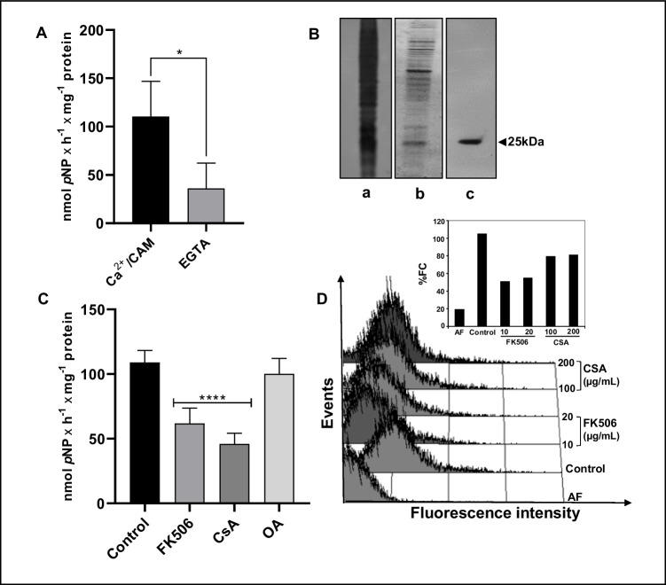
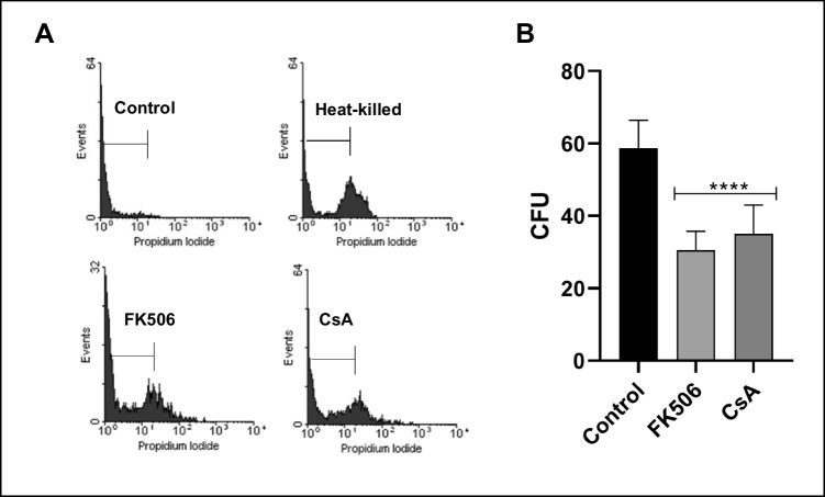
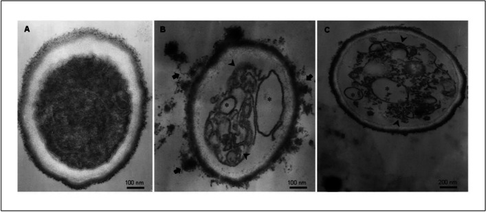
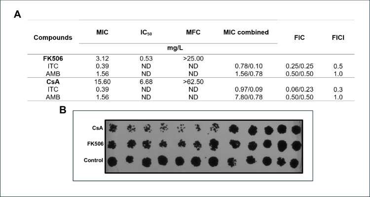
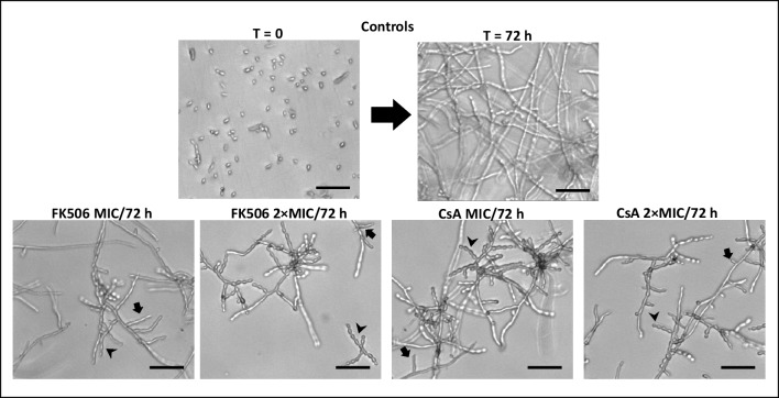
Fonsecaea pedrosoi is a melanized fungus that causes chromoblastomycosis (CBM), a tropical neglected disease responsible for chronic and disability-related subcutaneous mycosis. Given the challenging nature of CBM treatment, the study of new targets and novel bioactive drugs capable of improving patient life quality is urgent. In the present work, we detected a calcineurin activity in F. pedrosoi conidial form, employing primarily colorimetric, immunoblotting and flow cytometry assays. Our findings reveal that the calcineurin activity of F. pedrosoi was stimulated by Ca2+/calmodulin, inhibited by EGTA and specific inhibitors, such as tacrolimus (FK506) and cyclosporine A (CsA), and proved to be insensitive to okadaic acid. In addition, FK506 and CsA were able to affect the cellular viability and the fungal proliferation. This effect was corroborated by transmission electron microscopy that showed both calcineurin inhibitors promoted profound changes in the ultrastructure of conidia, causing mainly cytoplasm condensation and intense vacuolization that are clear indication of cell death. Our data indicated that FK506 exhibited the highest effectiveness, with a minimum inhibitory concentration (MIC) of 3.12 mg/L, whereas CsA required 15.6 mg/L to inhibit 100% of conidial growth. Interestingly, when both were combined with itraconazole, they demonstrated anti-F. pedrosoi activity, exhibiting a synergistic effect. Moreover, the fungal filamentation was affected after treatment with both calcineurin inhibitors. These data corroborate with other calcineurin studies in fungal cells and open up further discussions aiming to establish the role of this enzyme as a potential target for antifungal therapy against CBM infections.
Keywords: Antifungal drugs; Calcineurin inhibitors; Chromoblastomycosis; Combined therapy; Intracellular phosphatase activity.
© 2024. The Author(s) under exclusive licence to Sociedade Brasileira de Microbiologia.
Conflict of interest statement
Declarations. Conflict of interest: The authors declare that they have no conflict of interest.
Figures
Similar articles
-
Structure-guided design and synthesis of C22- and C32-modified FK520 analogs with enhanced activity against human pathogenic fungi.Proc Natl Acad Sci U S A. 2025 Jan 7;122(1):e2419883121. doi: 10.1073/pnas.2419883121. Epub 2024 Dec 31. Proc Natl Acad Sci U S A. 2025. PMID: 39739817 Free PMC article.
-
Topical tacrolimus for atopic dermatitis.Cochrane Database Syst Rev. 2015 Jul 1;2015(7):CD009864. doi: 10.1002/14651858.CD009864.pub2. Cochrane Database Syst Rev. 2015. PMID: 26132597 Free PMC article.
-
The role of melanin in Fonsecaea monophora pathogenicity: adhesion, fibroblast interactions, and implications for chromoblastomycosis.Future Microbiol. 2025 Apr;20(6):449-456. doi: 10.1080/17460913.2025.2480973. Epub 2025 Mar 20. Future Microbiol. 2025. PMID: 40109157
-
The Black Book of Psychotropic Dosing and Monitoring.Psychopharmacol Bull. 2024 Jul 8;54(3):8-59. Psychopharmacol Bull. 2024. PMID: 38993656 Free PMC article. Review.
-
Establishment of epidemiological cutoff values for Fonsecaea pedrosoi, the primary etiologic agent of chromoblastomycosis, and eight antifungal medications.J Clin Microbiol. 2025 May 14;63(5):e0190324. doi: 10.1128/jcm.01903-24. Epub 2025 Apr 4. J Clin Microbiol. 2025. PMID: 40183549 Free PMC article.
References
-
- GAFFI. Global Action for Fungal Infection (2017). https://gaffi.org/poor-farmers-fungal-skin-condition-gets-approval-from-.... Accessed 16 Jan 2024
MeSH terms
Substances
LinkOut - more resources
Full Text Sources
Miscellaneous