. 2024 Jul 23;19(1):276.
doi: 10.1186/s13023-024-03200-2.

A multi-step approach to overcome challenges in the management of head and neck lymphatic malformations, and response to treatment

Affiliations

A multi-step approach to overcome challenges in the management of head and neck lymphatic malformations, and response to treatment

Valentina Trevisan et al. Orphanet J Rare Dis. .

Abstract

Background: Lymphatic malformations are vascular developmental anomalies varying from local superficial masses to diffuse infiltrating lesions, resulting in disfigurement. Patients' outcomes range from spontaneous regression to severe sequelae notwithstanding appropriate treatment. The current classification guides, in part, clinicians through the decision-making process, prognosis prediction and choice of therapeutic strategies. Even though the understanding of molecular basis of the disease has been recently improved, a standardized management algorithm has not been reached yet.

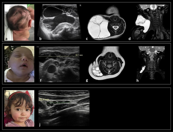
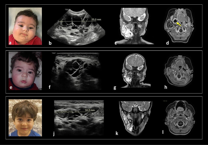
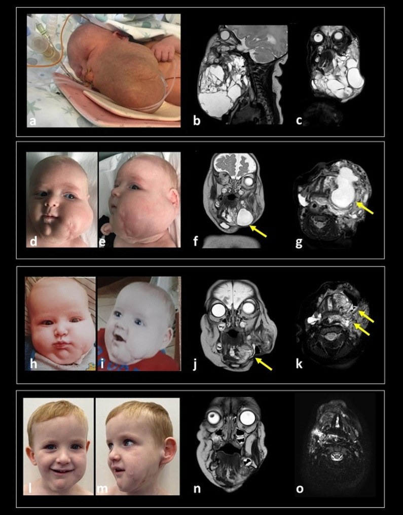
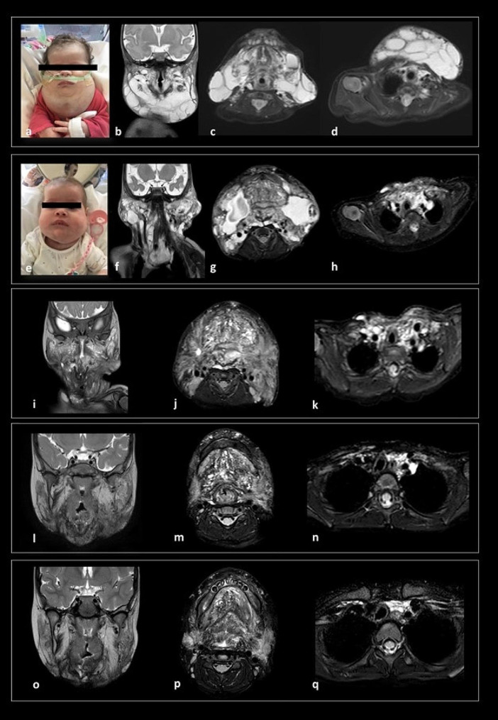
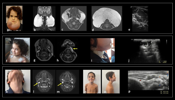
Results: Here, we report our experience on five children with different lymphatic anomalies of the head and neck region treated by applying a multidisciplinary approach reaching a consensus among specialists on problem-solving and setting priorities.

Conclusions: Although restitutio ad integrum was rarely achieved and the burden of care is challenging for patients, caregivers and healthcare providers, this study demonstrates how the referral to expert centres can significantly improve outcomes by alleviating parental stress and ameliorating patients' quality of life. A flow-chart is proposed to guide the multidisciplinary care of children with LMs and to encourage multidisciplinary collaborative initiatives to implement dedicated patients' pathways.

Keywords: Alpelisib; Lymphangioma; Lymphatic malformations; Personalized medicine; Sirolimus; mTOR.


Conflict of interest statement

Chiara Leoni and Nicoletta Resta belong to the advisory board for PROS by Novartis. The other authors declare no conflict of interest.

Figures

Graph 1
Graph 1
Systematic multistep approach applied to patients with isolated LMs of the head and neck region
Fig. 1
Fig. 1
(a) Clinical examination at 8 DOL showing a cervical swelling mass, extending from the posterior lateral area of the neck to the ipsilateral supraclavicular region. Pre-treatment US (b) and MRI (c, d - axial and coronal T2-w respectively) confirmed a lateral cervical multiloculated cystic mass in the posterior lateral triangle of the neck, extending inferiorly in the right infraclavicular space, without involvement of mediastinum and visceral space of the neck. One-month post-sclerotherapy, the patient presented a marked clinical reduction of the cervical mass (e). US examination confirmed a dimensional reduction of the lymphatic lesion, with maximal extension of 5 cm towards the deep tissues (f). Six months post-sclerotherapy, MRI (g, h - axial and coronal T2-w respectively) showed > 50% volumetric reduction with a residual macrocystic mass in the right posterior lateral neck, without infiltration of muscles and deep neck spaces. (i) Picture at last clinical evaluation (2 years and 2 months of age) did not document any visible mass, and US examination (j) showed resolution, with a residual hypo-/anechoic multiloculated component in the right supraclavicular area (24 × 5 mm)
Fig. 2
Fig. 2
Clinical examination at 40 DOL (a), showing a lateral cervical mass on the right side of the face and neck, overlain by hyperaemic and warm skin. US (b) and MRI (c, d coronal T2w and axial T1-w after contrast medium) confirmed a large, combined LM in the right upper neck with extensive multicompartmental involvement, infiltration of the parotid, masticatory, and parapharyngeal and retropharyngeal spaces; a parapharyngeal abscess (arrow, fluid collection with thick peripheral rim of contrast enhancement), compatible with the clinically suspected superinfection, was present. Seven weeks post-sclerotherapy (5 months of age) a clinical improvement of the lesion was detected (e). US (f) and MRI (g: coronal T2w, h: axial T1-w after contrast) showed slight volumetric reduction of the LMs. The clinical examination at 2 years and 5 months showed a very mild asymmetry of the right lateral neck (i). Concomitant US showed a further reduction of the multi-chambered lesion, from 5 cm to 3.4 cm (j). The 3 years follow-up MRI (k: coronal T2w, l: axial T1-w after contrast) showed residual microcystic components in the right parotid, parapharyngeal and submandibular spaces
Fig. 3
Fig. 3
A large mass with elastic consistency involving the neck and the left side of the face was confirmed at birth(a). MRI at the time showed a large multicompartmental mixed solid-cystic mass (mixed solid enhancing and cystic non-enhancing components) that involved the left face and the suprahyoid and infrahyoid neck superficial and deep spaces, including parapharyngeal and visceral spaces infiltration (b, c). At 3 months of life, a clinical rapid worsening of the swelling mass in the left cheek was detected (d, e). A follow-up MRI (f, g) confirmed increased volume of some macrocystic components along the left submandibular and parapharyngeal spaces (arrows). After the second surgical procedure, a clinical improvement of the facial asymmetry was evident with reduction of the left swelling cheek mass (h, i) and the MRI (j, k) showed residual microcystic components in the left sublingual and parapharyngeal spaces (arrows). At last evaluation, 4 years 6 months old (l, m), head and neck MRI (n, o) revealed a reduction of the residual lymphatic tissue located in the left sublingual and submandibular spaces and in the left neck (Left MRI column: sagittal T2-w; Right MRI column: coronal T2-w)
Fig. 4
Fig. 4
(a) Picture at birth showing a large soft swelling involving the neck, with tongue hypertrophy. MRI after birth (b, c,d) showed a voluminous macro- and microcystic LM involving the tongue, and with bilateral extensive involvement of the suprahyoid spaces (sublingual, submandibular and parotid spaces) and caudal spread up to the upper mediastinum, along with the parapharyngeal, retropharyngeal, vascular and visceral neck spaces. Persistent swelling of the neck was evident soon after surgery and sclerotherapy (e). MRI study (f, g, h) performed 10 days after these procedures documented a mild enlargement of the LM in the deep spaces of the neck and in the upper mediastinum, with microcystic involvement of the tongue and oropharyngeal walls causing pharyngeal air column obstruction needing tracheostomy placement. An increased volume of superficial macrocystic components was also detected. The serial follow-up MRI scans during sirolimus therapy (7 months 22 day – i, j, k, 2 year 8 months – l, m, n, 6 years – o, p, q) show progressive volumetric reduction of the LMs, especially the upper mediastinal components (left column, coronal view, T2-w; middle and right column, axial view, T2-w fat sat) respectively passing through the oropharynx and the upper mediastinum. The last US examination (j) shows absence of residual macrocystic components in the superficial neck spaces
Fig. 5
Fig. 5
A giant swelling of a soft-elastic consistency originating from the subcutaneous tissues of the mandibular angles bilaterally with downward extension anteriorly to the neck was evident at 6 years and 4 months. The overlying skin showed clinical signs of infection (a). MRI (b-d) and US (e) revealed a large bulky macro-cystic mass in the suprahyoid and infrahyoid neck involving bilaterally the submandibular, parapharyngeal, parotid and vascular spaces, without mediastinal extension and determining minimal deformation of pharyngeal air column, which maintained regular patency. After surgical debulking, the clinical examination revealed an apparent complete response to treatment (f). MRI showed grossly total excision of the LM with minimal fluid collections in the submandibular spaces (g, h). The arrow indicates the left drainage tube in the submandibular space. Swelling in the retromandibular right angle and submental region was progressively documented after removal of drains (2 weeks post-surgery) (i, lateral view). The US performed at that time revealed slight enlargement of some cystic components in the submandibular and submental spaces (j). Clinical evaluation at last follow-up (during Alpelisib treatment at 125 mg/day) documented no evidence of swelling mass; the skin in the submental area and in the superior part of the neck appeared thin but not atrophic (k). Frontal (n) and lateral (o) view of the patient confirmed the absence of persistent swelling. The clinically detected facial asymmetry involved the subcutaneous tissues, probably because of the surgical procedure. MR (l, m) and US (p) showed small residual cystic component in the posterior portion of the right submandibular space (arrow in l, m)

References

    1. Lee SY, Loll EG, Hassan AES, Cheng M, Wang A, Farmer DL. Genetic and molecular determinants of lymphatic malformations: potential targets for Therapy. J Dev Biol. 2022;10(1). 10.3390/JDB10010011. - PMC - PubMed
    1. ISSVA classification for vascular anomalies &#169.
    1. Liu X, Cheng C, Chen K, Wu Y, Wu Z. Recent progress in Lymphangioma. Front Pediatr. 2021;9(December):1–14. 10.3389/fped.2021.735832.10.3389/fped.2021.735832 - DOI - PMC - PubMed
    1. Snyder EJ, Sarma A, Borst AJ, Tekes A. Lymphatic anomalies in children: update on imaging diagnosis, Genetics, and treatment. AJR Am J Roentgenol. 2022;218(6):1089–101. 10.2214/AJR.21.27200. 10.2214/AJR.21.27200 - DOI - PubMed
    1. Pessanha I, Bravo M, Piedade C, Lopes MF. Complex lymphatic malformations in pediatrics: a review of treatment options. Minerva Pediatr. 2022;74(1):70–80. 10.23736/S2724-5276.20.06037-5.10.23736/S2724-5276.20.06037-5 - DOI - PubMed

LinkOut - more resources