Review
. 2024 Apr 17;24(14):6060-6080.
doi: 10.1021/acs.cgd.4c00260. eCollection 2024 Jul 17.

Emergent Properties from Three-Dimensional Assemblies of (Nano)particles in Confined Spaces

Affiliations
Review

Emergent Properties from Three-Dimensional Assemblies of (Nano)particles in Confined Spaces

Emanuele Marino et al. Cryst Growth Des. .

Abstract

The assembly of (nano)particles into compact hierarchical structures yields emergent properties not found in the individual constituents. The formation of these structures relies on a profound knowledge of the nanoscale interactions between (nano)particles, which are often designed by researchers aided by computational studies. These interactions have an effect when the (nano)particles are brought into close proximity, yet relying only on diffusion to reach these closer distances may be inefficient. Recently, physical confinement has emerged as an efficient methodology to increase the volume fraction of (nano)particles, rapidly accelerating the time scale of assembly. Specifically, the high surface area of droplets of one immiscible fluid into another facilitates the controlled removal of the dispersed phase, resulting in spherical, often ordered, (nano)particle assemblies. In this review, we discuss the design strategies, computational approaches, and assembly methods for (nano)particles in confined spaces and the emergent properties therein, such as trigger-directed assembly, lasing behavior, and structural photonic color. Finally, we provide a brief outlook on the current challenges, both experimental and computational, and farther afield application possibilities.


Conflict of interest statement

The authors declare no competing financial interest.

Figures

Figure 1
Figure 1
Assembly of (nano)particles. (A) Conventional 2D assembly through deposition and evaporation. (B) Confined 3D assembly discussed in this review. (C) Additional interparticle forces arising from the presence of an interface. (D) Confined assembly creates large interfacial areas. (E) Superparticles can be pipetted, transferred, and deployed. Topics discussed in this review: (F) Confined geometry simulation, (G) light triggered confined assembly, (H) magnetically triggered confined assembly, (I) anisotropic superparticles formed during confined assembly, (J) core/shell superparticles, (K) porous superparticles, (L) superparticles which exhibit structural coloration, and (M) superparticles with show lasing. Panel D is adapted with permission from ref (31). Copyright 2018 John Wiley and Sons. Panel F is adapted with permission from ref (32). Copyright 2018 Springer Nature. Panel G is adapted with permission from ref (33). Copyright 2023 The American Chemical Society. Panel H is adapted with permission from ref (34). Copyright 2019 The American Chemical Society. Panel I is adapted with permission from ref (35). Copyright 2012 The American Chemical Society. Panel J is adapted with permission from ref (7). Copyright 2022 The American Chemical Society. Panel K is adapted with permission from ref (36). Copyright 2018 The American Chemical Society. Panel L is adapted with permission from ref (37). Copyright 2019 John Wiley and Sons. Panel M is adapted with permission from ref (7). Copyright 2022 The American Chemical Society.
Figure 2
Figure 2
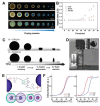
Light-activated self-assembly. (A) (Top) Ligand used for light-activated self-assembly and structural changes in the nanoparticle (NP) ligand corona upon photoexcitation with ultraviolet and visible light. (Bottom) Phase diagram and SEM images of suprastructures obtained by light-activated self-assembly for photoswitchable ligands density and composition of the dispersing solvent. Legend: NP, dispersed NPs; RC, light-reversible 3D superlattices; AP, amorphous precipitate; IC, irreversible 3D superlattices; SS, superparticles. (Scale bars are 100 nm.) (B) SEMs (low and high magnifications) of PS10k-b-P4VP10k superparticles prepared with (a) ring-opened form of the surfactant and (b) ring-closed form of the surfactant. (c) Photoluminescence (PL) spectra of spherical (orange) and ellipsoidal (blue) PS10k-b-P4VP10k superparticles. Insets: fluorescence optical images of the superparticle suspension during assembly. (C) (a) TEM image of three superparticles of 5.5 nm Au NPs functionalized with the thiolated spiropyran, as shown as inset. (b) Extinction spectra of 5.5 nm Au NPs in toluene before and after photoexcitation with ultraviolet light. Inset shows the TEM of the NPs, scale bar indicates 20 nm. (D) Optical micrographs of optically tweezed Ag nanocrystal superlattices (laser intensity = 3 × 10⁹ W/m2). The dashed hexagon represents the boundary between the central area and the edge of the superlattice, while the color of the hexagon and the dashed circle indicate lattice orientation. Scale bar indicates 1 μm. (E) Representative SEMs of the nanocrystal superlattices of Au-TMA/NBTS after ultraviolet exposure for the indicated times (scale bars indicate 0.5 μm). Panel A is adapted with permission from ref (1). Copyright 2007 The American Association for the Advancement of Science. Panel B is adapted with permission from ref (51). Copyright 2021 The American Chemical Society. Panel C is adapted with permission from ref (52). Copyright 2016 The Royal Society of Chemistry. Panel D is adapted with permission from ref (53). Copyright 2020 The American Chemical Society. Panel E is adapted with permission from ref (33). Copyright 2023 The American Chemical Society.
Figure 3
Figure 3
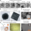
Magnetic-field-activated self-assembly. (A) Dark-field optical micrographs showing the evolutions of droplets at different drying stages: (a) absence of an external magnetic field, (b) under a horizontal magnetic field of 95 Oe, and (c) of 470 Oe. Scale bars indicate 100 mm. (B) Evolution of droplet aspect ratios with time corresponding to A. (C) Evolution of a droplet loaded with 3 wt % of Fe3O4 nanocrystals embedded in polystyrene during drying in the absence (top) and the presence (bottom) of an external magnetic field. Scale bars indicate 0.5 mm. (D) Magnetically anisotropic Janus superparticles; inset: magnified image of a single superparticle with the PNIPAm side attracted to a magnet. (E) Schematic of the experimental system composed of nanocrystal cores, polymer brush, and supramolecular binding groups that drive assembly. Assembly is directed via complementary hydrogen bonding moieties, strengthened via magnetic dipole coupling between aligned spins at short interparticle distances. (F) (Left) Melting profiles of 16 nm iron oxide nanocrystals functionalized with 13 kDa polymers and 15 nm Au nanocrystals functionalized with 14 kDa polymers. (Right) Melting profile of 16 nm iron oxide coated with 8 kDa polymers is shifted by 10 °C with respect to 15 nm Au nanocrystals coated with 9 kDa polymers. Panels A and B are adapted with permission from ref (62). Copyright 2019 The Royal Society of Chemistry. Panel C is adapted with permission from ref (34). Copyright 2019 The American Chemical Society. Panel D is adapted with permission from ref (63). Copyright 2009 John Wiley and Sons. Panels E and F are adapted with permission from ref (64). Copyright 2020 The American Chemical Society.
Figure 4
Figure 4
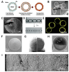
Anisotropic superparticles. (A) Scanning electron micrographs of superparticles obtained by the evaporation of aqueous droplets with different NaCl concentrations containing a dispersion of polystyrene spheres 440 nm in diameter at a volume fraction of 8%. (B) Transmission electron micrographs of superparticles consisting of 11 nm nanocubes of Fe3O4 assembled in the presence excess oleate ligands. (C) Scanning transmission electron micrographs of superparticles consisting of CsPbBr3 nanocrystals characterized by rounded (top, 14 nm diameter) and sharp (bottom, 10 nm diameter) edges. (D) (Left) Schematic of the stabilization of aqueous wires in a silicone oil by hydrophilic 20 nm SiO2 nanocrystals. (Right) Optical micrograph of a nanocrystal-stabilized aqueous spiral in silicone oil. Panel A is adapted with permission from ref (65). Copyright 2022 Elsevier B.V. Panel B is adapted with permission from ref (68). Copyright 2012 The American Chemical Society. Panel C is adapted with permission from ref (66). Copyright 2020 The American Chemical Society. Panel D is adapted with permission from ref (67). Copyright 2018 John Wiley and Sons.
Figure 5
Figure 5
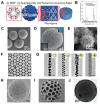
Core/shell superparticles. (A) Schematic for the formation of colloidosomes from nanocrystal-stabilized water-in-oil-in-water double emulsions. (B) SEM image of poly(d,l-lactic acid)/SiO2 dried colloidosomes. Inset shows the cross-section of the broken shell (scale bar indicates 500 nm). (C) SEM image of acetylated dextran colloidosomes. The broken colloidosome reveals the cavity and shell thickness. (D) (Top) Optical micrographs of CO2 bubbles generated in an aqueous dispersion of 3.5 μm polystyrene microparticles at a volume fraction of 1.5% w/w. (Bottom) The bubbles quickly decrease in size to lead to the formation of spherical colloidosomes. Scale bars indicate 200 μm. (E) The colloidosomes become fluorescent when using polystyrene particles loaded with CdSe/ZnS nanocrystals. Scale bar indicates 100 μm. (F) SEM image of a SiO2 microsphere uniformly coated with CdSe/CdS@Cd1–xZnxS core/shell nanoplatelets. (G) SEM image of a core/shell nanocrystal superparticle consisting of PbS/CdS spheres and NaGdF4 disks before (left) and after (right) focused-ion beam milling, revealing the phase separation of nanocrystals. (H) TEM image of a half-full colloidosome viewed along the [111] zone axis of the Fe3O4 nanocrystal superlattice. (I) Overlay of SEM image of the cross section of a superparticles consisting of 338 nm and 1430 nm polystyrene colloidal particles at initial volume fractions of 2.5% and 5.5%, respectively. Panels A and B are adapted with permission from ref (78). Copyright 2008 John Wiley and Sons. Panel C is adapted with permission from ref (79). Copyright 2015 The American Chemical Society. Panels D and E are adapted with permission from ref (80). Copyright 2009 John Wiley and Sons. Panel F is adapted with permission from ref (81). Copyright 2022 The American Chemical Society. Panel G is adapted with permission from ref (7). Copyright 2022 The American Chemical Society. Panel H is adapted with permission from ref (82). Copyright 2016 The American Chemical Society. Panel I is adapted with permission from ref (83). Copyright 2019 The American Chemical Society.
Figure 6
Figure 6
Porous superparticles. (A) Fabrication of hierarchically structured materials based on superparticles. ZIF-8 metal–organic framework (MOFs) particles form superparticles, which are further assembled into macroscopic pellets with structural hierarchy of pores. (B) Pore size distribution of pellets of ZIF-8 particles and superparticles. (C) SEM image of ZIF-8 superparticles. (D) SEM image of (UiO-66) MOFs superparticles which exhibit structural coloration (optical microscopy images in inset). (E) SEM image of complex nanostructured hedgehog superparticles from CdS assembled at unity water-to-ethylenediamine volume ratio and 160 °C for 20 h. (F) Network structure formed by a 1:1 mixture of Au and CdSe/CdS/ZnS nanocrystals passivated with a dendritic ligand drop-cast from chloroform at 15 mg/mL. (G) TEM image of Au nanocrystal assemblies with diverse structures at different diameter ratio (γ) between carbon nanotube hosts and Au nanocrystal guests: γ = 1.1, 1.5, 1.8, 2.3. Scale bars indicate 20 nm. (H) SEM image of porous CoFe2O4 superparticles. (I) SEM image of a porous silica SP after removal of the polystyrene nanoparticles by calcination. (J) TEM image of hybrid micromesoporous graphitic carbon superparticles. Panels A, B, and C are adapted with permission from ref (91). Copyright 2023 John Wiley and Sons. Panel D is adapted with permission from ref (73). Copyright 2022 John Wiley and Sons. Panel E is adapted with permission from ref (93). Copyright 2021 The American Chemistry Society. Panel F is adapted with permission from ref (94). Copyright 2022 The American Chemistry Society. Panel G is adapted with permission from ref (95). Copyright 2021 The American Chemistry Society. Panel H is adapted with permission from ref (5). Copyright 2022 The American Chemistry Society. Panel I is adapted with permission from ref (36). Copyright 2018 The American Chemistry Society. Panel J is adapted with permission from ref (96). Copyright 2017 The American Chemistry Society.
Figure 7
Figure 7
Structural color pigments. (A) 810 nm latex particles dried containing 0.21 wt % of 22 nm diameter Au nanocrystals (particle diameter is 50 μm). (B) A monolayer of microfluidically produced structural color spheres composed of 610 nm diameter latex particles. (C) Reflectance confocal microscopy (RCM) image of a structural color pigment at approximately the midplane of the particle, note the appearance of multiple crystalline domains. (D) SEM image of a structural pigment created with icosahedral cluster composed of 275 nm diameter particles (scale bar indicates 1 μm). Inset: A magnified SEM image of one of the hexagonal faces of the cluster. Scale bar indicates 200 nm. (E) Dark-field microscopy image of cellulose nanocrystal pigments dispersed in oil (refractive index 1.55). (F) Fluorescence microscopy image of silica-based inverse-opal structural pigments in a dried film of latex paint. Note: the fluorescence intensity is excluded from the round structural color particle; (scale bar indicates 10 μm). Panel A is adapted with permission from ref (112). Copyright 2008 John Wiley and Sons. Panel B is adapted with permission from ref (6). Copyright 2008 The American Association for the Advancement of Science. Panel C is adapted with permission from ref (115). Copyright 2021 The American Chemical Society. Panel D is adapted with permission from ref (117). Copyright 2020 The American Chemical Society. Panel E is adapted with permission from ref (111). Copyright 2022 Springer Nature Publishing Group. Panel F is adapted with permission from ref (110). Copyright 2019 John Wiley and Sons.
Figure 8
Figure 8
Lasing superparticles. (A) Rendering of a toroidal optical resonant cavity coated with Au nanorods. (B) Lasing spectra and threshold data (inset) of Au nanorod-coated microtoroid cavity. The quadratic relationship between the lasing intensity and the input power is due to the two-photon lasing mechanism. (C) Emission spectra for CdSe/ZnS nanorods loaded in the microcavity at different pump powers. (Inset) intensity of the lasing peak (filled squares) and the fluorescence peak (empty circles) versus the pump power. (D) SEM image of a CdSe/CdS/ZnS nanocrystal ring resonator with diameter and width of 5 μm and 500 nm, respectively. (E) Representative spectra showing the dependence of the emission spectra on excitation power. (F) Spectra of a single 7 μm silica-core CdSe/CdZnS-shell microsphere below (205 μW) and above (366 μW) laser threshold. Inset: Optical and fluorescence micrographs of the microsphere; scale bar indicates 15 μm. (G) SEM image of a single superparticle of CdSe/CdS nanocrystals. (H) Emission from a single superparticle of CdSe/CdS nanocrystals at different pump fluences (18–145 μJ/cm2) at low spectral resolution (300 lines/mm grating). Inset: High spectral resolution (1800 lines/mm grating), revealing peak substructure. (I) Time-dependent emission spectra for a superparticles of CdSe/CdS nanocrystals recorded over 15 min of continuous operation at an excitation fluence of 1.6 mJ/cm2 before (left) and after (right) light-soaking. (J) Monodisperse superparticles of CdSe nanocrystals generated using a source-sink emulsion system. (K) SEM image of a dimer of CdSe/CdS nanocrystal superparticles; (insets) dark-field optical micrographs of superparticles clusters. (L) Photoluminescence spectra of the clusters shown in (K). Panels A and B are adapted with permission from ref (123). Copyright 2013 The American Chemical Society. Panel C is adapted with permission from ref (124). Copyright 2002 John Wiley and Sons. Panels D and E are adapted with permission from ref (125). Copyright 2018 The American Chemical Society. Panel F is adapted with permission from ref (126). Copyright 2005 John Wiley and Sons. Panels G and H are adapted with permission from ref (127). Copyright 2018 The American Chemical Society. Panel I is adapted with permission from ref (128). Copyright 2023 The American Chemical Society. Panels J, K, and L are adapted with permission from ref (7). Copyright 2022 The American Chemical Society.

References

    1. Klajn R.; Bishop K. J. M.; Grzybowski B. A. Light-controlled self-assembly of reversible and irreversible nanoparticle suprastructures. Proc. Natl. Acad. Sci. U. S. A. 2007, 104, 10305–10309. 10.1073/pnas.0611371104. - DOI - PMC - PubMed
    1. Liu X.; Kent N.; Ceballos A.; Streubel R.; Jiang Y.; Chai Y.; Kim P. Y.; Forth J.; Hellman F.; Shi S.; Wang D.; Helms B. A.; Ashby P. D.; Fischer P.; Russell T. P. Reconfigurable ferromagnetic liquid droplets. Science 2019, 365, 264–267. 10.1126/science.aaw8719. - DOI - PubMed
    1. Velev O. D.; Lenhoff A. M.; Kaler E. W. A Class of Microstructured Particles Through Colloidal Crystallization. Science 2000, 287, 2240–2243. 10.1126/science.287.5461.2240. - DOI - PubMed
    1. Dinsmore A. D.; Hsu M. F.; Nikolaides M. G.; Marquez M.; Bausch A. R.; Weitz D. A. Colloidosomes: Selectively Permeable Capsules Composed of Colloidal Particles. Science 2002, 298, 1006–1009. 10.1126/science.1074868. - DOI - PubMed
    1. Yang Y.; Xiao J.; Xia Y.; Xi X.; Li T.; Yang D.; Dong A. Assembly of CoFe2O4 Nanocrystals into Superparticles with Tunable Porosities for Use as Anode Materials for Lithium-Ion Batteries. ACS Applied Nano Materials 2022, 5, 9698–9705. 10.1021/acsanm.2c01929. - DOI

LinkOut - more resources