

A systematic review and meta-analysis of transthoracic echocardiogram vs. cardiac magnetic resonance imaging for the detection of left ventricular thrombus
- PMID: 39045058
- PMCID: PMC11240154
- DOI: 10.1093/ehjimp/qyad041
A systematic review and meta-analysis of transthoracic echocardiogram vs. cardiac magnetic resonance imaging for the detection of left ventricular thrombus
Abstract
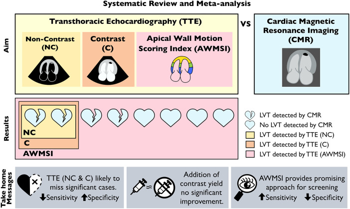
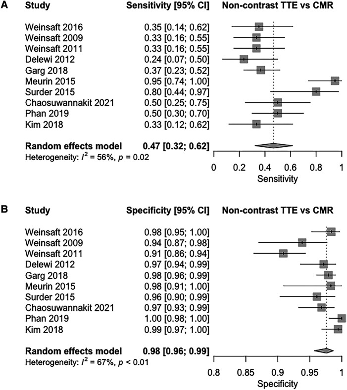
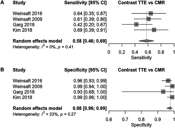
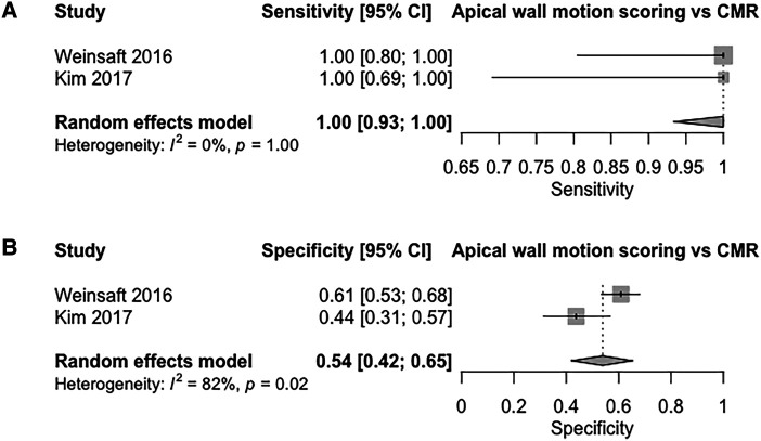
Transthoracic echocardiography (TTE) is the most commonly used imaging modality to diagnose left ventricular thrombus (LVT), however, cardiac magnetic resonance (CMR) remains the gold standard investigation. A comparison of the diagnostic performance between two modalities is needed to inform guidelines on a diagnostic approach towards LVT. We performed a systematic review and meta-analysis to investigate the diagnostic performance of three methods of TTE (non-contrast, contrast, and apical wall motion scoring) for the detection of LVT compared to CMR as a reference test. Studies comprising 2113 patients investigated for LVT using both TTE and CMR were included in the meta-analysis. For non-contrast TTE, pooled sensitivity and specificity were 47% [95% confidence interval (CI): 32-62%], and 98% (95% CI: 96-99%), respectively. In contrast, TTE pooled sensitivity and specificity values were 58% (95% CI: 46-69%), and 98% (95% CI: 96-99%), respectively. Apical wall motion scoring on non-contrast TTE yielded a sensitivity of 100% [95% CI: 93-100%] and a specificity of 54% (95% CI: 42-65%). The area under the curve (AUC) values from our summary receiver operating characteristic curve (SROC) for non-contrast and contrast TTE were 0.87 and 0.86 respectively, with apical wall motion studies having the highest AUC of 0.93. Despite high specificity, routine contrast and non-contrast TTE are likely to miss a significant number of LVT, making it a suboptimal screening tool. The addition of apical wall motion scoring provides a promising method to reliably identify patients requiring further investigations for LVT, whilst excluding others from unnecessary testing.
Keywords: cardiac magnetic resonance; diagnosis; echocardiography; left ventricular thrombus; screening; sensitivity and specificity.
Plain language summary
The formation of left ventricular thrombus (LVT), a blood clot in the left pumping chamber of the heart, can lead to serious complications such as a stroke. Whilst cardiac magnetic resonance (CMR) is the best imaging tool to detect these clots, the most used tool is a transthoracic echocardiogram (TTE), which visualizes the heart by placing an ultrasound on the chest. This is due to the affordability and widespread availability of TTE. Thus, it is important to know how TTE compares to CMR when it comes to detecting LVT. This study pools the results of previous research to compare the diagnostic performance of three different methods of TTE compared to CMR for detecting LVT. Non-contrast TTE.Contrast TTE: The addition of an enhancing dye is thought to improve imaging.Apical wall motion scoring: Evaluating the movement of the heart’s walls using TTE. Our results show that current methods of TTE may miss half of the patients with LVT and that the use of contrast did not provide significant improvement. Interestingly, the use of apical wall motion scoring was able to accurately detect all the patients with LVT. This shows promise as a future tool to reliably exclude patients from unnecessary testing, whilst identifying those who need further investigations.
© The Author(s) 2023. Published by Oxford University Press on behalf of the European Society of Cardiology.
Conflict of interest statement
Conflict of interest: Nothing to disclose.
Figures
Similar articles
-
Improved detection of echocardiographically occult left ventricular thrombi following ST-elevation myocardial infarction.Eur Heart J Acute Cardiovasc Care. 2023 Oct 25;12(10):703-710. doi: 10.1093/ehjacc/zuad069. Eur Heart J Acute Cardiovasc Care. 2023. PMID: 37348047
-
Elusive left ventricular thrombus: Diagnostic role of cardiac magnetic resonance imaging-A case report and review of the literature.World J Clin Cases. 2018 Jun 16;6(6):127-131. doi: 10.12998/wjcc.v6.i6.127. World J Clin Cases. 2018. PMID: 29988880 Free PMC article.
-
LV thrombus detection by routine echocardiography: insights into performance characteristics using delayed enhancement CMR.JACC Cardiovasc Imaging. 2011 Jul;4(7):702-12. doi: 10.1016/j.jcmg.2011.03.017. JACC Cardiovasc Imaging. 2011. PMID: 21757159 Free PMC article.
-
Emerging Trends in Left Ventricular Thrombus: A Comprehensive Review of Non-Ischemic and Ischemic Cardiopathies, Including Eosinophilic Myocarditis, Chagas Cardiomyopathy, Amyloidosis, and Innovative Anticoagulant Approaches.Diagnostics (Basel). 2024 Apr 30;14(9):948. doi: 10.3390/diagnostics14090948. Diagnostics (Basel). 2024. PMID: 38732361 Free PMC article. Review.
-
Echocardiography versus computed tomography and cardiac magnetic resonance for the detection of left heart thrombosis: a systematic review and meta-analysis.Clin Res Cardiol. 2021 Nov;110(11):1697-1703. doi: 10.1007/s00392-020-01741-7. Epub 2020 Sep 13. Clin Res Cardiol. 2021. PMID: 32920662
Cited by
-
Left Ventricular Thrombosis in Ischemic and Non-Ischemic Cardiomyopathies: Focus on Evidence-Based Treatment.J Clin Med. 2025 Feb 27;14(5):1615. doi: 10.3390/jcm14051615. J Clin Med. 2025. PMID: 40095541 Free PMC article. Review.
-
Direct oral anticoagulants or warfarin in patients with left ventricular thrombus after ST-elevation myocardial infarction: a pilot trial and a prespecified meta-analysis of randomised trials.EuroIntervention. 2025 Jan 6;21(1):82-92. doi: 10.4244/EIJ-D-24-00527. EuroIntervention. 2025. PMID: 39773831
-
[Clinical, diagnostic and therapeutic profile of patients with left intraventricular thrombus in three high-complexity centers during the period 2000-2022].Arch Peru Cardiol Cir Cardiovasc. 2024 Dec 11;5(4):207-214. doi: 10.47487/apcyccv.v5i4.430. eCollection 2024 Oct-Dec. Arch Peru Cardiol Cir Cardiovasc. 2024. PMID: 39850346 Free PMC article. Spanish.
-
Evolution of left ventricular thrombus on serial cardiovascular magnetic resonance imaging.Eur Heart J Cardiovasc Imaging. 2025 Jan 31;26(2):349-358. doi: 10.1093/ehjci/jeae271. Eur Heart J Cardiovasc Imaging. 2025. PMID: 39437323
References
-
- Vaitkus PT, Barnathan ES. Embolic potential, prevention and management of mural thrombus complicating anterior myocardial infarction: a meta-analysis. J Am Coll Cardiol 1993;22:1004–9. - PubMed
-
- Jungehülsing M, Sechtem U, Theissen P, Hilger HH, Schicha H. Left ventricular thrombi: evaluation with spin-echo and gradient-echo MR imaging. Radiology 1992;182:225–9. - PubMed
-
- Ibanez B, James S, Agewall S, Antunes MJ, Bucciarelli-Ducci C, Bueno Het al. . 2017 ESC guidelines for the management of acute myocardial infarction in patients presenting with ST-segment elevation: the task force for the management of acute myocardial infarction in patients presenting with ST-segment elevation of the European society of cardiology (ESC). Eur Heart J 2017;39:119–77. - PubMed
Publication types
LinkOut - more resources
Full Text Sources