

Quantification of myocardial extracellular volume without blood sampling
- PMID: 39045067
- PMCID: PMC11195702
- DOI: 10.1093/ehjimp/qyad022
Quantification of myocardial extracellular volume without blood sampling
Abstract
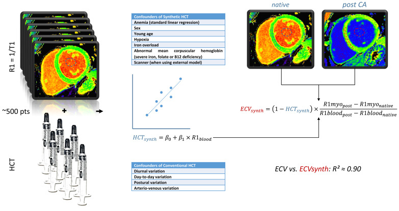
Aims: Cardiac magnetic resonance (CMR) T1 relaxation time mapping is an established technique primarily used to identify diffuse interstitial fibrosis and oedema. The myocardial extracellular volume (ECV) can be calculated from pre- and post-contrast T1 relaxation times and is a reproducible parametric index of the proportion of volume occupied by non-cardiomyocyte components in myocardial tissue. The conventional calculation of the ECV requires blood sampling to measure the haematocrit (HCT). Given the high variability of the HCT, the blood collection is recommended within 24 h of the CMR scan, limiting its applicability and posing a barrier to the clinical routine use of ECV measurements. In recent years, several research groups have proposed a method to determine the ECV by CMR without blood sampling. This is based on the inverse relationship between the T1 relaxation rate (R1) of blood and the HCT. Consequently, a 'synthetic' HCT could be estimated from the native blood R1, avoiding blood sampling.
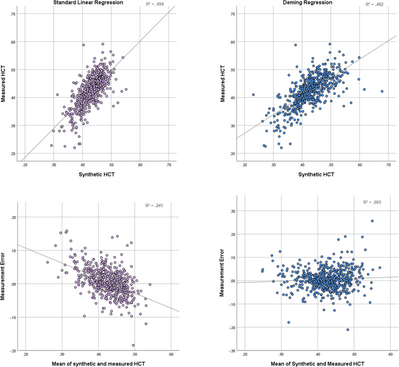
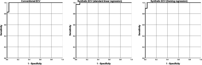
Methods and results: We performed a review and meta-analysis of published studies on synthetic ECV, as well as a secondary analysis of previously published data to examine the effect of the chosen regression modell on bias. While, overall, a good correlation and little bias between synthetic and conventional ECV were found in these studies, questions regarding its accuracy remain.
Conclusion: Synthetic HCT and ECV can provide a 'non-invasive' quantitative measurement of the myocardium's extracellular space when timely HCT measurements are not available and large alterations in ECV are expected, such as in cardiac amyloidosis. Due to the dependency of T1 relaxation times on the local setup, calculation of local formulas using linear regression is recommended, which can be easily performed using available data.
Keywords: CMR; ECV classification description; T1 mapping; cardiac magnetic resonance (CMR); synthetic HCT; tissue characterization.
© The Author(s) 2023. Published by Oxford University Press on behalf of the European Society of Cardiology.
Conflict of interest statement
Conflict of interest: P.D. owns stock of Siemens and Bayer and received a travel grant from the Berlin University Alliance. A.F. is a shareholder of BOCAhealthcare GmbH. S.K. received funding from the DZHK (German Centre for Cardiovascular Research) and by the BMBF (German Ministry of Education and Research) and personal fees from Servier, outside of the current work. S.K. received an unrestricted research grant from Philips Healthcare and received lecture honoraria from Medis, NL. The remaining authors declare that the research was conducted in the absence of any commercial or financial relationships that could be construed as a potential conflict of interest.
Figures
References
-
- Aretz HT, Billingham ME, Edwards WD, Factor SM, Fallon JT, Fenoglio JJ Jret al. . Myocarditis. A histopathologic definition and classification. Am J Cardiovasc Pathol 1987;1:3–14. - PubMed
-
- Beltrami CA, Finato N, Rocco M, Feruglio G, Puricelli C, Cigola Eet al. . The cellular basis of dilated cardiomyopathy in humans. J Mol Cell Cardiol 1995;27:291–305. - PubMed
-
- Verdonschot JAJ, Hazebroek MR, Derks KWJ, Barandiarán Aizpurua A, Merken JJ, Wang Pet al. . Titin cardiomyopathy leads to altered mitochondrial energetics, increased fibrosis and long-term life-threatening arrhythmias. Eur Heart J 2018;39:864–73. - PubMed
LinkOut - more resources
Full Text Sources