. 2024 Jul 9:4:1415393.
doi: 10.3389/fopht.2024.1415393. eCollection 2024.

Retinal vascular reactivity in carriers of X-linked inherited retinal disease - a study using optical coherence tomography angiography

Affiliations

Retinal vascular reactivity in carriers of X-linked inherited retinal disease - a study using optical coherence tomography angiography

Sena Ayse Gocuk et al. Front Ophthalmol (Lausanne). .

Abstract

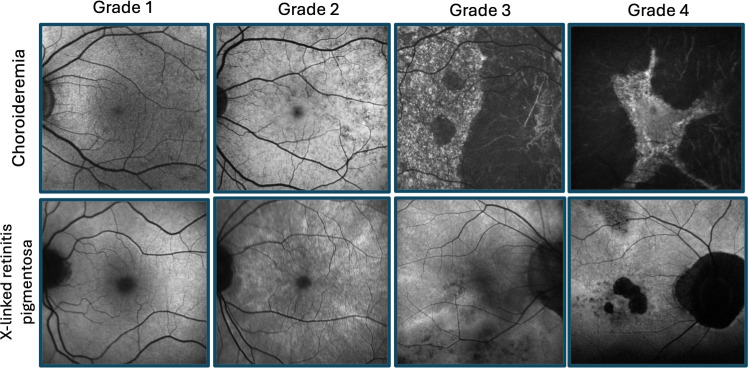
Purpose: Female carriers of X-linked inherited retinal diseases (IRDs) can show highly variable phenotypes and disease progression. Vascular reactivity, a potential disease biomarker, has not been investigated in female IRD carriers. In this study, functional optical coherence tomography angiography (OCT-A) was used to dynamically assess the retinal microvasculature of X-linked IRD carriers.

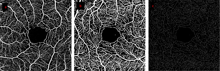
Methods: Genetically confirmed female carriers of IRDs (choroideremia or X-linked retinitis pigmentosa), and healthy women were recruited. Macular angiograms (3x3mm, Zeiss Plex Elite 9000) were obtained in 36 eyes of 15 X-linked IRD female carriers and 21 age-matched control women. Two tests were applied to test vascular reactivity: (i) mild hypoxia and (ii) handgrip test, to induce a vasodilatory or vasoconstrictive response, respectively. Changes to vessel density (VD) and vessel length density (VLD) were independently evaluated during each of the tests for both the superficial and deep capillary plexuses.

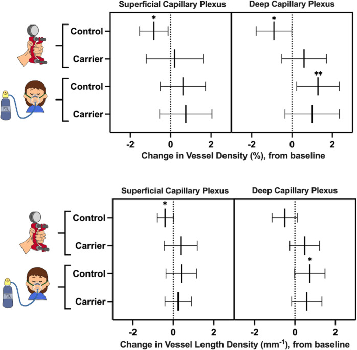
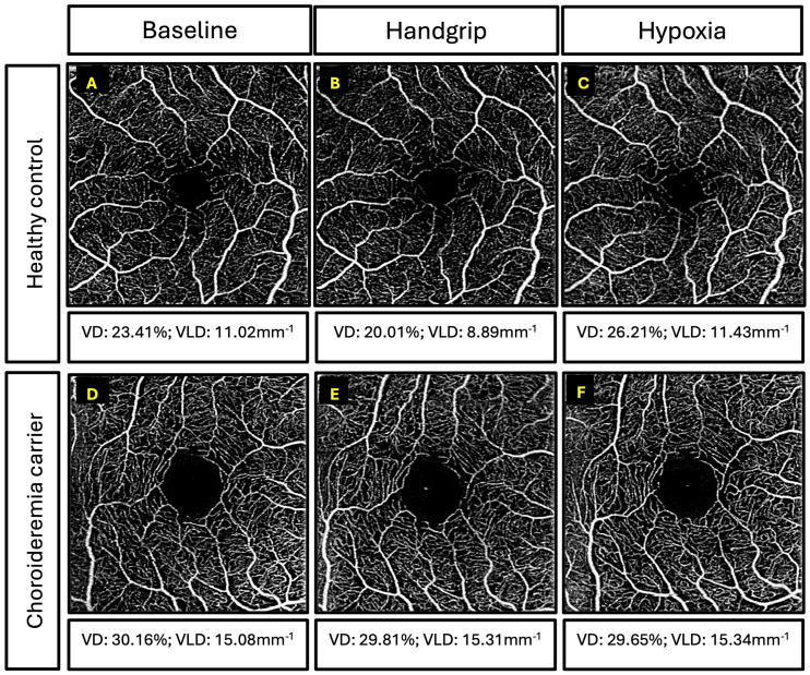
Results: In the control group, the superficial and deep VD decreased during the handgrip test (p<0.001 and p=0.037, respectively). Mean superficial VLD also decreased during the handgrip test (p=0.025), while the deep plexus did not change significantly (p=0.108). During hypoxia, VD and VLD increased in the deep plexus (p=0.027 and p=0.052, respectively) but not in the superficial plexus. In carriers, the physiologic vascular responses seen in controls were not observed in either plexus during either test, with no difference in VD or VLD noted (all p>0.05).

Conclusions: Functional OCT-A is a useful tool to assess dynamic retinal microvascular changes. Subclinical impairment of the physiological vascular responses seen in carriers of X-linked IRDs may serve as a valuable clinical biomarker.

Keywords: OCT-A; X-linked; carrier; females; inherited retinal disease; retinal vasculature.


Conflict of interest statement

The authors declare that the research was conducted in the absence of any commercial or financial relationships that could be construed as a potential conflict of interest. The author(s) declared that they were an editorial board member of Frontiers, at the time of submission. This had no impact on the peer review process and the final decision.

Figures

Figure 1
Figure 1
Retinal disease spectrum of female carriers of X-linked retinitis pigmentosa and choroideremia. Fundus autofluorescence imaging illustrating grades 1-4 for each condition, as previously described by Edwards et al (1) (fine, coarse, geographic, and male pattern phenotypes, respectively) and Nanda et al (2) (normal, radial, focal pigmentary retinopathy, and male pattern phenotypes, respectively).
Figure 2
Figure 2
Study protocol. Baseline OCT-A images (*) were captured before each challenge test. Participants completed both challenge tests in the following order: (i) Handgrip test (green arrows) was performed first: OCT-A images were captured following 90 seconds of gripping the hydraulic dynamometer. A 10-minute break for recovery was provided after the handgrip test, and a second set of images were taken, to account for any residual variation in the vasculature following the rest period. (ii) Hypoxia challenge test (blue, dotted arrows): OCT-A images captured following 30 minutes of breathing fraction of inspired oxygen (FiO2).
Figure 3
Figure 3
Step by step processing of OCT-A images. Images from a representative healthy control participant: (A) retinal angiogram of the superficial capillary plexus, (B) binarized image (used for VD calculation), (C) skeletonized image (used for VLD calculation).
Figure 4
Figure 4
Change in vessel density (top) and vessel length density (bottom) of female carriers and healthy controls during the handgrip test and hypoxia challenge test, compared to baseline. Lines represent mean values and 95% confidence intervals. Asterisks represent statistically significant changes in vessel density and vessel length density after the challenge test, compared to baseline: * p<0.05; ** p<0.01.
Figure 5
Figure 5
Examples of superficial capillary plexus images for participants. Top three panels are retinal angiography images of a 39-year-old healthy control and vessel density (VD) and vessel length density (VLD) measurements for: (A) baseline, (B) handgrip, and (C) hypoxia. Bottom three panels are retinal angiography images of a 55-year-old female carrier with coarse retinal phenotype and VD and VLD measurements for: (D) baseline, (E) handgrip, and (F) hypoxia.

Similar articles

Cited by

References

    1. Edwards TL, Groppe M, Jolly JK, Downes SM, MacLaren RE. Correlation of retinal structure and function in choroideremia carriers. Ophthalmology. (2015) 122:1274–6. doi: 10.1016/j.ophtha.2014.12.036 - DOI - PubMed
    1. Nanda A, Salvetti AP, Clouston P, Downes SM, MacLaren RE. Exploring the variable phenotypes of RPGR carrier females in assessing their potential for retinal gene therapy. Genes (Basel). (2018) 9. doi: 10.3390/genes9120643 - DOI - PMC - PubMed
    1. Gocuk SA, Jolly JK, Edwards TL, Ayton LN. Female carriers of X-linked inherited retinal diseases – Genetics, diagnosis, and potential therapies. Prog Retinal Eye Res. (2023) 96:101190. doi: 10.1016/j.preteyeres.2023.101190 - DOI - PubMed
    1. Gocuk SA, Lancaster J, Su S, Jolly JK, Edwards TL, Hickey DG, et al. . Measuring X inactivation skew for retinal diseases with adaptive nanopore sequencing. bioRxiv. (2024). doi: 10.1101/2024.03.20.585856 - DOI
    1. Lyon MF. Gene action in the X-chromosome of the mouse (Mus musculus L.). Nature. (1961) 190:372–3. doi: 10.1038/190372a0 - DOI - PubMed

LinkOut - more resources