. 2024 May 24;27(7):110103.
doi: 10.1016/j.isci.2024.110103. eCollection 2024 Jul 19.

Inhibiting retinoic acid signaling in dendritic cells suppresses respiratory syncytial virus infection through enhanced antiviral immunity

Affiliations

Inhibiting retinoic acid signaling in dendritic cells suppresses respiratory syncytial virus infection through enhanced antiviral immunity

Mohammad Farazuddin et al. iScience. .

Abstract

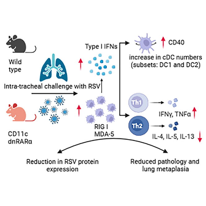
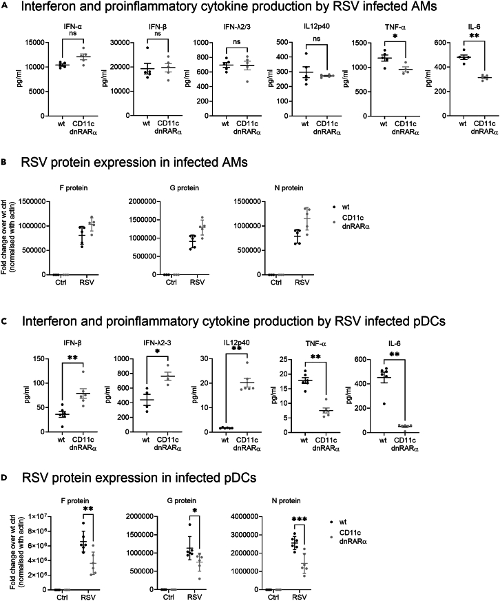
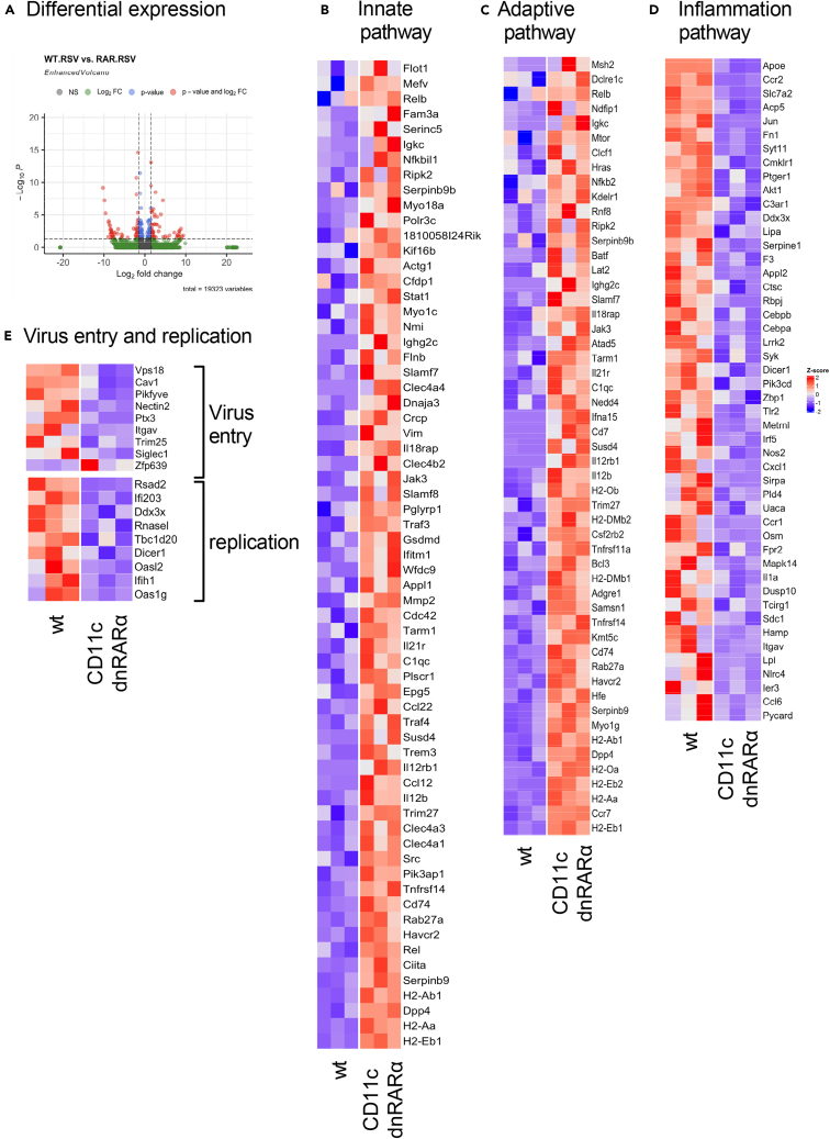
Retinoic acid (RA), controls the immunoregulatory functions of many immune cells, including dendritic cells (DCs), and is important for mucosal immunity. In DCs, RA regulates the expression of pattern recognition receptors and stimulates interferon production. Here, we investigated the role of RA in DCs in mounting immunity to respiratory syncytial virus (RSV). To abolish RA signaling in DCs, we used mice expressing a dominant negative form of retinoic acid receptor-α (RARα) under the CD11c promoter (CD11c-dnRARα). Paradoxically, upon RSV challenge, these animals had lower viral burden, reduced pathology, and greater Th1 polarized immunity than wild-type (WT) mice. Moreover, CD11c-dnRARα DCs infected with RSV showed enhancement in innate and adaptive immunity genes, while genes associated with viral replication were downregulated. These findings suggest that the absence of RA signaling in DCs enhances innate immunity against RSV infection leading to decreased viral load and reduced pathogenicity.

Keywords: Immunology; Virology; cell biology.


Conflict of interest statement

The authors declare no competing interests.

Figures

None
Graphical abstract
Figure 1
Figure 1
CD11c-dnRARα animals had reduced inflammation and mucus secretion Lungs were isolated from naive CD11c-dnRARα and wt littermates as well as RSV (3 × 105 pfu) infected animals on day 8. (A and B) Lungs were embedded in paraffin; PAS was performed to stain mucus secreting cells and H&E was used to examine inflammatory cell infiltration. Inflammation and PAS staining were more severe in WT than CD11c-dnRARα animals. (C) Lungs were homogenized in Trizol and RNA was extracted from RSV infected and naive control animals. Gene expression for mucus-associated genes (muc5AC and Gob5) was determined using qPCR. Mucus production was greater after infection in the WT animals. (D) RSV viral load was determined by gene expression of RSV F, G, and N proteins. qPCR data shown in the figure are normalized against rRNA and fold changes are calculated over naive WT littermate controls. RSV protein expression was greater in the WT animals. (E) RSV infection induced greater numbers of activated DCs in the lungs of CD11c-dnRARα animals. Lungs were processed into single cell suspension. Cell staining was done as described in the STAR methods section. Data shown here are the absolute total number of CD11c+ MHC II + CD11b+ and CD11c+ MHC II + CD103+ DCs and CD40-MFI on these two populations. The gating strategy is presented in Figure S1. Data shown represents n = 5 (naive CD11c-dnRARα and wt littermate) and n = 6 for RSV infected mice (CD11c-dnRARα and WT littermate). Data were analyzed by paired t test or Tukey’s multiple comparison test using GraphPad Prism. Significance is shown as ∗p < 0.05, ∗∗p < 0.01, ∗∗∗p < 0.001, and ∗∗∗∗p < 0.0001.
Figure 2
Figure 2
RA signaling in DCs regulates T cell immunity to RSV (A) CD11c-dnRARα and WT littermate controls were infected with RSV (3 × 105 pfu), and 8 days post challenge, LDLN were isolated and processed into single cells suspension. Cells were re-stimulated with RSV ex vivo for 48 h. Cells were centrifuged and supernatants were collected to determine secreted cytokine. (B) Lungs were isolated from RSV infected animals and homogenized in Trizol. RNA was extracted and the expression level of cytokines was determined using qPCR. Expression data are normalized against 18s rRNA and fold changes are calculated over naive WT littermate controls with n = 5 (for naive CD11c-dnRARα and WT littermates) and n = 6 for RSV infected mice (CD11c-dnRARα and WT littermate). Data were analyzed with paired t test and Tukey’s multiple comparison test using GraphPad Prism and significance is shown as ∗p < 0.05, ∗∗p < 0.01, ∗∗∗p < 0.001, and ∗∗∗∗p < 0.0001.
Figure 3
Figure 3
RA signaling in DCs enhances RSV gene expression DCs from WT animals were infected with RSV in the presence or absence of RA. (A and B) RA activity was measured by aldh1a2 expression in DCs. RSV F, G, and N proteins expression was quantified using qPCR after 24 h. (C and D) In another set of experiments, DCs were infected with RSV in the presence of RA, either with or without the RARα inhibitor (Ro41-5253). The addition of the inhibitor reduced the expression of aldh1a2 and reduced viral protein production. Aldh1a2 as well as RSV F, G, and N proteins expression were analyzed with qPCR. Data presented here are from experimental technical replicates, n = 3, mean ± SD. Statistical analysis was performed with Prism GraphPad paired t test and significance is shown as ∗p < 0.05, ∗∗p < 0.01, ∗∗∗p < 0.001, and ∗∗∗∗p < 0.0001.
Figure 4
Figure 4
RA signaling in DCs regulates innate immunity to RSV infection (A) CD11c-dnRARα BMDCs show greater expression of the innate immune-associated genes aldh1a2, MDA 5 ad RIG-I after RSV infection. Genes expression was quantified with qPCR at 0, 4, 8, and 12 h time points. (B) Production of interferons and other cytokines after RSV infection (1:1 MOI) for 24 h. (C) Decreased RSV protein expression in RSV infected CD11c-dnRARα BMDCs 24 h post stimulation. (D) Dose dependent effect in IFN-β production from RSV infected wt BMDCs cultured with increasing concentration of RA (10, 50, and 100 nM) in combination with RARα inhibitor (RO41-5253-500 nM). Representative data of two independent experiments performed in triplicates, mean ± SD. Statistical analysis was performed using GraphPad Prism and significance is shown as ∗p < 0.05, ∗∗p < 0.01, ∗∗∗p < 0.001, and ∗∗∗∗p < 0.0001.
Figure 5
Figure 5
RA signaling in pDCs but not alveolar macrophages regulates innate immunity to RSV BAL AMs and splenic pDCs were sorted as described in the STAR Methods. 105-2 × 105 cells were infected/stimulated with RSV. Interferons and other cytokines were measured in the supernatants of the cells 24 h post infection. Interferons (IFN-α, IFN-β, and IFN-ƛ2-3) and proinflammatory cytokines (TNF-α and IL-6) production by RSV infected AMs (A) and pDCs (C). RSV gene expression levels were determined by lysing the cells with TriZol and extracting the RNA from infected cells and analyzed with qPCR. Expression data are normalized with β-actin and fold changes were calculated over the respective uninfected controls. RSV proteins expressed from infected AMs (B) and pDCs (D) 24 h post infection. Ctrl represents untreated samples. Representative data of two independent experiments with the cells pooled from 5 to 6 mice each time, mean ± SD. Statistical analysis was performed using GraphPad Prism and significance is shown as ∗p < 0.05, ∗∗p < 0.01, ∗∗∗p < 0.001, and ∗∗∗∗p < 0.0001.
Figure 6
Figure 6
DCs with deficient RA signaling have enhanced antiviral response to RSV BMDCs cultured from WT and CD11c-dnRARα animals were infected with RSV for 24 h and RNA sequencing was performed on total RNA. (A) Differentially expressed genes between WT and CD11c-dnRARα BMDCs. Specific pathways were analyzed in CD11c-dnRARα BMDCs in comparison to BMDCs from WT animals. Heatmaps show genes associated with innate immunity (B), adaptive immunity (C), inflammatory pathway (D), and virus entry and replication (E). Data presented here are from experimental technical replicates, n = 3.
Figure 7
Figure 7
RA signaling differentially regulate TLR-3 and RIG-I responses in DCs (A) 3p-hpRNA (500 ng/ml) was transfected into DCs using lipofectamine 3000 and cultured for 24 h or (B) stimulated with polyIC (1 μg/mL) for 24 h. Post incubation, cells were centrifuged, and the supernatant was collected to analyze for secreted cytokine. Representative data are from 3 independent experiments performed in technical triplicates presented here as mean ± SD (n = 3). Statistical analysis was performed using GraphPad Prism and significance is shown as ∗p, 0.05, ∗∗p, 0.01, ∗∗∗p, 0.001, and #p, 0.0001.

Similar articles

Cited by

References

    1. Smyth R.L., Openshaw P.J.M. Bronchiolitis. Lancet. 2006;368:312–322. doi: 10.1016/S0140-6736(06)69077-6. - DOI - PubMed
    1. Meissner H.C. Viral Bronchiolitis in Children. N. Engl. J. Med. 2016;374:62–72. doi: 10.1056/NEJMra1413456. - DOI - PubMed
    1. Nair H., Nokes D.J., Gessner B.D., Dherani M., Madhi S.A., Singleton R.J., O'Brien K.L., Roca A., Wright P.F., Bruce N., et al. Global burden of acute lower respiratory infections due to respiratory syncytial virus in young children: a systematic review and meta-analysis. Lancet. 2010;375:1545–1555. doi: 10.1016/S0140-6736(10)60206-1. - DOI - PMC - PubMed
    1. Openshaw P.J.M., Chiu C., Culley F.J., Johansson C. Protective and Harmful Immunity to RSV Infection. Annu. Rev. Immunol. 2017;35:501–532. doi: 10.1146/annurev-immunol-051116-052206. - DOI - PubMed
    1. Zhang L., Peeples M.E., Boucher R.C., Collins P.L., Pickles R.J. Respiratory syncytial virus infection of human airway epithelial cells is polarized, specific to ciliated cells, and without obvious cytopathology. J. Virol. 2002;76:5654–5666. doi: 10.1128/jvi.76.11.5654-5666.2002. - DOI - PMC - PubMed

LinkOut - more resources