Review
. 2024 Jul 8:12:1363811.
doi: 10.3389/fcell.2024.1363811. eCollection 2024.

Molecular pathophysiology of secondary lymphedema

Affiliations
Review

Molecular pathophysiology of secondary lymphedema

Sang-Oh Lee et al. Front Cell Dev Biol. .

Abstract

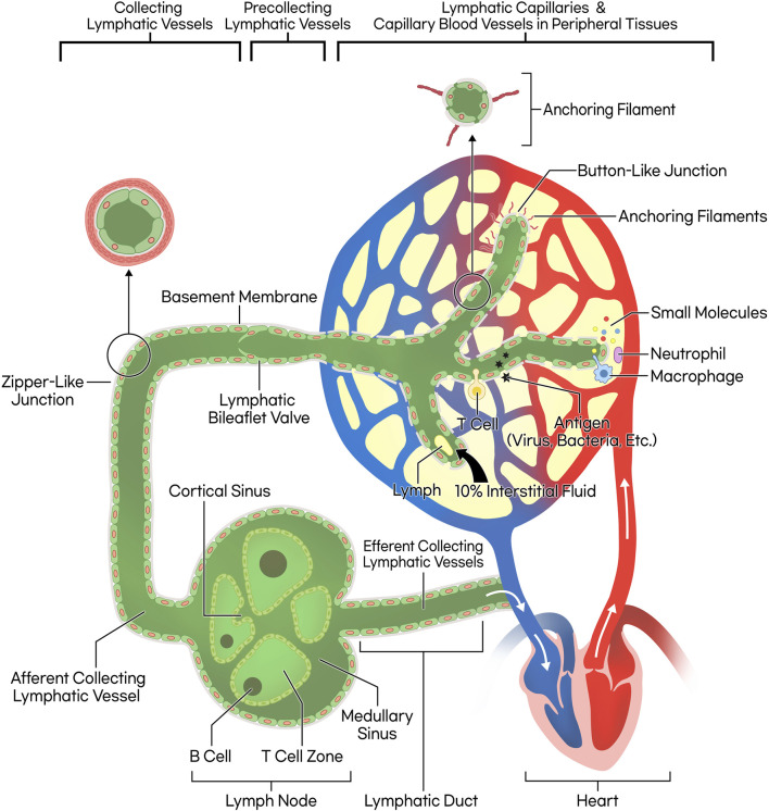
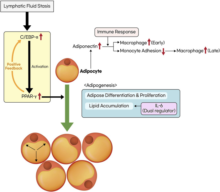
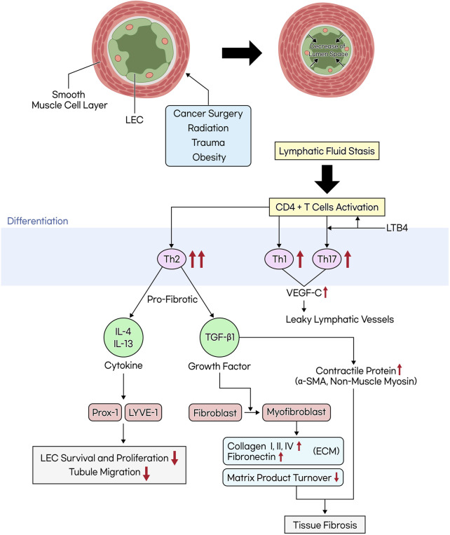
Lymphedema occurs as a result of lymphatic vessel damage or obstruction, leading to the lymphatic fluid stasis, which triggers inflammation, tissue fibrosis, and adipose tissue deposition with adipocyte hypertrophy. The treatment of lymphedema is divided into conservative and surgical approaches. Among surgical treatments, methods like lymphaticovenular anastomosis and vascularized lymph node transfer are gaining attention as they focus on restoring lymphatic flow, constituting a physiologic treatment approach. Lymphatic endothelial cells form the structure of lymphatic vessels. These cells possess button-like junctions that facilitate the influx of fluid and leukocytes. Approximately 10% of interstitial fluid is connected to venous return through lymphatic capillaries. Damage to lymphatic vessels leads to lymphatic fluid stasis, resulting in the clinical condition of lymphedema through three mechanisms: Inflammation involving CD4+ T cells as the principal contributing factor, along with the effects of immune cells on the VEGF-C/VEGFR axis, consequently resulting in abnormal lymphangiogenesis; adipocyte hypertrophy and adipose tissue deposition regulated by the interaction of CCAAT/enhancer-binding protein α and peroxisome proliferator-activated receptor-γ; and tissue fibrosis initiated by the overactivity of Th2 cells, leading to the secretion of profibrotic cytokines such as IL-4, IL-13, and the growth factor TGF-β1. Surgical treatments aimed at reconstructing the lymphatic system help facilitate lymphatic fluid drainage, but their effectiveness in treating already damaged lymphatic vessels is limited. Therefore, reviewing the pathophysiology and molecular mechanisms of lymphedema is crucial to complement surgical treatments and explore novel therapeutic approaches.

Keywords: adipose tissue; fibrosis; inflammation; lymphatic system; lymphedema; molecular biology; physiopathology.


Conflict of interest statement

The authors declare that the research was conducted in the absence of any commercial or financial relationships that could be construed as a potential conflict of interest.

Figures

FIGURE 1
FIGURE 1
Schematic illustration of lymphatic circulation and lymphatic vessel structure. The lymphatic capillary possesses a discontinuous basement membrane and lacks pericytes that typically envelop blood endothelial cells, resulting in a button-like junction pattern. This structural characteristic facilitates the ingress of small molecules, fluid, and leukocytes. The collecting lymphatic vessel, on the other hand, features a smooth muscle layer, bileaflet valves, and zipper-like junctions, allowing lymph propulsion forward through wall contraction. Consequently, the lymph progresses through lymph nodes and lymphatic ducts, ultimately merging into venous return, completing systemic circulation.
FIGURE 2
FIGURE 2
The process of adipose tissue deposition and remodeling. Lymphatic fluid stasis leads to an upregulation in the expression of CCAAT/enhancer-binding protein α (C/EBP-α) and peroxisome proliferator-activated receptor-γ (PPAR-γ). C/EBP-α is a prerequisite for the activation of PPAR-γ, and they mutually establish a positive feedback loop. The activation of PPAR-γ facilitates adipogenesis, which includes processes such as adipose differentiation, proliferation, and lipid accumulation. Interleukin-6 (IL-6) possesses a dual role in regulating lipid accumulation. In addition, the activation of adipocytes triggers the secretion of adiponectin, which exhibits distinct effects on inflammatory responses at both the early and late stages.
FIGURE 3
FIGURE 3
The mechanism of tissue fibrosis occurring in lymphedema. When lymphatic vessels are damaged due to cancer surgery, radiation, trauma, or obesity, the smooth muscle cells within these vessels thicken and transform. This transformation leads to the narrowing of the lymphatic lumen. Additionally, an increase in inner pressure within the lymphatic vessels causes the junctions between lymphatic endothelial cells to weaken, which in turn leads to increased lymph leakage and exacerbates lymphatic fluid stasis. This stasis activates CD4+ T cells, favoring differentiation into Th2 cells over Th1 cells. Th2 cells secrete profibrotic cytokines (IL-4, IL-13) and growth factors (TGF-β1). TGF-β1 induces the differentiation of fibroblasts into myofibroblasts, promoting the accumulation of the extracellular matrix (ECM) and the production of contractile proteins, while reducing matrix product turnover, ultimately leading to tissue fibrosis.

Similar articles

Cited by

References

    1. Alitalo K. (2011). The lymphatic vasculature in disease. Nat. Med. 17 (11), 1371–1380. 10.1038/nm.2545 - DOI - PubMed
    1. Allen R. J., Cheng M.-H. (2016). Lymphedema surgery: patient selection and an overview of surgical techniques. J. Surg. Oncol. 113 (8), 923–931. 10.1002/jso.24170 - DOI - PubMed
    1. Angeli V., Lim H. Y. (2023). Biomechanical control of lymphatic vessel physiology and functions. Cell Mol. Immunol. 20 (9), 1051–1062. 10.1038/s41423-023-01042-9 - DOI - PMC - PubMed
    1. Aschen S., Zampell J. C., Elhadad S., Weitman E., De Brot Andrade M., Mehrara B. J. (2012). Regulation of adipogenesis by lymphatic fluid stasis: part II. Expression of adipose differentiation genes. Plast. Reconstr. Surg. 129 (4), 838–847. 10.1097/PRS.0b013e3182450b47 - DOI - PMC - PubMed
    1. Aschen S. Z., Farias-Eisner G., Cuzzone D. A., Albano N. J., Ghanta S., Weitman E. S., et al. (2014). Lymph node transplantation results in spontaneous lymphatic reconnection and restoration of lymphatic flow. Plast. Reconstr. Surg. 133 (2), 301–310. 10.1097/01.prs.0000436840.69752.7e - DOI - PMC - PubMed

LinkOut - more resources