Review
. 2024 Nov 4;147(11):3665-3680.
doi: 10.1093/brain/awae251.

The ageing central nervous system in multiple sclerosis: the imaging perspective

Affiliations
Review

The ageing central nervous system in multiple sclerosis: the imaging perspective

Massimo Filippi et al. Brain. .

Abstract

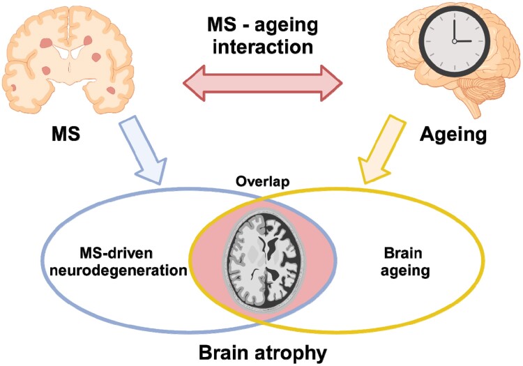
The interaction between ageing and multiple sclerosis is complex and carries significant implications for patient care. Managing multiple sclerosis effectively requires an understanding of how ageing and multiple sclerosis impact brain structure and function. Ageing inherently induces brain changes, including reduced plasticity, diminished grey matter volume, and ischaemic lesion accumulation. When combined with multiple sclerosis pathology, these age-related alterations may worsen clinical disability. Ageing may also influence the response of multiple sclerosis patients to therapies and/or their side effects, highlighting the importance of adjusted treatment considerations. MRI is highly sensitive to age- and multiple sclerosis-related processes. Accordingly, MRI can provide insights into the relationship between ageing and multiple sclerosis, enabling a better understanding of their pathophysiological interplay and informing treatment selection. This review summarizes current knowledge on the immunopathological and MRI aspects of ageing in the CNS in the context of multiple sclerosis. Starting from immunosenescence, ageing-related pathological mechanisms and specific features like enlarged Virchow-Robin spaces, this review then explores clinical aspects, including late-onset multiple sclerosis, the influence of age on diagnostic criteria, and comorbidity effects on imaging features. The role of MRI in understanding neurodegeneration, iron dynamics and myelin changes influenced by ageing and how MRI can contribute to defining treatment effects in ageing multiple sclerosis patients, are also discussed.

Keywords: MRI; ageing; comorbidities; diagnosis; multiple sclerosis; progression.


Conflict of interest statement

The authors declare that they have no competing interests in relation to this work. Potential conflicts of interest outside the submitted work are as follows: M.F. is Editor-in-Chief of the Journal of Neurology, Associate Editor of Human Brain Mapping, Neurological Sciences and Radiology; received compensation for consulting services from Alexion, Almirall, Biogen, Merck, Novartis, Roche, Sanofi; speaking activities from Bayer, Biogen, Celgene, Chiesi Italia SpA, Eli Lilly, Genzyme, Janssen, Merck-Serono, Neopharmed Gentili, Novartis, Novo Nordisk, Roche, Sanofi, Takeda, and TEVA; participation in Advisory Boards for Alexion, Biogen, Bristol-Myers Squibb, Merck, Novartis, Roche, Sanofi, Sanofi-Aventis, Sanofi-Genzyme, Takeda; scientific direction of educational events for Biogen, Merck, Roche, Celgene, Bristol-Myers Squibb, Lilly, Novartis, Sanofi-Genzyme; he receives research support from Biogen Idec, Merck-Serono, Novartis, Roche, the Italian Ministry of Health, the Italian Ministry of University and Research, and Fondazione Italiana Sclerosi Multipla. P.P. received speaker honoraria from Roche, Biogen, Novartis, Merck Serono, Bristol Myers Squibb and Genzyme; he has received research support from Italian Ministry of Health and Fondazione Italiana Sclerosi Multipla. F.B. acts in Steering committee or Data Safety Monitoring Board member for Biogen, Merck, ATRI/ACTC and Prothena. Consultant for Roche, Celltrion, Rewind Therapeutics, Merck, IXICO, Jansen, Combinostics. Research agreements with Merck, Biogen, GE Healthcare, Roche. Co-founder and shareholder of Queen Square Analytics LTD. F.B. is supported by the NIHR Biomedical Research Centre at UCLH. O.C. is NIHR Research Professor (RP-2017-08-ST2-004); over the last 2 years she has been a member of independent DSMB for Novartis, gave a teaching talk in a Merck local symposium, and contributed to an Advisory Board for Biogen; she is Deputy Editor of Neurology, for which she receives an honorarium; she has received research grant support from the MS Society of Great Britain and Northern Ireland, the NIHR UCLH Biomedical Research Centre, the Rosetree Trust, the National MS Society, and the NIHR-HTA.

A.C. is supported by FISM, Bando Ricerca COVID, Project 2021/C19-R-Single/011. N.D.S. is Associate Editor of Neurological Sciences; received compensation for speaking activities from Biogen, Bristol-Myers Squibb, Merck-Serono, Novartis, Roche, and Sanofi-Genzyme; participation in Advisory Boards for Bristol-Myers Squibb, Merck, Roche, and Sanofi-Genzyme; receives research support from the Italian Ministry of Health, the Italian Ministry of University and Research, and Fondazione Italiana Sclerosi Multipla; co-founder of SIENA Imaging. C.G. has received speaker honoraria and/or travel expenses for attending meeting from Bayer Schering Pharma, Sanofi-Aventis, Merck, Biogen, Novartis and Almirall. R.G. has received support for scientific meetings and courses and honoraria for advisory work from Bayer, Biogen, Merck, Novartis, Janssen, MIAC. The university hospital Basel and the research center for clinical neuroimmunology and neuroscience (RC2NB), as the employers of C.G., have received the following fees which were used exclusively for research support: advisory boards and consultancy fees from Actelion, Novartis, Genzyme-Sanofi, GeNeuro, Hoffmann La Roche, Merck and Siemens Healthineers; speaker fees from Biogen, Hoffmann La Roche, Teva, Novartis, Janssen, Merck and Genzyme-Sanofi; and research grants from Hoffmann La Roche, GeNeuro, Genzyme, Novartis and Biogen. CG is supported by the Swiss National Fund n. PP00P3_206151, the Hasler Foundation and the Stiftung zur Förderung der gastroenterologischen und allgemeinen klinischen Forschung. H.L. has received honoraria for lectures from Novartis, Biogen, ROCHE, Merck, and Sanofi-Aventis. M.M. reports grants and personal fees from Sanofi-Genzyme, Merck-Serono, Novartis and Almiral. She was awarded a MAGNIMS-ECTRIMS fellowship in 2020. G.P. was supported by MAGNIMS/ECTRIMS research fellowship program in 2020 and ESNR research fellowship program in 2021. A.R. serves/ed on scientific advisory boards for Novartis, Sanofi-Genzyme, Synthetic MR, TensorMedical, Roche, and Biogen, and has received speaker honoraria from Bayer, Sanofi-Genzyme, Merck-Serono, Teva Pharmaceutical Industries Ltd, Novartis, Roche, Bristol-Myers and Biogen, is CMO and co-founder of TensorMedical, and receives research support from Fondo de Investigación en Salud (PI19/00 950 and PI22/01589) from Instituto de Salud Carlos III, Spain. J.S-G. declares fees from Sanofi, Biogen, Celgene, Merck, Biopass, Novartis and Roche and receives research support from Fondo de Investigatiòn en Salud (PI19/00950), from Instituo de Salud Carlos III, Spain. M.A.R. received consulting fees from Biogen, Bristol Myers Squibb, Eli Lilly, Janssen, Roche; and speaker honoraria from AstraZaneca, Biogen, Bristol Myers Squibb, Bromatech, Celgene, Genzyme, Horizon Therapeutics Italy, Merck Serono SpA, Novartis, Roche, Sanofi and Teva. She receives research support from the MS Society of Canada, the Italian Ministry of Health, the Italian Ministry of University and Research, and Fondazione Italiana Sclerosi Multipla. She is Associate Editor for Multiple Sclerosis and Related Disorders.

Figures

Figure 1
Figure 1
The ageing perivascular compartment in multiple sclerosis. Perivascular spaces (PVS), which are implicated in brain waste removal, are involved in ageing and multiple sclerosis (MS) at different levels. CSF, produced in the choroid plexus, exchanges with brain interstitial fluid. In addition to the established CSF exit pathways along the spinal subarachnoid space, cranial nerves and arachnoid granulations, a portion of CSF flows into the brain parenchyma via the periarterial space. This flow is part of the glymphatic drainage pathway, illustrated along the arteries and through pial fenestrations. Concurrently, protein degradation products are conveyed within the muscularis of arteries, moving counter to the direction of blood flow, into the subarachnoid arteries. This process is part of the intramural peri-arterial drainage pathway, represented in blue along the artery (A). At the arterial and arteriolar level, cross-sectional views reveal that the perivascular space comprises the astrocytic end-feet processes (including their corresponding basement membrane), the pia mater (which becomes increasingly fenestrated closer to the capillary level), smooth muscle cells and the endothelium (each with their respective basement membranes). Within capillaries, the perivascular space is defined by the shared basement membranes of the astrocytic end-feet processes and the endothelium. The CSF-filled subarachnoid spaces are also evident along veins and venules, where the layers of smooth muscle cells are largely absent (B). Age-related factors, such as atherosclerosis/arteriolosclerosis, elastin dysfunction and periarterial collagen deposition, have been implicated in vascular stiffness, diminished debris transport capacity, and an increased barrier to oxygen delivery. In multiple sclerosis, perivascular changes include collagen deposition and perivenous inflammatory infiltrates that come into contact with CSF (C).
Figure 2
Figure 2
Summary of the typical lesional MRI findings in multiple sclerosis compared to ageing and cerebral small vessel disease. Typical multiple sclerosis (MS) lesions include (A) periventricular lesions, (B) juxtacortical and cortical lesions, (C) white matter (WM) lesions showing the central vein sign (CVS), (E) paramagnetic rim lesion (PRLs), (F) infratentorial lesions mainly located at the periphery, close to the CSF, and (G) spinal cord lesions. Typical lesions occurring with ageing and cerebral small vessel disease (cSVD) include (H) subcortical WM lesions, (I) deep WM lesions, (J) periventricular lesions and ‘capping’, (K) cortical microinfarcts, (L) central pontine lesions and (L) no spinal cord lesions. See text for further details.
Figure 3
Figure 3
Schematic representation of the interplay between the effects of multiple sclerosis-related neurodegeneration and ageing on brain atrophy. Both ageing and multiple sclerosis (MS) are associated with brain atrophy, with partially overlapping patterns (blue arrow). Rather than being simply additive, the effects of ageing and multiple sclerosis on brain atrophy are linked by a complex interaction (red arrow): the relationship between age and brain volume is influenced by multiple sclerosis and encodes disease-related information; ageing shapes multiple sclerosis-related brain atrophy by modifying the disease course and the response to treatment. Created with http://www.biorender.com/.

References

    1. Filippi M, Bar-Or A, Piehl F, et al. Multiple sclerosis. Nat Rev Dis Primers. 2018;4:43. - PubMed
    1. Lee J, Kim H-J. Normal aging induces changes in the brain and neurodegeneration progress: Review of the structural, biochemical, metabolic, cellular, and molecular changes. Front Aging Neurosci. 2022;14:931536. - PMC - PubMed
    1. Marrie RA, Yu N, Blanchard J, Leung S, Elliott L. The rising prevalence and changing age distribution of multiple sclerosis in Manitoba. Neurology. 2010;74:465–471. - PubMed
    1. Vaughn CB, Jakimovski D, Kavak KS, et al. Epidemiology and treatment of multiple sclerosis in elderly populations. Nat Rev Neurol. 2019;15:329–342. - PubMed
    1. Prosperini L, Lucchini M, Ruggieri S, et al. Shift of multiple sclerosis onset towards older age. J Neurol Neurosurg Psychiatry. 2022;93:1137–1139. - PubMed

MeSH terms