. 2024 Oct;119(5):751-772.
doi: 10.1007/s00395-024-01067-9. Epub 2024 Jul 24.

Empagliflozin prevents heart failure through inhibition of the NHE1-NO pathway, independent of SGLT2

Affiliations

Empagliflozin prevents heart failure through inhibition of the NHE1-NO pathway, independent of SGLT2

Sha Chen et al. Basic Res Cardiol. 2024 Oct.

Abstract

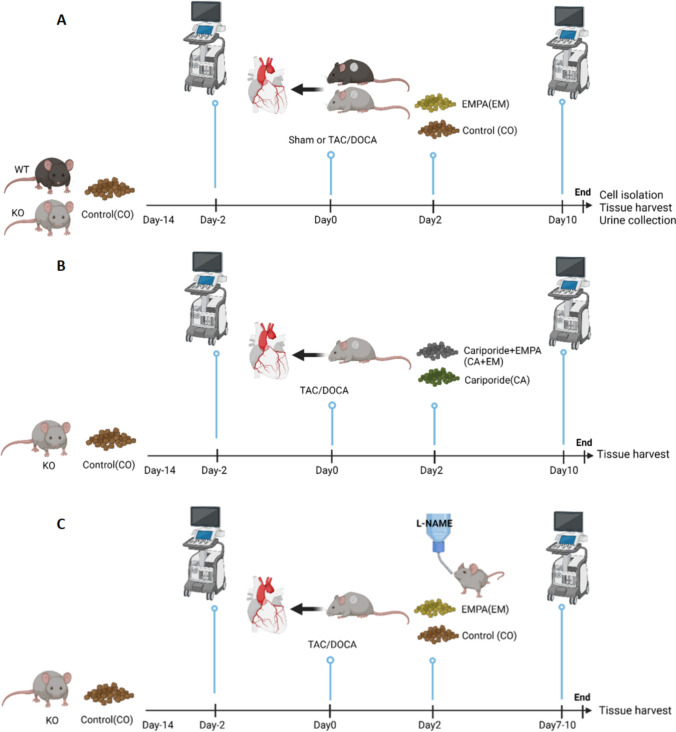
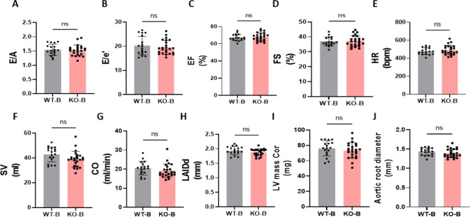
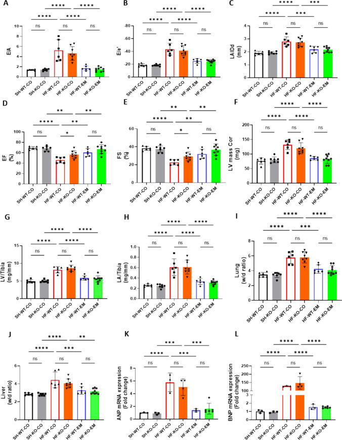
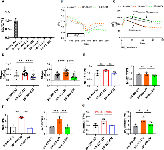
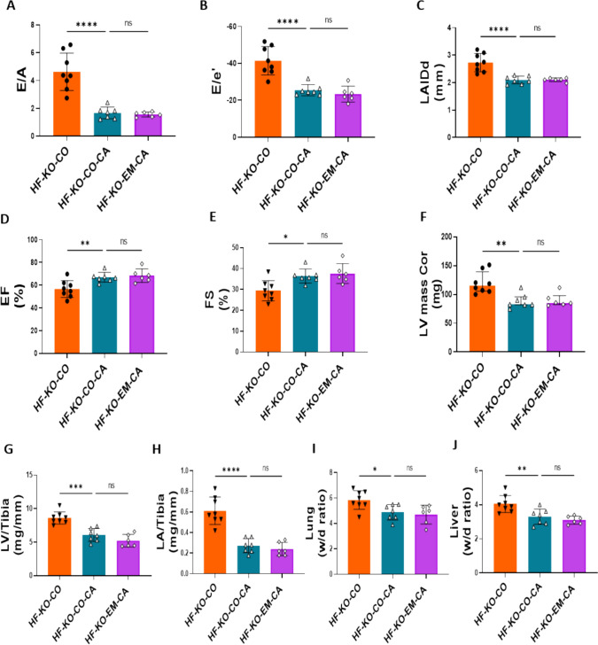
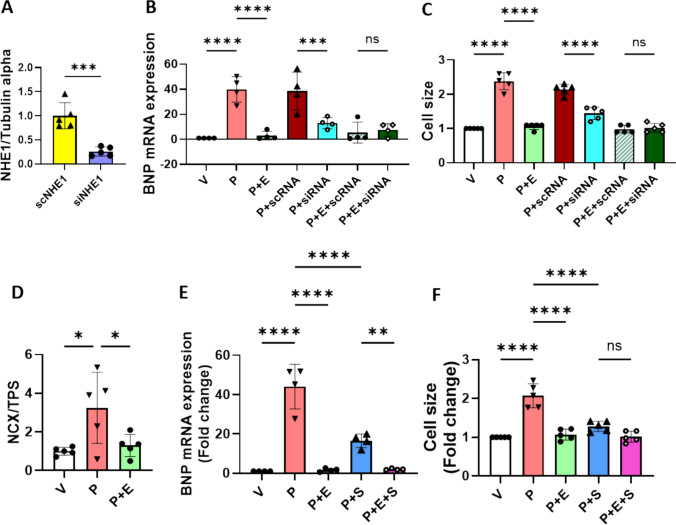
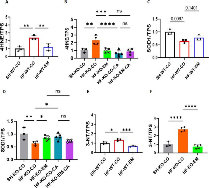
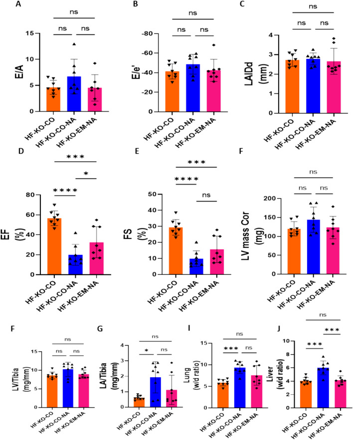
Sodium glucose cotransporter 2 inhibitors (SGLT2i) constitute the only medication class that consistently prevents or attenuates human heart failure (HF) independent of ejection fraction. We have suggested earlier that the protective mechanisms of the SGLT2i Empagliflozin (EMPA) are mediated through reductions in the sodium hydrogen exchanger 1 (NHE1)-nitric oxide (NO) pathway, independent of SGLT2. Here, we examined the role of SGLT2, NHE1 and NO in a murine TAC/DOCA model of HF. SGLT2 knockout mice only showed attenuated systolic dysfunction without having an effect on other signs of HF. EMPA protected against systolic and diastolic dysfunction, hypertrophy, fibrosis, increased Nppa/Nppb mRNA expression and lung/liver edema. In addition, EMPA prevented increases in oxidative stress, sodium calcium exchanger expression and calcium/calmodulin-dependent protein kinase II activation to an equal degree in WT and SGLT2 KO animals. In particular, while NHE1 activity was increased in isolated cardiomyocytes from untreated HF, EMPA treatment prevented this. Since SGLT2 is not required for the protective effects of EMPA, the pathway between NHE1 and NO was further explored in SGLT2 KO animals. In vivo treatment with the specific NHE1-inhibitor Cariporide mimicked the protection by EMPA, without additional protection by EMPA. On the other hand, in vivo inhibition of NOS with L-NAME deteriorated HF and prevented protection by EMPA. In conclusion, the data support that the beneficial effects of EMPA are mediated through the NHE1-NO pathway in TAC/DOCA-induced heart failure and not through SGLT2 inhibition.

Keywords: Diastolic dysfunction; Heart failure; NHE1; Nitric oxide; Oxidative stress; SGLT2 inhibitors.


Conflict of interest statement

PF is the founder and CEO of Pharmahungary Group, a group of R&D companies.

Figures

Fig. 1
Fig. 1
Timeline of interventions. A Series #1: Baseline cardiac function was evaluated using ultrasound two days prior surgery to induce HF. Two days post-surgery, oral treatment (CO and EM) [67] was started by switching to treatment-enriched chows. Then, 10 days after Sham or TAC/DOCA surgery, cardiac function was re-evaluated to determine parameters of cardiac HF. Finally, at days 10–12 after start HF surgery, animals were sacrificed. Hearts were used either for cell isolation to perform NHE1 activity assays or immunoblotting. Other organs and urine were collected for further measurements. B Series #2: Two days post-surgery, oral treatment (CA and CA + EM) was started by switching to treatment-enriched chows. Then, 10 days after Sham or TAC/DOCA surgery, cardiac function was re-evaluated. Finally, at days 10–12 after start HF surgery, animals were sacrificed. Hearts were used for immunoblotting. Other organs were collected for further measurements. C Series #3: Two days post-surgery, oral treatment (CO and EM) was started by switching to treatment-enriched chows and/or L-NAME in drinking water [39]. Then, 7–10 days after Sham or TAC/DOCA surgery, cardiac function was re-evaluated. Finally, at days 8–11 after start HF surgery, animals were sacrificed. Organs were collected for further measurements. CO control, EM EMPA, CA Cariporide, CA + EM Cariporide + EMPA, L-NAME L-nitro-arginine-methyl-esther, TAC/DOCA Transverse aortic constriction and implantation of a deoxycorticosterone acetate pellet. (Created with BioRender.com)
Fig. 2
Fig. 2
Baseline systolic and diastolic cardiac function in WT and SGLT2 KO mice, before surgery. All of the parameters are measured 2 days before TAC/DOCA surgery. A, The ratio of early diastolic mitral inflow velocity to late diastolic mitral inflow velocity (E/A ratio). B, The ratio of early diastolic mitral inflow velocity to early diastolic mitral annulus velocity (E/e' ratio). C, Ejection fraction (EF). D, Fractional shortening (FS). E, Heart rate (HR). F, Stroke volume (SV). G, Cardiac output (CO). H, Left atria internal diameter end diastole (LAIDd). I, Left ventricle mass (LV mass Cor). J, Aortic root diameter. Data are presented as mean ± SD. ns: not significant. WT-B, wild-type baseline (n = 18; 9 M (male), 9F (female)), KO-B, SGLT2 KO baseline (n = 24; 10 M/14F). AF and HJ: unpaired t test; G: Mann–Whitney test. B: Baseline
Fig. 3
Fig. 3
EMPA ameliorated HF independent of SGLT2, whereas SGLT2 deficiency partly protected against HF-induced systolic dysfunction. A The ratio of early diastolic mitral inflow velocity to late diastolic mitral inflow velocity (E/A ratio) measured at 10 days after TAC/DOCA surgery. B The ratio of early diastolic mitral inflow velocity to early diastolic mitral annulus velocity (E/e' ratio) measured at 10 days after TAC/DOCA surgery. C Left atria internal diameter end diastole (LAIDd) measured at 10 days after TAC/DOCA surgery. D Ejection fraction (EF) measured at 10 days after TAC/DOCA surgery. E Fractional shortening (FS) measured at 10 days after TAC/DOCA surgery. F Left ventricle mass (LV mass Cor) measured at 10 days after TAC/DOCA surgery. G Left ventricle weight to tibia length(LV/tibia). H Left atria weight to tibia length (LA/tibia). I Lung wet/dry ratio (Lung w/d ratio). J, Liver wet/dry ratio (Liver w/d ratio). K, L Nppa and Nppb mRNA expression. Data are presented as mean ± SD. *p < 0.05, **p < 0.01, ***p < 0.001, ****p < 0.0001. ns: not significant. AJ SH-WT-CO, sham-wild-type-control chow (n = 6; 3 M (male), 3F(female)), SH-KO-CO, sham-SGLT2 KO-control chow (n = 7; 4 M/3F), HF-WT-CO, heart failure-wild-type-control chow (n = 6; 3 M/3F), HF-KO-CO, heart failure-SGLT2 KO-control chow (n = 8; 3 M/5F), HF-WT-EM, heart failure-wild-type-EMPA chow (n = 6; 3 M/3F) and HF-KO-EM, heart failure-SGLT2KO-EMPA chow (n = 9; 4 M/5F). K, L SH-WT-CO (n = 3; 1 M/2F), SH-KO-CO (n = 3; 1 M/2F), HF-WT-CO (n = 3; 2 M/1F), HF-KO-CO (n = 4; 2 M/2F), HF-WT-EM (n = 3; 2 M/1F) and HF-KO-EM (n = 5; 2 M/3F). A–L One-way ANOVA with Holm-Šídák's multiple comparisons test
Fig. 4
Fig. 4
EMPA reduced HF-induced activation of NHE1, NCX expression and CamKII phosphorylation, independently of SGLT2. A Sodium glucose cotransporter 2 protein expression/total protein staining (SGLT2/TPS). B Typical example of the curve changes of intracellular pH during the whole measurement, showing the slope only for KO mice; C Typical example of the curve changes of intracellular [H+] during the first 200 s after NH4+ wash-out, showing the slope only for KO mice. D Chronic effects of EMPA on NHE1 activity as reflected by the; E Sodium hydrogen exchanger 1 protein expression/total protein staining (NHE1/TPS). F sodium calcium exchanger 1 protein expression/total protein staining (NCX/TPS). G Phosphorylation of calcium/calmodulin-dependent protein kinase expression/total protein staining (pCamkII/TPS). Data are presented as mean ± SD. *p < 0.05, **p < 0.01, *** p < 0.001, **** p < 0.0001. ns: not significant. A and E–G and G-J: SH-WT-CO (n = 3; 1 M/2F), SH-KO-CO (n = 3; 1 M/2F), HF-WT-CO (n = 3; 2 M/1F), HF-KO-CO (n = 4; 2 M/2F), HF-WT-EM (n = 3; 2 M/1F) and HF-KO-EM (n = 5; 2 M/3F). B–D SH-WT-CO (15 cells from 3 mice, 2 M/1F), SH-KO-CO (39 cells from 4 mice, 3 M/1F), HF-WT-CO (29 cells from 3 mice, 1 M/2F), HF-KO-CO (37 cells from 4 mice, 1 M/3F), HF-WT-EM(33 cells from 3 mice, 1 M/2F) and HF-KO-EM (34 cells from 3 mice, 1 M/2F). D–G One-way ANOVA with Holm-Šídák's multiple comparisons test
Fig. 5
Fig. 5
NHE1 inhibitor Cariporide abolished EMPA’s protection against HFmrEF. A The ratio of early diastolic mitral inflow velocity to late diastolic mitral inflow velocity (E/A ratio) measured at 10 days after TAC/DOCA surgery. B The ratio of early diastolic mitral inflow velocity to early diastolic mitral annulus velocity (E/e' ratio) measured at 10 days after TAC/DOCA surgery. C Left atria internal diameter end diastole (LAIDd) measured at 10 days after TAC/DOCA surgery. D Ejection fraction (EF) measured at 10 days after TAC/DOCA surgery. E Fractional shortening (FS) measured at 10 days after TAC/DOCA surgery. F Left ventricle mass (LV mass Cor) measured at 10 days after TAC/DOCA surgery. G Left ventricle weight to tibia length (LV/tibia). H Left atria weight to tibia length (LA/tibia). I Lung wet/dry ratio (Lung w/d ratio). J Liver wet/dry ratio (Liver w/d ratio). Data are presented as mean ± SD. *p < 0.05, **p < 0.01, ***p < 0.001, ****p < 0.0001. ns: not significant. KT HF-KO-CO (n = 8; 3 M/5F), HF-KO-CO-CA (n = 7; 3 M/4F) and HF-KO-EM-CA (n = 6; 3 M/3F). AJ One-way ANOVA with Holm-Šídák's multiple comparisons test
Fig. 6
Fig. 6
NHE1 downregulation and NCX inhibition partly reversed PE-induced cellular hypertrophy and abolished EMPA’s protection on hypertrophy. A Representative immunoblots and analysis of NHE1 protein in H9c2 cells with scRNA and siRNA treatment (5 individual experiments). Unpaired t test. B BNP mRNA expression was measured by RT-PCR after 48 h treatment under each condition. One way ANOVA with Holm-Šídák's multiple comparisons test (4 individual experiments). C Cell surface area was measured at 48 h. One way ANOVA with Holm-Šídák's multiple comparisons test (5 individual experiments). D Representative immunoblots and analysis of NCX protein in H9c2 cells with PE and PE plus EMPA treatments. One way ANOVA with Holm-Šídák's multiple comparisons test (5 individual experiments). E BNP mRNA expression was measured by RT-PCR after 48 h treatment under each condition. One way ANOVA with Holm-Šídák's multiple comparisons test (4 individual experiments). F Cell surface area was measured at 48 h. One way ANOVA with Holm-Šídák's multiple comparisons test (5 individual experiments). Data are presented as mean ± SD. *p < 0.05, **p < 0.01, ***p < 0.001, ****p < 0.0001. ns: not significant
Fig. 7
Fig. 7
EMPA reduced HF-induced elevations oxidative/nitrosative stress independent of SGLT2. A, B, 4-hydroxynonenal protein expression/total protein staining (4-HNE/TPS). C, D Superoxide dismutase 1 protein expression/total protein staining (SOD1/TPS). E, F, 3-nitrotyrosine protein expression/total protein staining (3-NT/TPS). Data are presented as mean ± SD. *p < 0.05, **p < 0.01, ***p < 0.001, ****p < 0.0001. ns: not significant. SH-WT-CO (n = 3; 1 M/2F), SH-KO-CO (n = 3; 1 M/2F), HF-WT-CO (n = 3; 2 M/1F), HF-KO-CO (n = 4; 2 M/2F), HF-WT-EM(n = 3; 2 M/1F) and HF-KO-EM(n = 5; 2 M/3F), HF-KO-CO-CA (n = 4; 2 M/3F) and HF-KO-EM-CA (n = 5; 2 M/2F). AF One-way ANOVA with Holm-Šídák's multiple comparisons test
Fig. 8
Fig. 8
Nitric oxide synthase (NOS) inhibition abolished HF protection by EMPA. A The ratio of early diastolic mitral inflow velocity to late diastolic mitral inflow velocity (E/A ratio) measured at 10 days after TAC/DOCA surgery. B The ratio of early diastolic mitral inflow velocity to early diastolic mitral annulus velocity (E/e' ratio) measured at 10 days after TAC/DOCA surgery. C Left atria internal diameter end diastole (LAIDd) measured at 10 days after TAC/DOCA surgery. D Ejection fraction (EF) measured at 10 days after TAC/DOCA surgery. E Fractional shortening (FS) measured at 10 days after TAC/DOCA surgery. F Left ventricle mass (LV mass Cor) measured at 10 days after TAC/DOCA surgery. G Left ventricle weight to tibia length(LV/tibia). H, Left atria weight to tibia length (LA/tibia). I, Lung wet/dry ratio (Lung w/d ratio). J, Liver wet/dry ratio (Liver w/d ratio). Data are presented as mean ± SD. * p < 0.05, *** p < 0.001, **** p < 0.0001. ns: not significant. A-K: HF-KO-CO (n = 8; 3 M/5F), HF-KO-CO-NA (n = 8; 4 M/4F) and HF-KO-EM-NA (n = 8; 4 M/4F). A-J: One-way ANOVA with Holm-Šídák's multiple comparisons test

References

    1. Anker SD, Khan MS, Butler J, Ofstad AP, Peil B, Pfarr E, Doehner W, Sattar N, Coats AJS, Filippatos G, Ferreira JP, Zannad F, Pocock S, Packer M (2023) Weight change and clinical outcomes in heart failure with reduced ejection fraction: insights from EMPEROR-Reduced. Eur J Heart Fail 25:117–127. 10.1002/ejhf.2728 - PMC - PubMed
    1. Baartscheer A, Hardziyenka M, Schumacher CA, Belterman CN, van Borren MM, Verkerk AO, Coronel R, Fiolet JW (2008) Chronic inhibition of the Na+/H+ - exchanger causes regression of hypertrophy, heart failure, and ionic and electrophysiological remodelling. Br J Pharmacol 154:1266–1275. 10.1038/bjp.2008.189 - PMC - PubMed
    1. Baartscheer A, Schumacher CA, van Borren MM, Belterman CN, Coronel R, Fiolet JW (2003) Increased Na+/H+-exchange activity is the cause of increased [Na+]i and underlies disturbed calcium handling in the rabbit pressure and volume overload heart failure model. Cardiovasc Res 57:1015–1024. 10.1016/s0008-6363(02)00809-x - PubMed
    1. Baartscheer A, Schumacher CA, Wust RC, Fiolet JW, Stienen GJ, Coronel R, Zuurbier CJ (2017) Empagliflozin decreases myocardial cytoplasmic Na(+) through inhibition of the cardiac Na(+)/H(+) exchanger in rats and rabbits. Diabetologia 60:568–573. 10.1007/s00125-016-4134-x - PMC - PubMed
    1. Bay J, Kohlhaas M, Maack C (2013) Intracellular Na(+) and cardiac metabolism. J Mol Cell Cardiol 61:20–27. 10.1016/j.yjmcc.2013.05.010 - PubMed

Publication types

MeSH terms