. 2024 Sep 20;5(3):103199.
doi: 10.1016/j.xpro.2024.103199. Epub 2024 Jul 22.

Protocol for assessing GD2 on formalin-fixed paraffin-embedded tissue sections using immunofluorescence staining

Affiliations

Protocol for assessing GD2 on formalin-fixed paraffin-embedded tissue sections using immunofluorescence staining

Sareetha Kailayangiri et al. STAR Protoc. .

Abstract

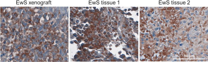
The detection of disialoganglioside GD2 on tumor biopsies, especially in paraffin-embedded tissues, has been challenging due to the glycolipid structure of GD2 and its membrane anchorage. Here, we present an immunofluorescence protocol for the reliable assessment of GD2 on formalin-fixed paraffin-embedded (FFPE) tissues. We describe steps for antigen retrieval with Tris-EDTA buffer and staining with unconjugated anti-GD2 antibody (clone 14.G2a) and horse radish peroxidase (HRP)-conjugated secondary antibody. We then detail procedures for signal amplification using the tyramide signal amplification technique. For complete details on the use and execution of this protocol, please refer to Fischer-Riepe et al.1.

Keywords: antibody; cancer; immunology; microscopy.


Conflict of interest statement

Declaration of interests The authors declare no competing interests.

Figures

None
Graphical abstract
Figure 1
Figure 1
Validation of GD2 expression by immunofluorescence analysis on tissue sections from a GD2-positive xenograft (Kelly, upper panel) and a GD2-negative xenograft (Raji, lower panel), respectively FFPE tissues were stained with the GD2-specific antibody 14.G2a, followed by immunofluorescence analysis according to the protocol (left panels). The presence and absence of GD2 surface expression in the two cell lines, respectively, was confirmed by flow cytometry with the same antibody clone by membraneous staining (right panels). Figure reprinted with permission from Fischer-Riepe et al. Immunofluorescence images were taken with a 4x objective (200 μM) and a 20x objective (50 μM).
Figure 2
Figure 2
Validation of GD2 immunofluorescence staining on EwS xenograft tissues FFPE tissue of EwS xenografts from cell lines with heterogeneous expression of GD2 at variable levels were stained with the GD2-specific antibody 14.G2a according to the protocol (upper panel). GD2 expression of the cell lines used to establish the xenografts were also analyzed by flow cytometry with the same antibody clone (lower panel). Immunofluorescence images were taken with a 20x objective (50 μM).
Figure 3
Figure 3
GD2 immunofluorescence staining of tumor tissues from EwS, breast cancer, and lung cancer patients FFPE tissues from tumors were stained with the GD2-specific protocol. Immunofluorescence images were taken with a 20x objective (50 μM).
Figure 4
Figure 4
Inter-assay precision GD2 staining of two different EwS tissues (patient 1, upper panel; patient 2, lower panel) was performed on different days (A, D, E = day 1; B,C,F = day 2) and operators (A, C, E, F = operator 1; B, D = operator 2). Immunofluorescence images were taken with a 20x objective (50 μM).
Figure 5
Figure 5
Immunohistochemistry staining of GD2 on EwS xenografts and patient-derived tumor tissues FFPE tissues were stained with the GD2-specific antibody 14.G2a by immunohistochemistry using DAB. Images were taken with a 20x objective (50 μM).
Figure 6
Figure 6
GD2 staining on EwS xenograft with different H2O2 solutions An FFPE EwS xenograft was stained with the GD2-specific 14.G2a antibody according to the protocol using decomposed H2O2 solution (right panel) and non-decomposed H2O2 solution (left panel) for endogenous peroxidase blocking. Immunofluorescence images were taken with a 20x objective (50 μM).

References

    1. Fischer-Riepe L., Kailayangiri S., Zimmermann K., Pfeifer R., Aigner M., Altvater B., Kretschmann S., Volkl S., Hartley J., Dreger C., et al. Preclinical Development of CAR T Cells with Antigen-Inducible IL18 Enforcement to Treat GD2-Positive Solid Cancers. Clin. Cancer Res. 2024:OF1–OF14. doi: 10.1158/1078-0432.CCR-23-3157. - DOI - PubMed
    1. Schulz G., Cheresh D.A., Varki N.M., Yu A., Staffileno L.K., Reisfeld R.A. Detection of ganglioside GD2 in tumor tissues and sera of neuroblastoma patients. Cancer Res. 1984;44:5914–5920. - PubMed
    1. Yanagisawa M., Yoshimura S., Yu R.K. Expression of GD2 and GD3 gangliosides in human embryonic neural stem cells. ASN Neuro. 2011;3 doi: 10.1042/AN20110006. - DOI - PMC - PubMed
    1. Yu A.L., Gilman A.L., Ozkaynak M.F., London W.B., Kreissman S.G., Chen H.X., Smith M., Anderson B., Villablanca J.G., Matthay K.K., et al. Anti-GD2 antibody with GM-CSF, interleukin-2, and isotretinoin for neuroblastoma. N. Engl. J. Med. 2010;363:1324–1334. - PMC - PubMed
    1. Ladenstein R., Potschger U., Valteau-Couanet D., Luksch R., Castel V., Yaniv I., Laureys G., Brock P., Michon J.M., Owens C., et al. Interleukin 2 with anti-GD2 antibody ch14.18/CHO (dinutuximab beta) in patients with high-risk neuroblastoma (HR-NBL1/SIOPEN): a multicentre, randomised, phase 3 trial. Lancet Oncol. 2018;19:1617–1629. doi: 10.1016/S1470-2045(18)30578-3. - DOI - PubMed

Publication types

LinkOut - more resources