. 2024 Jul 26;10(30):eadp2679.
doi: 10.1126/sciadv.adp2679. Epub 2024 Jul 24.

A water-resistant, ultrathin, conformable organic photodetector for vital sign monitoring

Affiliations

A water-resistant, ultrathin, conformable organic photodetector for vital sign monitoring

Baocai Du et al. Sci Adv. .

Abstract

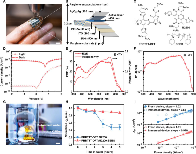
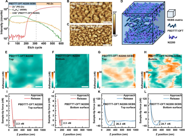
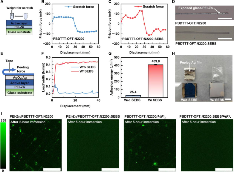
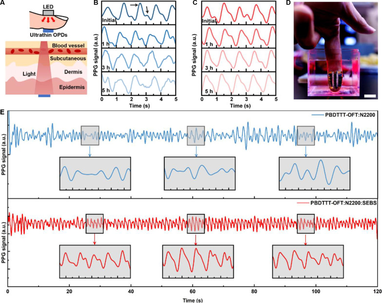
Ultrathin flexible photodetectors can be conformably integrated with the human body, offering promising advancements for emerging skin-interfaced sensors. However, the susceptibility to degradation in ambient and particularly in aqueous environments hinders their practical application. Here, we report a 3.2-micrometer-thick water-resistant organic photodetector capable of reliably monitoring vital sign while submerged underwater. Embedding the organic photoactive layer in an adhesive elastomer matrix induces multidimensional hybrid phase separation, enabling high adhesiveness of the photoactive layer on both the top and bottom surfaces with maintained charge transport. This improves the water-immersion stability of the photoactive layer and ensures the robust sealing of interfaces within the device, notably suppressing fluid ingression in aqueous environments. Consequently, our fabricated ultrathin organic photodetector demonstrates stability in deionized water or cell nutrient media over extended periods, high detectivity, and resilience to cyclic mechanical deformation. We also showcase its potential for vital sign monitoring while submerged underwater.


Figures

Fig. 1.
Fig. 1.. Device structure, performance, and water resistance of the ultrathin organic photodiodes.
(A) Photograph of a fingertip with the ultrathin conformable organic photodiode attached to it, being washed with water (scale bar, 1 cm). (B) Schematic of the structure of an ultrathin organic photodiode device. (C) Chemical structures of active layer components, including polymer donor PBDTTT-OFT, polymer acceptor N2200, and elastomer SEBS. (D) J-V characteristics, (E) EQE and responsivity spectra at −2 V, and (F) specific detectivity at −2 V of the PBDTTT-OFT:N2200:SEBS–based device. (G) Photograph of an ultrathin OPD device immersed in deionized water for water resistance evaluation (scale bar, 1 cm). (H) Change in photocurrent density over time at −2 V for ultrathin OPDs immersed in deionized water. (I) Power dependence of the devices before and after 5 hours of water immersion.
Fig. 2.
Fig. 2.. Morphology and interfacial adhesiveness of the photoactive layer.
(A) TOF-SIMS depth profiles of the SEBS-containing active layer on an electron transport layer (PEI-Zn). (B and C) AFM phase images of the top and bottom interfaces of the SEBS-containing active layer (scale bar, 1 μm). (D) An illustration of the morphology of the SEBS-containing active layer. AFM adhesion force mapping of (E) top and (F) bottom surfaces of PBDTTT-OFT:N2200 and (G) top and (H) bottom surfaces of PBDTTT-OFT:N2200:SEBS (scale bar, 100 nm). (I to L) Typical AFM force spectroscopy of the AFM tip on different surfaces extracted from the mappings.
Fig. 3.
Fig. 3.. Mechanism of water resistance.
(A) Schematic of nano scratch test. Scratching forces applied on (B) PBDTTT-OFT:N2200/PEI-Zn/glass and (C) PBDTTT-OFT:N2200:SEBS/PEI-Zn/glass. (D) Representative optical images of the scratches after tests for active layer coating on PEI-Zn (scale bar, 10 mm). (E) Schematic of 180° peeling test. (F) Interfacial adhesion strength of the top Ag electrode on active layer with and without SEBS. (G) Interfacial adhesion energy of Ag electrode on SEBS-containing active layer (409.8 ± 5.3 J/m2) is 1613% higher than that of the active layer without SEBS (25.4 ± 1.8 J/m2). (H) Photographs of the samples after 180° peeling off test for Ag deposited on active layer (scale bar, 1 cm). (I) Confocal laser scanning microscopy depicts photoluminescence at the interfaces of the devices after 5 hours of water immersion (incident laser wavelength, 405 nm; scale bar, 50 μm).
Fig. 4.
Fig. 4.. Applicability of water-resistant ultrathin OPD to PPG signal detection.
(A) Schematic of PPG signal detection along with transmission mode. Change in PPG signals with time during water immersion for (B) PBDTTT-OFT:N2200–and (C) PBDTTT-OFT:N2200:SEBS–based devices. The inset arrows indicate systolic and diastolic peaks. (D) Ultrathin water-resistant OPD attached to the fingertip and illuminated by a red LED for underwater PPG signal detection (scale bar, 1 cm). PPG signals acquired underwater from (E) PBDTTT-OFT:N2200–(top) and PBDTTT-OFT:N2200:SEBS–based (bottom) devices after 5 hours of immersion.

Similar articles

Cited by

References

    1. Lee G.-H., Moon H., Kim H., Lee G. H., Kwon W., Yoo S., Myung D., Yun S. H., Bao Z., Hahn S. K., Multifunctional materials for implantable and wearable photonic healthcare devices. Nat. Rev. Mater. 5, 149–165 (2020). - PMC - PubMed
    1. Yun S. H., Kwok S. J. J., Light in diagnosis, therapy and surgery. Nat. Biomed. Eng. 1, 0008 (2017). - PMC - PubMed
    1. van Soest G., Regar E., van der Steen A. F. W., Photonics in cardiovascular medicine. Nat. Photonics 9, 626–629 (2015).
    1. Ollearo R., Ma X., Akkerman H. B., Fattori M., Dyson M. J., van Breemen A. J. J. M., Meskers S. C. J., Dijkstra W., Janssen R. A. J., Gelinck G. H., Vitality surveillance at distance using thin-film tandem-like narrowband near-infrared photodiodes with light-enhanced responsivity. Sci. Adv. 9, eadf9861 (2023). - PMC - PubMed
    1. Tuchin V. V., Polarized light interaction with tissues. J. Biomed. Opt. 21, 071114 (2016). - PubMed

LinkOut - more resources