. 2024 Jul 26;10(30):eadk9878.
doi: 10.1126/sciadv.adk9878. Epub 2024 Jul 24.

Macrophage depletion protects against cisplatin-induced ototoxicity and nephrotoxicity

Affiliations

Macrophage depletion protects against cisplatin-induced ototoxicity and nephrotoxicity

Cathy Yea Won Sung et al. Sci Adv. .

Abstract

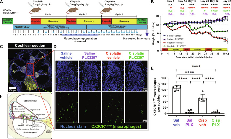
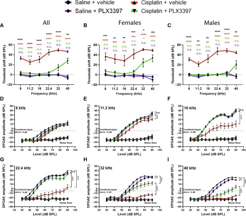
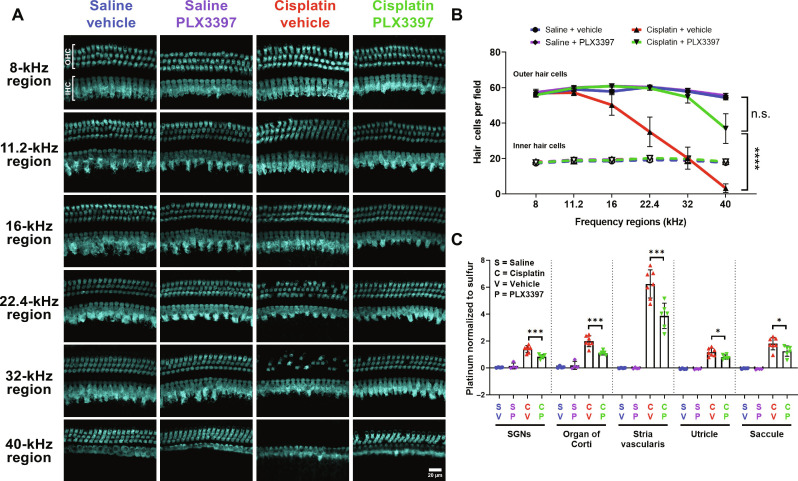
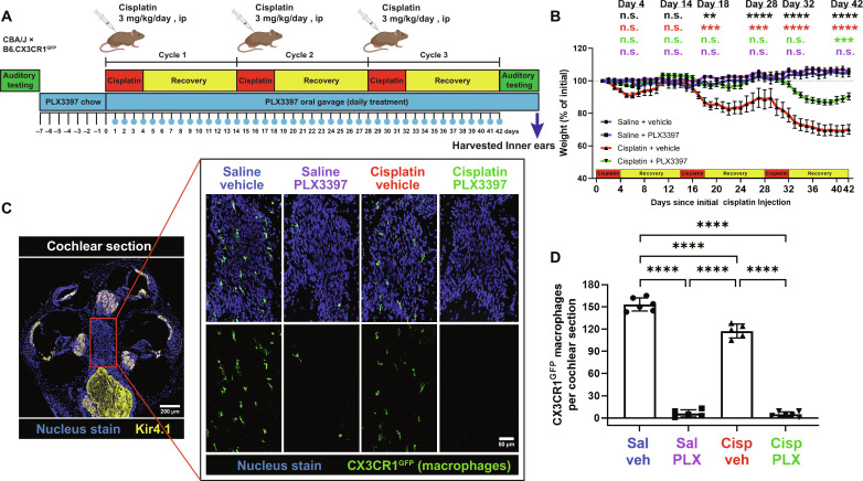
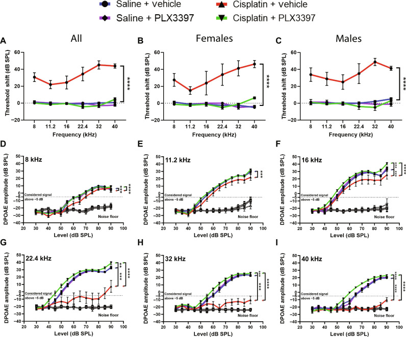
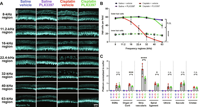
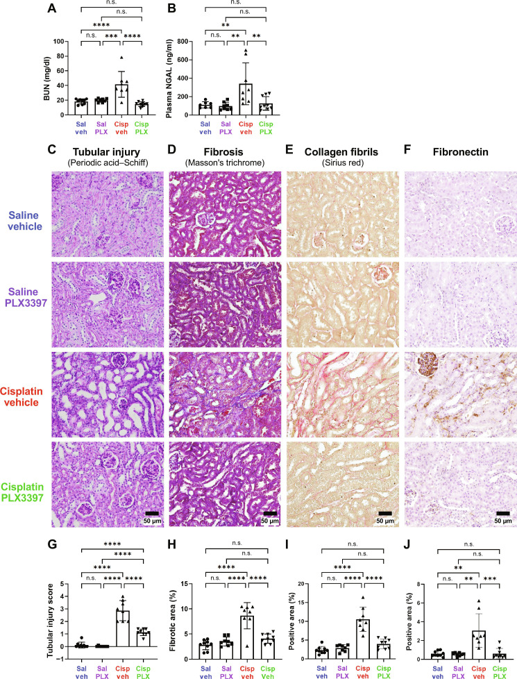
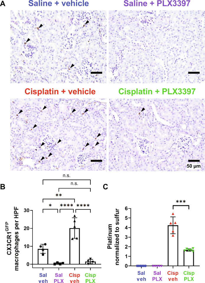
Cisplatin is a widely used anticancer drug with notable side effects including ototoxicity and nephrotoxicity. Macrophages, the major resident immune cells in the cochlea and kidney, are important drivers of both inflammatory and tissue repair responses. To investigate the roles of macrophages in cisplatin-induced toxicities, we used PLX3397, a U.S. Food and Drug Administration-approved inhibitor of the colony-stimulating factor 1 receptor, to eliminate tissue-resident macrophages. Mice treated with cisplatin alone had considerable hearing loss (ototoxicity) and kidney injury (nephrotoxicity). Macrophage ablation resulted in significantly reduced hearing loss and had greater outer hair cell survival. Macrophage ablation also protected against cisplatin-induced nephrotoxicity, as evidenced by markedly reduced tubular injury and fibrosis. Mechanistically, our data suggest that the protective effect of macrophage ablation against cisplatin-induced ototoxicity and nephrotoxicity is mediated by reduced platinum accumulation in both the inner ear and the kidney. Together, our data indicate that ablation of tissue-resident macrophages represents an important strategy for mitigating cisplatin-induced ototoxicity and nephrotoxicity.


Figures

Fig. 1.
Fig. 1.. Initial protocol in experiment 1 resulted in macrophage ablation via PLX3397, followed by partial macrophage repopulation.
(A) Experimental design showing auditory tests, PLX3397 treatment, and 3 cycles of cisplatin administration. Mice received PLX3397-formulated chow for 7 days to facilitate macrophage depletion. During cisplatin administration, mice received PLX3397 via oral gavage once every 3 days. Upon observation of macrophage repopulation on day 14 (see fig. S1), daily treatment of PLX3397 via oral gavage was initiated. The days on which mice received PLX3397 via oral gavage are denoted with blue circles. ip, intraperitonally. (B) Changes in mouse weight are shown throughout the 3 cycles of cisplatin administration protocol. Means ± SEM, n = 10 to 16 mice per experimental group (total 49 mice; 24 females and 25 males). Statistical comparisons (asterisks or n.s.) are color-coded as described in Methods. (C) Cochleae harvested after end-point auditory tests were immunolabeled for Kir4.1 (yellow) to visualize cochlear structures and green fluorescent protein (GFP) to visualize CX3CR1GFP-positive macrophages (green). Statistical significance is indicated as the following: *P < 0.05, **P < 0.01, ***P < 0.001, and ****P < 0.0001. P value above 0.05 (P > 0.05) was considered nonsignificant (n.s.). (D) Representative confocal images [cochlear modiolus; from red box in (C)] and (E) quantification of macrophages per cochlear section. PLX3397 resulted in >93% depletion of all macrophages. (F) Schematic diagram of macrophage distribution in the middle cochlear turn [from green box in (C)]. Scale bars, 200 μm (cochlear section) and 50 μm (mid-modiolar section). Means ± SD, n = 6 cochleae (3 females and 3 males) per experimental group (total 24 cochleae; 12 females and 12 males). One-way analysis of variance (ANOVA) with Tukey’s multiple comparisons test.
Fig. 2.
Fig. 2.. Macrophage ablation followed by partial repopulation protected against cisplatin-induced hearing loss and OHC dysfunction (experiment 1).
(A to C) Auditory sensitivity was measured via ABRs at baseline and end point. Threshold shifts are reported as the difference between baseline and end point ABR thresholds [saline/vehicle (blue line), saline/PLX3397 (purple line), cisplatin/vehicle (red line), and cisplatin/PLX3397 (green line)]. (B and C) PLX3397 protected against cisplatin-induced hearing loss in both (B) female and (C) male mice, with better protection in males than in female (*P = 0.0277). P values were calculated using one-way ANOVA with Tukey’s multiple comparisons test. Means ± SEM, n = 10 to 16 mice per experimental group (5 to 7 females and 5 to 9 males). Statistical comparisons (asterisks or n.s.) are color-coded as described in Methods. (D to I) OHC function was measured via DPOAEs. An emission at 2f1f2 was considered present when its amplitude was greater than −5 dB (dotted lines). Statistical analyses were performed using two-way ANOVA with Tukey’s multiple comparisons test (main column effect). Means ± SEM, n = 10 to 16 mice (5 to 7 females and 5 to 9 males) per experimental group (total 49 mice; 24 females and 25 males).
Fig. 3.
Fig. 3.. Macrophage ablation followed by partial repopulation via PLX3397 increased OHC survival and decreased cisplatin accumulation in the cochleae of cisplatin-treated mice (experiment 1).
(A) Microdissected cochlear turns were immunostained for Myosin 7a (cyan) and imaged to assess OHC and IHC across the cochlea. Scale bar, 20 μm. (B) Quantification of OHC and IHC numbers. The solid lines (blue, purple, red, and green) represent OHC counts, and dotted lines represent IHC counts. Means ± SEM, n = 6 cochleae (3 females and 3 males) per experimental group (total 24 cochleae; 12 females and 12 males). Statistical analysis was performed using two-way ANOVA with Tukey’s multiple comparisons test (main column effect). (C) Platinum concentrations in microdissected inner ear tissues were measured using ICP-MS. Means ± SD, n = 4 to 7 inner ears (2 to 4 females and 2 to 4 males) per experimental group (total 22 cochleae; 11 females and 11 males). Statistical analysis was performed using one-way ANOVA with Tukey’s multiple comparisons test.
Fig. 4.
Fig. 4.. PLX3397 in rodent chow followed by daily oral gavage resulted in sustained macrophage ablation and a significant reduction in weight loss induced by cisplatin (experiment 2).
(A) Experimental design of experiment 2. Mice received either control chow or PLX3397-formulated chow for 7 days to facilitate macrophage depletion. Following initiation of cisplatin administration, mice received daily PLX3397 treatment via oral gavage, which continued until euthanasia, ensuring sustained macrophage ablation. The days on which mice received PLX3397 through oral gavage are indicated with blue circles. (B) Changes in mouse weight are indicated across the 3 cycles of cisplatin administration protocol. Means ± SEM, n = 8 to 9 mice per experimental group. Statistical analysis was performed using one-way ANOVA with Tukey’s multiple comparisons test. Statistical comparisons (asterisks or n.s.) are color-coded as described in Methods. (C and D) Macrophages expressing CX3CR1GFP (green) are visualized in the cochlear modiolus. (C) Scale bars, 200 μm (cochlear section) and 50 μm (modiolus, magnified inset). (D) Quantification of macrophages in whole cochlear sections. Means ± SD, n = 5 to 6 cochleae (2 females and 4 males) per experimental group (total 22 cochleae; 8 females and 16 males). Statistical analysis was performed using one-way ANOVA with Tukey’s multiple comparisons test.
Fig. 5.
Fig. 5.. Sustained depletion of macrophages via PLX3397 provided complete protection against cisplatin-induced hearing loss and OHC dysfunction (experiment 2).
(A to C) Hearing loss was assessed by ABRs before (baseline) PLX3397 treatment and after (end point) completion of the cisplatin administration protocol. Hearing loss is reported as threshold shifts (the difference between baseline and end point ABR thresholds). ABR threshold shifts were assessed in both (B) female and (C) male mice. (D to I) OHC function was evaluated using DPOAEs. An emission at 2f1f2 was considered present when its amplitude exceeded the threshold of −5 dB (dotted lines). The gray line represents the biological noise floor. For both ABRs and DPOAEs, groups include saline/vehicle-treated mice (blue line), saline/PLX3397-treated mice (purple line), cisplatin/vehicle-treated mice (red line), and cisplatin/PLX3397-treated mice (green line). Data are shown as means ± SEM, n = 8 to 9 mice (4 to 5 females and 4 to 5 males) per experimental group (total 34 mice; 16 females and 16 males). Statistical analysis was performed using two-way ANOVA with Tukey’s multiple comparisons test (main column effect).
Fig. 6.
Fig. 6.. Sustained macrophage ablation protected against cisplatin-induced OHC death and resulted in reduced cisplatin accumulation in the cochlea (experiment 2).
(A and B) Cochlear wholemounts were stained for Myosin 7a (cyan) to visualize hair cells. (A) Representative images and (B) quantitative analysis of Myosin 7a–positive hair cells. Scale bar, 100 μm. (B) Data are shown as means ± SEM, n = 6 cochleae (3 females and 3 males) per experimental group (total 24 cochleae; 12 females and 12 males). Statistical analysis was performed using two-way ANOVA with Tukey’s multiple comparisons test (main column effect). (C) Platinum levels were measured using ICP-MS in microdissected inner ear tissues. Data are shown as means ± SD, n = 4 to 6 cochleae (2 to 3 females and 2 to 3 males) per experimental group (total 22 cochleae; 12 females and 10 males). Statistical analysis was performed using one-way ANOVA with Tukey’s multiple comparisons test.
Fig. 7.
Fig. 7.. Sustained depletion of macrophages protects against cisplatin-induced kidney damage (experiment 2).
Kidney function, tubular injury, and fibrosis were evaluated after end point auditory testing. Macrophage ablation using PLX3397 protected against cisplatin-induced increases in (A) plasma BUN levels and (B) NGAL levels. (C to F) Representative images of corticomedullary junction are presented. Scale bars, 50 μm. (C) and (G) Tubular injury was semiquantitatively assessed using periodic acid–Schiff staining, and tubular injury scores were assigned (see Methods). (D) and (H) Fibrosis was assessed using Masson’s trichrome staining, and the percentage of fibrotic area in the entire surface area of the section was calculated. (E) and (I) Fibrillar collagen–positive areas were assessed by Sirius red staining and (F) and (J) fibronectin-positive areas shown and evaluated. (G to J) Data are expressed as means ± SD, n = 8 to 9 blood and kidney samples (4 females and 4 to 5 males) per experimental group (total 34 mice; 16 females and 18 males). P values were calculated using one-way ANOVA with Tukey’s multiple comparisons test.
Fig. 8.
Fig. 8.. PLX3397 results in ablation of CX3CR1GFP-positive cells and significant reduction in cisplatin accumulation in the kidney (experiment 2).
(A) Representative images of renal CX3CR1GFP-positive cells (arrowheads), identified through immunohistochemical staining. Scale bars, 50 μm. (B) The number of CX3CR1GFP-positive cells in each field was quantified. Data are expressed as means ± SD, n = 4 to 5 kidney samples (2 females and 2 to 3 males) per experimental group (total 17 mice; 8 females and 9 males). Compared to control mice (saline/vehicle-treated mice), PLX3397 treatment in saline-treated mice and cisplatin-treated mice ablated 95.63 and 82.52% of CX3CR1GFP-positive cells, respectively. HPF, high-power field (the area of a slide visible under ×400 magnification of corticomedullary junction). (C) Platinum levels in the kidney tissues were analyzed by ICP-MS. Data are expressed as means ± SD, n = 5 to 6 blood and kidney samples (2 to 3 females and 2 to 3 males) per experimental group (total 22 mice; 11 females and 11 males). Cisplatin resulted in increased platinum in the kidney, while PLX3397 significantly reduced cisplatin accumulation. Platinum levels were normalized to sulfur contained in the kidney. Data are expressed as means ± SE. P values were calculated using one-way ANOVA with Tukey’s multiple comparisons test.
Fig. 9.
Fig. 9.. Working model of the mechanisms underlying the protective effects of macrophage ablation against cisplatin-induced hearing loss and kidney injury.
(A) In the absence of PLX3397, macrophages are present, and the cochlear BLB is permeable enough to permit cisplatin to cross the BLB and enter the cochlea, where it results in the death of OHCs, loss of ribbon synapses and SGNs, and hearing loss. In the presence of PLX3397, PVMs are ablated, and BLB permeability is reduced, resulting in reduced cisplatin entry and protection against cisplatin-induced hearing loss. (B) In the absence of PLX3397, renal macrophages are present and may promote cisplatin entry into proximal tubule cells causing renal fibrosis and kidney dysfunction. In the presence of PLX3397, macrophages are ablated in the kidney, and cisplatin accumulation in the kidney is reduced, thus providing protection against cisplatin-induced tubular injury and interstitial fibrosis.

Update of

References

    1. Dasari S., Tchounwou P. B., Cisplatin in cancer therapy: Molecular mechanisms of action. Eur. J. Pharmacol. 740, 364–378 (2014). - PMC - PubMed
    1. Zhang J., Ye Z. W., Tew K. D., Townsend D. M., Cisplatin chemotherapy and renal function. Adv. Cancer Res. 152, 305–327 (2021). - PMC - PubMed
    1. Rybak L. P., Mukherjea D., Ramkumar V., Mechanisms of cisplatin-induced ototoxicity and prevention. Semin. Hear. 40, 197–204 (2019). - PMC - PubMed
    1. Frisina R. D., Wheeler H. E., Fossa S. D., Kerns S. L., Fung C., Sesso H. D., Monahan P. O., Feldman D. R., Hamilton R., Vaughn D. J., Beard C. J., Budnick A., Johnson E. M., Ardeshir-Rouhani-Fard S., Einhorn L. H., Lipshultz S. E., Dolan M. E., Travis L. B., Comprehensive audiometric analysis of hearing impairment and tinnitus after cisplatin-based chemotherapy in survivors of adult-onset cancer. J. Clin. Oncol. 34, 2712–2720 (2016). - PMC - PubMed
    1. Rademaker-Lakhai J. M., Crul M., Zuur L., Baas P., Beijnen J. H., Simis Y. J. W., van Zandwijk N., Schellens J. H. M., Relationship between cisplatin administration and the development of ototoxicity. J. Clin. Oncol. 24, 918–924 (2006). - PubMed

Publication types

MeSH terms

LinkOut - more resources