Review
. 2024 Aug 23;9(8):3828-3839.
doi: 10.1021/acssensors.4c00602. Epub 2024 Jul 24.

Levodopa: From Biological Significance to Continuous Monitoring

Affiliations
Review

Levodopa: From Biological Significance to Continuous Monitoring

David Probst et al. ACS Sens. .

Abstract

A continuous levodopa sensor can improve the quality of life for patients suffering with Parkinson's disease by enhancing levodopa titration and treatment effectiveness; however, its development is currently hindered by the absence of a specific levodopa molecular recognition element and limited insights into how real-time monitoring might affect clinical outcomes. This gap in research contributes to clinician uncertainty regarding the practical value of continuous levodopa monitoring data. This paper examines the current state of levodopa sensing and the inherent limitations in today's methods. Further, these challenges are described, including aspects such as interference from the metabolic pathway and adjunct medications, temporal resolution, and clinical questions, with a specific focus on a comprehensive selection of molecules, such as adjunct medications and structural isomers, as an interferent panel designed to assess and validate future levodopa sensors. We review insights and lessons from previously reported levodopa sensors and present a comparative analysis of potential molecular recognition elements, discussing their advantages and drawbacks.

Keywords: Parkinson’s disease; and sensor specificity; biosensor development; continuous levodopa monitoring; electrochemical detection; levodopa therapy; molecular recognition elements.


Conflict of interest statement

The authors declare the following competing financial interest(s): K.B., D.P., and K.S. are inventors of three patents related to this work filed by the University of North Carolina and Chapel Hill. (1) U.S. Provisional Patent Application No. 63/505,267, filed May 31, 2023, (2) U.S. Provisional Patent Application No. 63/505,267, filed May 31, 2023, and (3) U.S. Provisional Patent Application No. 63/505,259, filed May 31, 2023. The authors declare no other competing interests.

Figures

Figure 1
Figure 1
Proposed clinical pathway for a Parkinson’s disease (PD) patient alongside a representation of PD progression across five stages. The top portion highlights the pathological progression of PD with the healthy brain showing melanin-pigmented substantia nigra (SN) neurons, while the PD-affected brain shows a loss of SN neurons and the presence of Lewy bodies. The middle portion shows a conceptual PD clinical management pathway employing a levodopa sensor which monitors and transmits levodopa levels to movement disorder specialists or neurologists who then analyze the data to inform treatment decisions and adjustments, with the goal of providing patient feedback. The lower portion shows the progression of PD from stages I to V, with the increase in “off-time” and levodopa-induced “dyskinesia” as PD progresses as the therapeutic window progressively narrows. Figure 1 was created with BioRender.com.
Figure 2
Figure 2
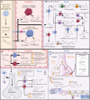
(A) Levodopa metabolic pathway and commonly administered (B) adjunct medication for PD treatment. Panel (A) outlines the absorption and metabolic conversion of orally administered Sinemet, a combined levodopa and carbidopa therapy, in the proximal jejunum, illustrating the metabolism of levodopa and its derivatives, detailing their transport across the jejunum, capillaries, and the blood-brain barrier. Panel (B) demonstrates the regulation of levodopa’s metabolic pathway by AADC inhibitors like carbidopa and COMT inhibitors such as entacapone and tolcapone, both peripherally and within the brain. It also highlights the mechanism of action of selective serotonin reuptake inhibitors and medications for dysautonomia, conditions frequently associated with PD. Adapted from refs (−, , and −37). Figure 2 was created with BioRender.com.
Figure 3
Figure 3
Mechanisms of levodopa sensing via tyrosinase-catalyzed and direct levodopa electrooxidation reactions. The top portion displays the mechanism of levodopa detection via tyrosinase, which exhibits monophenolase and diphenolase activities. Tyrosinase can catalyze the hydroxylation of l-tyrosine into levodopa which is paired with the reduction of oxygen to water. Subsequently, tyrosinase can then facilitate the oxidation of levodopa to o-dopaquinone, again reducing oxygen to water, which is acted on for secondary sensing. The bottom portion shows the generalized scheme for the direct electrooxidation of levodopa which can be oxidized to o-dopaquinone, which is then again reduced back to levodopa at potentials below −0.2 V vs a silver/silver chloride (Ag/AgCl) reference electrode. Alternatively, o-dopaquinone can form cyclo-dopa which at potentials greater than 0.1 V vs Ag/AgCl can be converted into dopachrome which can be acted on for secondary sensing. Figure 3 was created with BioRender.com.
Figure 4
Figure 4
Representative pathways for levodopa detection via (1) antibody, (2) molecularly imprinted polymer (MIP), (3) enzymatic, and (4) aptamer-based mechanisms. The first section demonstrates how levodopa interacts with the variable region of a levodopa antibody through noncovalent bonds, allowing for detection. The second section depicts the MIP approach where linkers polymerize around a levodopa template; following template removal, the imprint can be used for subsequent detection. The third section outlines the enzymatic detection strategy where the enzyme produces o-dopaquinone, which can be sensed through three distinct mechanisms. The fourth section highlights how an aptamer would bind with levodopa, with the conformational change allowing for an increase in relative signal compared to the prebinding signal. Figure 4 was created with BioRender.com.

References

    1. Damier P.; Hirsch E. C.; Agid Y.; Graybiel A. M. The Substantia Nigra of the Human Brain. II. Patterns of Loss of Dopamine-Containing Neurons in Parkinson’s Disease. Brain J. Neurol. 1999, 122 (Pt 8), 1437–1448. 10.1093/brain/122.8.1437. - DOI - PubMed
    1. Samii A.; Nutt J. G.; Ransom B. R. Parkinson’s Disease. Lancet London Engl. 2004, 363 (9423), 1783–1793. 10.1016/S0140-6736(04)16305-8. - DOI - PubMed
    1. Hawkes C. H.; Del Tredici K.; Braak H. A Timeline for Parkinson’s Disease. Parkinsonism Relat. Disord. 2010, 16 (2), 79–84. 10.1016/j.parkreldis.2009.08.007. - DOI - PubMed
    1. Olanow C. W.; Gauger L. L.; Cedarbaum J. M. Temporal Relationships between Plasma and Cerebrospinal Fluid Pharmacokinetics of Levodopa and Clinical Effect in Parkinson’s Disease. Ann. Neurol. 1991, 29 (5), 556–559. 10.1002/ana.410290516. - DOI - PubMed
    1. Wade L. A.; Katzman R. SYNTHETIC AMINO ACIDS AND THE NATURE OF L-DOPA TRANSPORT AT THE BLOOD-BRAIN BARRIER. J. Neurochem. 1975, 25 (6), 837–842. 10.1111/j.1471-4159.1975.tb04415.x. - DOI - PubMed

Publication types