. 2024 Jul 24;14(1):17044.
doi: 10.1038/s41598-024-67298-x.

Detailed three-dimensional analyses of tyloses in oak used for bourbon and wine barrels through X-ray computed tomography

Affiliations

Detailed three-dimensional analyses of tyloses in oak used for bourbon and wine barrels through X-ray computed tomography

Donghyun Kim et al. Sci Rep. .

Abstract

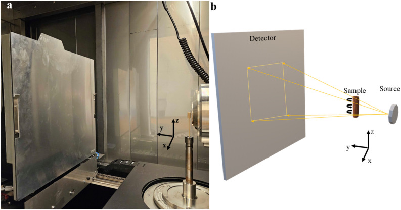
American white (Quercus alba L.) oak casks have been used for liquid storage for centuries. Their use in aged spirits is critical to imparting flavor and mouthfeel to the final product. The reason that barrels retain liquid has been hypothesized to be the result of abundant physiological structures called tyloses in parenchyma tissues and medullary rays in white oak. Using non-destructive X-ray computed tomography (XRCT) imaging, we reveal an unprecedented view of tylose structure and quantify the pore-filling capacity of tyloses in white oak that underscores the liquid retention we observe in casks. We show that pores of white oaks are filled with sevenfold higher tylose volume compared to northern red oak (Q. rubra), consistent with prior literature that casks made from white oak retain liquid while red oak fails to do so. We propose that XRCT represents a methodological standard for observing these complex structures and should be employed to understand the many questions related to liquid losses from casks, cultural treatment of casks, and the influence of climate change on oak tyloses in the future.


Conflict of interest statement

The authors declare no competing interests.

Figures

Figure 1
Figure 1
XRCT configuration. (a) A sample of white oak loaded into the HeliScan X-ray computed tomographic (XRCT) instrument. The three principle Cartesian axes are shown in (a) near the sample. (b) Schematic of the XRCT imaging process used to study oak cores.
Figure 2
Figure 2
Different cross-sectional views of the scanned white oak sample. Two-dimensional views of the three-dimensional reconstructed volume. The pith-to-bark direction lies in the xz plane, and the root-to-shoot direction lies along the z-axis.
Figure 3
Figure 3
White Oak’s high tylose content is visually apparent. Volumes of different species of Oak (Quercus): (a) white (alba), (b) French (robur), and (c) red (rubra). The volumes shown in a–c are 5003 voxels, and the viewing direction is along the z-axis, i.e., the viewing plane is the xy plane. The z-axis aligns with the root-to-shoot direction. The structure of vessel elements space (left) and tylose (right) in the perspective of the xz plane for white, French, and red oak is shown in d–i, respectively. A scale bar of 0.3 mm is added to provide a perspective on all dimensions. The volume of a–c is 1.33, 1.03, and 1.13 mm3, respectively, and the height of d–i is 4.2, 3.3, and 3.7 mm, respectively.
Figure 4
Figure 4
Segmented volume view of a region of Quercus alba. 3D visualization of Quercus alba data obtained from the XRCT scans. This volume is a cube with an edge length of 0.936 mm. The red material in the pores represents tylose, while the blue material depicts the base wood material. The cross-section cut of the rightmost pore provides an informative cross-sectional view of the tylose structure.
Figure 5
Figure 5
Average value of the vascular tissue element parameter reveals the variability across white (alba), french(robur), and red oak (rubra). The vessel elements are space volume per slice (a), Tylose volume per slice (b), pore volume per slice (c), Tylose percent of a pore (d), and pore diameter (e). Pore volume is the summation of vessel elements space and Tylose volume, and Tylose percent of a pore is the division of Tylose volume by pore volume. All volume measurements are divided by the slice length (height of the pore) because of the differences in the slice length across the species. The average values are obtained across 35 pores for white oak and 33 pores for French and red oak. The pore diameter is approximated using the measurement tool in the analysis software Avizo. A Tukey–Kramer multiple comparison test was used to determine statistical significance (n = 33 pores; different letters denote statistically significant differences at P < 0.05; error bars indicate a standard error).
Figure 6
Figure 6
Pore size affects the amount of tylose present in white (alba) Oak Data distribution of the Tylose percentage of a pore for different pore volumes per slice and diameter. Each data point here represents a pore that was analyzed. The dashed line represents the fit of each species. The data point of white (alba) oak is symbolized as a green circle, French (robur) as a blue triangle, and red (rubra) as a red diamond. Thirty-five pores of white oak and 33 pores of French and red oak are quantified in this figure.

References

    1. Leśniewska, J. et al. Defense responses in aspen with altered pectin methylesterase activity reveal the hormonal inducers of tyloses. Plant Physiol.173, 1409–1419. 10.1104/pp.16.01443 (2017). 10.1104/pp.16.01443 - DOI - PMC - PubMed
    1. Kirsch, S. The Origin and Development of Resin Canals in the Coniferae with Special Reference to the Development of Thyloses and their Correlation with the Thylosal Strands of the Pteridophytes (McGill University, 1910).
    1. Sachs, I., Kuntz, J., Ward, J., Nair, G. & Schultz, N. Tyloses structure. Wood Fiber Sci., 259–268 (1970).
    1. De Micco, V., Balzano, A., Wheeler, E. A. & Baas, P. Tyloses and gums: A review of structure, function and occurrence of vessel occlusions. IAWA J.37, 186–205. 10.1163/22941932-20160130 (2016).10.1163/22941932-20160130 - DOI
    1. HAMPSHIRE, N. & DAKOTA, S. The Discovery of Tylose Formation by a Viennese Lady in 1845 MH Zimmerman, Harvard Forest Petersham, Massachusetts 01366 USA.

MeSH terms