. 2024 Jul 24;14(1):17112.
doi: 10.1038/s41598-024-68126-y.

The effects of mitral stenosis on right ventricular mechanics assessed by three-dimensional echocardiography

Affiliations

The effects of mitral stenosis on right ventricular mechanics assessed by three-dimensional echocardiography

Zsuzsanna Ladányi et al. Sci Rep. .

Abstract

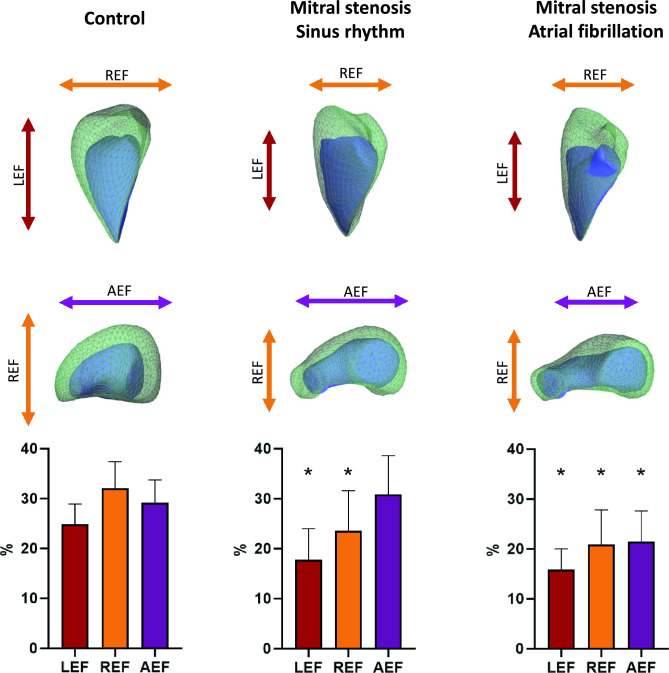
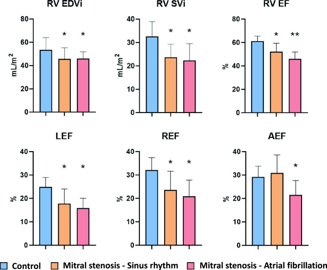
Mitral stenosis (MS) is a complex valvular pathology with significant clinical burden even today. Its effect on the right heart is often overlooked, despite it playing a considerable part in the symptomatic status. We enrolled 39 mitral valve stenosis patients and 39 age- and gender-matched healthy controls. They underwent conventional, speckle-tracking and 3D echocardiographic examinations. The 3D data was analyzed using the ReVISION software to calculate RV functional parameters. In the MS group, 3D RV ejection fraction (EF) (49 ± 7% vs. 61 ± 4%; p < 0.001), global circumferential (GCS) (- 21.08 ± 5.64% vs. - 25.07 ± 4.72%; p = 0.001) and longitudinal strain (GLS) (- 16.60% ± 4.07% vs. - 23.32 ± 2.82%; p < 0.001) were reduced. When comparing RV contraction patterns between controls, MS patients in sinus rhythm and those with atrial fibrillation, radial (REF) (32.06 ± 5.33% vs. 23.62 ± 7.95% vs. 20.89 ± 6.92%; p < 0.001) and longitudinal ejection fraction (LEF) (24.85 ± 4.06%; 17.82 ± 6.16% vs. 15.91 ± 4.09%; p < 0.001) were decreased in both MS groups compared to controls; however, they were comparable between the two MS subgroups. Anteroposterior ejection fraction (AEF) (29.16 ± 4.60% vs. 30.87 ± 7.71% vs. 21.48 ± 6.15%; p < 0.001) showed no difference between controls and MS patients in sinus rhythm, while it was lower in the MS group with atrial fibrillation. Therefore, utilizing 3D echocardiography, we found distinct morphological and functional alterations of the RV in MS patients.

Keywords: Atrial fibrillation; Contraction pattern; Mitral stenosis; Right ventricle; Speckle-tracking echocardiography; Three-dimensional echocardiography.


Conflict of interest statement

AK serves as Chief Medical Officer of Argus Cognitive, Inc, and receives financial compensation for his work, outside the submitted manuscript. BKL, and AF receive modest honoraria as medical consultants of Argus Cognitive, Inc., outside the submitted work. Other authors have no competing interests.

Figures

Figure 1
Figure 1
Graphical representation of a healthy control, a mitral stenosis patient in sinus rhythm and a mitral stenosis patient with atrial fibrillation in terms of three-dimensional right ventricular volumes and mechanics. The mitral stenosis patients in sinus rhythm (MS-SR) and the mitral stenosis patients with atrial fibrillation (MS-AF) had smaller end-diastolic volumes than the controls (CTR), while the MS-AF patients had larger end-systolic volumes than both the CTR and MS-SR patients (green mesh—right ventricular end-diastolic volume; blue surface—right ventricular end-systolic volume). Concerning right ventricular motion decomposition, both MS groups had lower LEF and REF values than the CTR group, whereas AEF remained comparable between the CTR and MS-SR patients, while it was lower in the MS-AF group. LEF, longitudinal ejection fraction; REF, radial ejection fraction; AEF, anteroposterior ejection fraction; *, significant difference compared to healthy contols.
Figure 2
Figure 2
The comparison of right ventricular morphological and functional parameters in healthy controls, mitral stenosis patients in sinus rhythm and mitral stenosis patients with atrial fibrillation. Both the mitral stenosis patients in sinus rhythm (MS-SR) and the mitral stenosis patients with atrial fibrillation (MS-AF) had lower RV EDVi and RV SVi compared to healthy controls (CTR). The MS-SR group had lower RV-EF compared to the CTR group, while the MS-AF patients presented with even lower values. LEF and REF were lower in both the MS-SR and the MS-AF groups compared to the CTR group, while AEF was lower only in the MS-AEF group. RV EDVi, right ventricular end-diastolic volume index; RV SVi, right ventricular stroke volume index; RV EF, right ventricular ejection fraction; LEF, longitudinal ejection fraction; REF, radial ejection fraction; AEF, anteroposterior ejection fraction; *, significant difference compared to healthy contols, **, significant difference compared to healthy contols and mitral stenosis patients in sinus rhythm.

References

    1. Chandrashekhar, Y., Westaby, S. & Narula, J. Mitral stenosis. Lancet374, 1271–1283. 10.1016/S0140-6736(09)60994-6 (2009). 10.1016/S0140-6736(09)60994-6 - DOI - PubMed
    1. Vahanian, A. et al. 2021 ESC/EACTS Guidelines for the management of valvular heart disease. Eur. Heart J.43, 561–632. 10.1093/eurheartj/ehab395 (2022). 10.1093/eurheartj/ehab395 - DOI - PubMed
    1. Marijon, E. et al. Prevalence of rheumatic heart disease detected by echocardiographic screening. N. Engl. J. Med.357, 470–476. 10.1056/NEJMoa065085 (2007). 10.1056/NEJMoa065085 - DOI - PubMed
    1. Iung, B. et al. A prospective survey of patients with valvular heart disease in Europe: The Euro heart survey on valvular heart disease. Eur. Heart J.24, 1231–1243. 10.1016/s0195-668x(03)00201-x (2003). 10.1016/s0195-668x(03)00201-x - DOI - PubMed
    1. Kingue, S. et al. The VALVAFRIC study: A registry of rheumatic heart disease in Western and Central Africa. Arch. Cardiovasc. Dis.109, 321–329. 10.1016/j.acvd.2015.12.004 (2016). 10.1016/j.acvd.2015.12.004 - DOI - PubMed

LinkOut - more resources