. 2024 Jul 24;7(1):898.
doi: 10.1038/s42003-024-06590-y.

Identification of new correctors for traffic-defective ABCB4 variants by a high-content screening approach

Affiliations

Identification of new correctors for traffic-defective ABCB4 variants by a high-content screening approach

Mounia Lakli et al. Commun Biol. .

Abstract

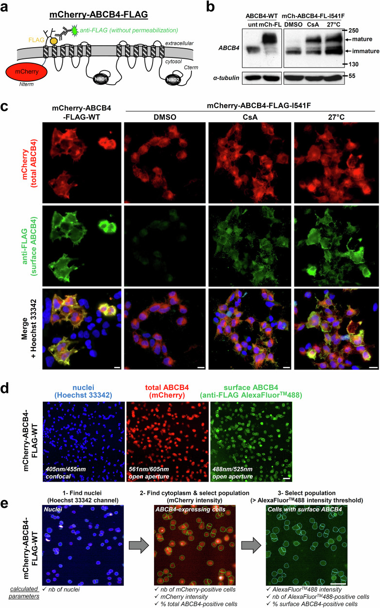
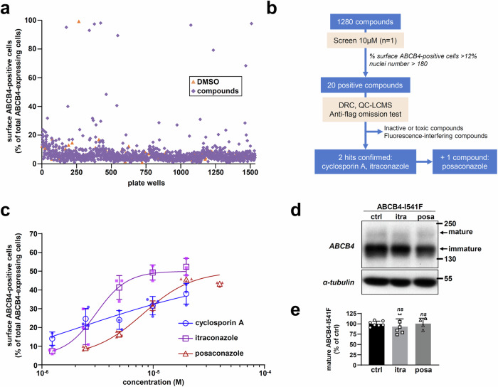
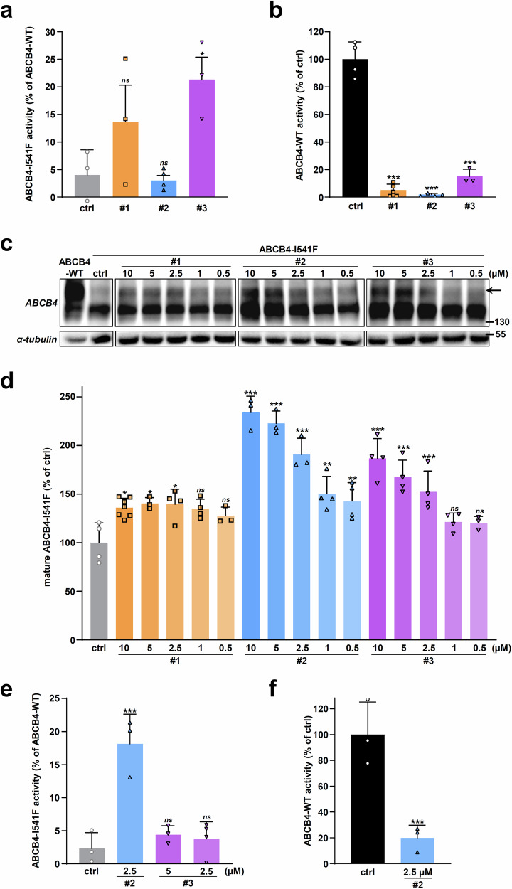
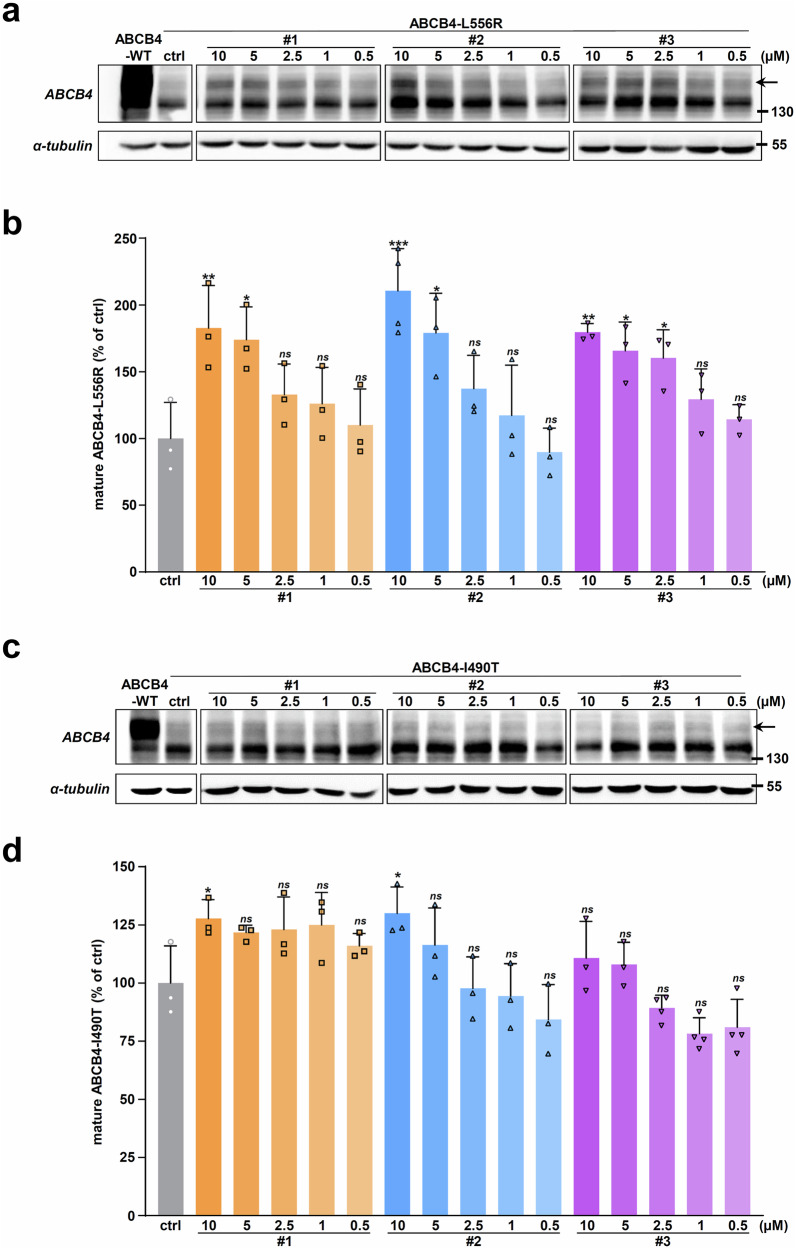
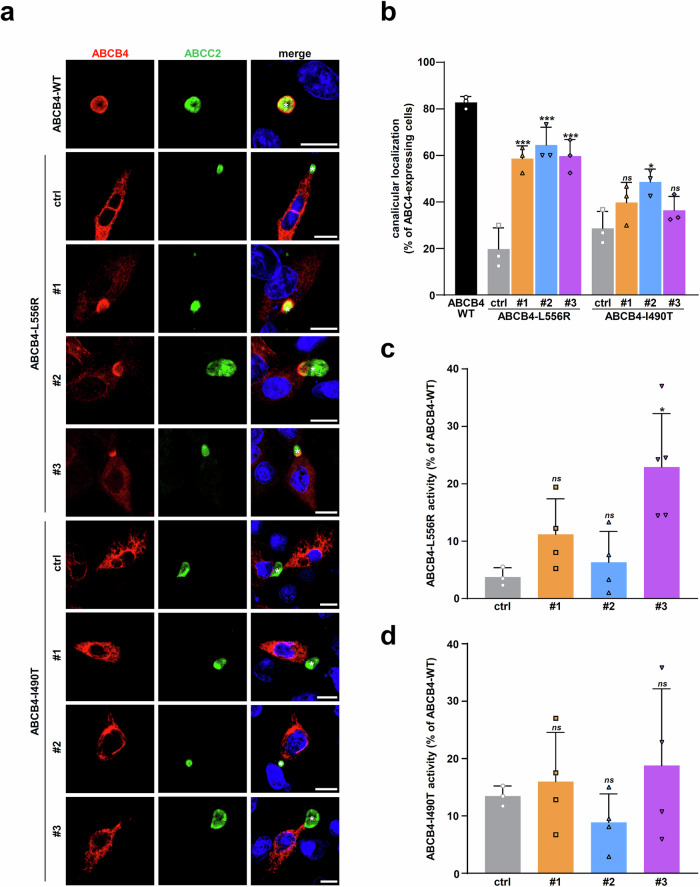
ABCB4 is located at the canalicular membrane of hepatocytes and is responsible for the secretion of phosphatidylcholine into bile. Genetic variations of this transporter are correlated with rare cholestatic liver diseases, the most severe being progressive familial intrahepatic cholestasis type 3 (PFIC3). PFIC3 patients most often require liver transplantation. In this context of unmet medical need, we developed a high-content screening approach to identify small molecules able to correct ABCB4 molecular defects. Intracellularly-retained variants of ABCB4 were expressed in cell models and their maturation, cellular localization and function were analyzed after treatment with the molecules identified by high-content screening. In total, six hits were identified by high-content screening. Three of them were able to correct the maturation and canalicular localization of two distinct intracellularly-retained ABCB4 variants; one molecule was able to significantly restore the function of two ABCB4 variants. In addition, in silico molecular docking calculations suggest that the identified hits may interact with wild type ABCB4 residues involved in ATP binding/hydrolysis. Our results pave the way for their optimization in order to provide new drug candidates as potential alternative to liver transplantation for patients with severe forms of ABCB4-related diseases, including PFIC3.


Conflict of interest statement

The authors declare no competing interests.

Figures

Fig. 1
Fig. 1. Development of a modified version of ABCB4 for HCS.
a The membrane topology of the mCherry-ABCB4-FLAG construct is represented. NBD, nucleotide-binding domain. b ABCB4-WT without tag (unt) or double tagged (mCh-FL: mCherry-ABCB4-FLAG), or mCherry-ABCB4-FLAG-I541F (mCh-ABCB4-FL-I541F) were transiently expressed in HEK cells. After the indicated treatment (16 h with 10 µM CsA or at 27 °C), cell lysates were prepared and analyzed by immunoblot using the indicated antibodies. ABCB4-WT is shown as reference. The mature and immature forms of ABCB4 are indicated, as well as molecular weight markers. This panel is representative of three independent experiments. Full immunoblots are shown in Supplementary Fig. 14a. c mCherry-ABCB4-FLAG-WT or mCherry-ABCB4-FLAG-I541F were transiently expressed in HEK cells. Cells were treated 16 h as in (b), then fixed but not permeabilized. After anti-FLAG immunolabeling, total (red) vs plasma membrane (green) ABCB4 were visualized by confocal microscopy. Nuclei shown in the merged images were labeled with Hoechst 33342 (blue). This panel is representative of three independent experiments. Bar: 10 μm. d Same as (c), except that constructs were stably expressed in HEK cells, sample processing was miniaturized and image acquisition was performed using an automated confocal microscope. Then, images were batch analyzed using a script written with Columbus™ (e). Bar: 50 µm. e Software-assisted image analysis. After nuclei detection with Hoechst 33342 blue channel (1), ABCB4-expressing cells were selected with the mCherry red channel (2). The cell surface ABCB4 subpopulation was detected using anti-FLAG (and AlexaFluorTM488) labeling (3). The number of total, ABCB4-positive and surface ABCB4-expressing cells were determined; red and green signal intensities were measured. Bar: 50 µm.
Fig. 2
Fig. 2. A first HCS with an FDA-approved chemical library.
a Overview of the screening with the percentage of surface ABCB4-expressing cells reported for each molecule: HEK cells stably expressing mCherry-ABCB4-FLAG-I541F were incubated for 16 h with 1280 compounds from the Prestwick chemical library® at 10 µM. DMSO was used as a vehicle negative control. b Workflow of the HCS campaign. DRC: dose-response curve; QC-LCMS: quality control by liquid chromatography-mass spectrometry. c Dose-response curves of the three compounds of interest using the primary screening model. Percentages of cells with surface ABCB4 were determined and reported as means (± SD, n = 3). d HEK cells transiently expressing mCherry-ABCB4-FLAG-I541F were treated for 16 h with DMSO (ctrl), 10 µM itraconazole (itra) or 10 µM posaconazole (posa). ABCB4-I541F maturation was assessed by immunoblot as in Fig. 1b. Molecular weight markers (in kDa) are indicated. This panel is representative of four independent experiments. Full immunoblots are shown in Supplementary Fig. 14b. e Densitometry analysis of (d). The amount of ABCB4-I541F mature form was quantified, normalized to the amount of tubulin and then expressed as a percentage of vehicle-treated cells (ctrl). Means (± SD) of at least four independent experiments per condition are shown.
Fig. 3
Fig. 3. Identification of new ABCB4 correctors through additional HCS.
a Overview of the screening in HEK cells stably expressing mCherry-ABCB4-FLAG-I541F, as in Fig. 2a: 3200 compounds were tested at 10 µM for 16 h. DMSO was used as a vehicle negative control. b Workflow of the HCS campaign, as in Fig. 2b. c Dose-response curves of the three hits, performed as in Fig. 2c. d Structure of ABCB4 correctors identified by HCS.
Fig. 4
Fig. 4. The HCS hits partially correct the maturation and canalicular targeting of ABCB4-I541F.
a HEK cells transiently expressing ABCB4-I541F were treated for 16 h with vehicle (ctrl, DMSO) or 10 μM of the three indicated hits. ABCB4-I541F maturation was assessed by immunoblot as in Fig. 1b. The arrow indicates mature ABCB4. This panel is representative of at least six independent experiments for each condition. Full immunoblots are shown in Supplementary Fig. 14c. b Densitometry analysis of (a). The amount of ABCB4-I541F mature form was quantified, normalized to the amount of tubulin and then expressed as a percentage of vehicle-treated cells (ctrl). Means (± SD) of at least six independent experiments per condition are shown. c ABCB4-WT or ABCB4-I541F were transiently expressed in HepG2 cells. After 16 h of treatment with vehicle (ctrl, DMSO) or 10 µM of the indicated hits, cells were fixed and permeabilized. Cellular localization of ABCB4 (red) and the canalicular transporter ABCC2 (green) was then visualized by confocal microscopy after indirect immunofluorescence labeling. Nuclei shown in the merged images were labeled with Hoechst 33342 (blue). White asterisks indicate bile canaliculi. This panel is representative of three independent experiments. Bars: 10 µm. d Quantification of ABCB4-I541F canalicular localization from c. ABCB4 colocalization with ABCC2 was determined in 300 cells for each condition. Means (± SD) of three independent experiments are shown.
Fig. 5
Fig. 5. Effect of the hits on the function of ABCB4-I541F and ABCB4-WT.
a, b HEK cells expressing ABCB4-I541F (a) or ABCB4-WT (b) were treated with vehicle (ctrl, DMSO) or 10 µM of the indicated hits. ABCB4-mediated PC secretion was measured, normalized to ABCB4 expression levels, and expressed as a percentage of the activity of vehicle-treated cells expressing ABCB4-WT. Means (± SD) of at least three independent experiments performed in triplicate for each tested condition are shown. c After transient expression of ABCB4-I541F, HEK cells were treated with decreasing doses of the indicated drugs for 16 h. Then, immunoblot analyses were performed to assess ABCB4-I541F maturation as in Fig. 4a. This panel is representative of three independent experiments. Full immunoblots are shown in Supplementary Fig. 14d. d Densitometry analysis of (c). The percentage of ABCB4-I541F mature form was determined and represented as in Fig. 4b. Means (± SD) of at least three independent experiments per condition are shown. e, f ABCB4-mediated PC secretion was determined as in (a, b) for ABCB4-I541F (e) and ABCB4-WT (f) -expressing cells treated with 2.5 or 5 µM of the indicated hits. Means (± SD) of at least three independent experiments performed in triplicate for each tested condition are shown.
Fig. 6
Fig. 6. Effect of the HCS hits on the maturation of two other ER-retained ABCB4 variants.
a ABCB4-L556R-expressing HEK cells were treated with vehicle (ctrl, DMSO) or decreasing doses of the indicated hits for 16 h. Then, immunoblot analyses were performed to assess ABCB4 maturation as in Fig. 4a. b Densitometry analysis of (a). The percentage of ABCB4-L556R mature form was determined and represented as in Fig. 4b. c, d The same analyses as in (a, b) were performed on ABCB4-I490T-expressing HEK cells. a, c are each representative of three independent experiments. For (a, c), full immunoblots are shown in Supplementary Fig. 14e, f. (b, d) represent means (± SD) of at least three independent experiments per condition.
Fig. 7
Fig. 7. Effect of the hits on the traffic and function of ABCB4-L556R and ABCB4-I490T variants.
a The subcellular localization of ABCB4-L556R and ABCB4-I490T was analyzed in HepG2 cells as in Fig. 4c, after treatment with vehicle (ctrl, DMSO) or 10 μM of the indicated hits. White asterisks indicate bile canaliculi. This panel is representative of three independent experiments. Bars: 10 μm. b The canalicular localization of ABCB4-L556R and ABCB4-I490T was determined from a, as in Fig. 4d. Means ( ± SD) of three independent experiments are shown. c, d The PC secretion activity of ABCB4-L556R (c) and ABCB4-I490T (d) expressed in HEK cells was determined as in Fig. 5a, b, after treatment with 10 µM of the indicated hits. Means ( ± SD) of at least three independent experiments performed in triplicate for each tested condition are shown.
Fig. 8
Fig. 8. Potential interaction of the correcting hits with ABCB4 revealed by molecular docking calculations.
a The selected centers of mass for compounds #1, #2 and #3 were obtained from brute force molecular docking calculations for which cutoff of 2.5 kcal.mol-1 was applied. Centers of mass were colored considering the lowest score affinity at 0.0 kcal.mol-1. b The most frequent contact residues were obtained by considering all selected poses displayed in (a). Given the large variability between selected number of poses, contact fractions were normalized as 1.0 for the most frequently observed residue per conformation. ABCB4if and ABCB4cc residues were depicted in orange and purple, respectively. c Examples of the lowest affinity scores for compounds #1 (left), #2 (center) and #3 (right) in the ATP-binding sites. Small molecules and key residues reported in b are shown in balls-and-sticks. Other residues with a contact distance lower than 4 Å are shown in licorice representation. For all panels, ABCB4 color code refers to as TMHs and NBDs.

References

    1. Smith, A. J. et al. The human MDR3 P-glycoprotein promotes translocation of phosphatidylcholine through the plasma membrane of fibroblasts from transgenic mice. FEBS Lett.354, 263–266 (1994). 10.1016/0014-5793(94)01135-4 - DOI - PubMed
    1. Dean, M., Rzhetsky, A. & Allikmets, R. The human ATP-binding cassette (ABC) transporter superfamily. Genome Res.11, 1156–1166 (2001). 10.1101/gr.184901 - DOI - PubMed
    1. Kroll, T., Prescher, M., Smits, S. H. J. & Schmitt, L. Structure and Function of Hepatobiliary ATP Binding Cassette Transporters. Chem. Rev.121, 5240–5288 (2021). 10.1021/acs.chemrev.0c00659 - DOI - PubMed
    1. Smit, J. J. et al. Tissue distribution of the human MDR3 P-glycoprotein. Lab Invest71, 638–649 (1994). - PubMed
    1. van Helvoort, A. et al. MDR1 P-glycoprotein is a lipid translocase of broad specificity, while MDR3 P-glycoprotein specifically translocates phosphatidylcholine. Cell87, 507–517 (1996). 10.1016/S0092-8674(00)81370-7 - DOI - PubMed

Publication types

MeSH terms

Substances

Supplementary concepts

LinkOut - more resources