. 2024 Jul 24;7(1):901.
doi: 10.1038/s42003-024-06584-w.

A ligand discovery toolbox for the WWE domain family of human E3 ligases

Affiliations

A ligand discovery toolbox for the WWE domain family of human E3 ligases

Lena Münzker et al. Commun Biol. .

Abstract

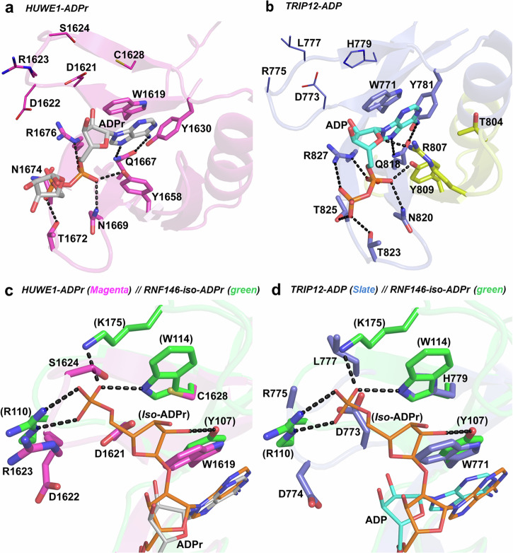
The WWE domain is a relatively under-researched domain found in twelve human proteins and characterized by a conserved tryptophan-tryptophan-glutamate (WWE) sequence motif. Six of these WWE domain-containing proteins also contain domains with E3 ubiquitin ligase activity. The general recognition of poly-ADP-ribosylated substrates by WWE domains suggests a potential avenue for development of Proteolysis-Targeting Chimeras (PROTACs). Here, we present novel crystal structures of the HUWE1, TRIP12, and DTX1 WWE domains in complex with PAR building blocks and their analogs, thus enabling a comprehensive analysis of the PAR binding site structural diversity. Furthermore, we introduce a versatile toolbox of biophysical and biochemical assays for the discovery and characterization of novel WWE domain binders, including fluorescence polarization-based PAR binding and displacement assays, 15N-NMR-based binding affinity assays and 19F-NMR-based competition assays. Through these assays, we have characterized the binding of monomeric iso-ADP-ribose (iso-ADPr) and its nucleotide analogs with the aforementioned WWE proteins. Finally, we have utilized the assay toolbox to screen a small molecule fragment library leading to the successful discovery of novel ligands targeting the HUWE1 WWE domain.


Conflict of interest statement

The authors declare no competing interests.

Figures

Fig. 1
Fig. 1. The WWE domains of human E3 ubiquitin ligases.
a Domain architecture of the six known WWE domain-bearing human E3 ligases, derived from the available structures and AlphaFold prediction models. b Sequence alignment and c the phylogenetic tree of the WWE domains in the six human E3 ligases. The tree is displayed without considering branch lengths. The sequence of TRIP12 isoform 2 (Uniprot ID: Q14669-2) was used in the alignment. d The structure of the HUWE1 WWE domain (PDB ID: 6MIW) showing the overall fold (left) and the strictly conserved residues (sticks) corresponding to red-highlighted residues in the sequence alignment presented in panel c. Most of the strictly conserved residues are in the ADPr/PAR binding site. e Structural units of PAR. ADPr, the PAR building block and iso-ADPr, the internal PAR structural unit containing the ribose-ribose glycosidic bond, are highlighted.
Fig. 2
Fig. 2. Binding interactions of PAR and ATP-analogs to the E3 ligase WWE domains.
a Fluorescence-polarization-based binding of fluorescein-labeled 11-mer of PAR (FAM-PAR) to the WWE domains of HUWE1, RNF146, TRIP12, DTX1, and DTX2. Fluorescence polarization percentage (% FP) of the reference is plotted as a function of WWE protein concentration in µM using a logarithmic scale. b Competitive displacement of FAM-labeled 11-mer PAR from the HUWE1 WWE domain by an unlabeled 11-mer PAR. Fluorescence polarization percentage (% FP) of the reference is plotted as a function of unlabeled PAR 11-mer in µM using a logarithmic scale. For the FP assays, all experiments were conducted in triplicate with three experimental repeats for direct PAR binding to HUWE1, TRIP12, DTX1, and RNF146, and two experimental repeats for direct PAR binding to DTX2, as well as FAM-PAR displacement in HUWE1. Samples from each experimental repeat were processed independently to ensure reproducibility and minimize bias. The plotted values represent the averages from the experimental repeats, and the statistical analyses conducted using GraphPad Prism Version 9.1.0 represent the standard deviations resulting from the analyzed repeats. c, d 15N-HSQC spectra of c HUWE1 and d DTX1 overlaid with protein in the presence of 1 mM 2F-ATP (1). e, f 15N-HSQC NMR Kd titration assay and zoom-in on a peak upon a two-fold titration of 2F-ATP (1) to e HUWE1 and f DTX1 using a concentration range of 62.5 µM to 2 mM. NMR Kd values originate from distinct samples (n = 1) measured for each concentration, mean Kds are obtained from curves of selected cross peaks ± standard deviations, ligand concentrations are plotted on the x-axis and the Δδ chemical shifts on the y-axis. g Chemical structure of 19F reporter 2F-ATP (1) 2'F-ATP (2) and competitor (ATP). h 19F-Displacement assay with HUWE1 and DTX1. In red lines, the 19F-NMR of 2F-ATP (1) reporter in the presence of protein is shown. In green, brown, and yellow, the titration of ATP at 250 mM, 500 mM, and 1000 mM concentration, respectively, to the protein in presence of 2F-ATP (1) is plotted. In blue lines, the 19F-NMR of the reporter 2F-ATP (1) reporter without protein is shown. 19F signals are displayed at an offset of 0.01 ppm to enhance clarity.
Fig. 3
Fig. 3. The WWE domain fold conservation in our natural ligands-complexed structures.
a A superposition of the reported RNF146-iso-ADPr structure (3V3L; green), the mouse RNF146-ATP structure (2RSF; ensemble 1; cyan) and the structures of HUWE1-ADPr (magenta), TRIP12-ADP (slate blue), DTX1-WWE1-ADP (gray), and DTX1-WWE2-ADP (yellow). The ligands are shown as thin sticks. b A close-up view of the ligand-binding site in the superimposed structure in panel a showing the conserved interaction of the ligands (lines) with the strictly conserved residues (thick and thin sticks). Residues are labeled based on HUWE1 WWE domain and the potential hydrogen bonds in the HUWE1-ADPr are shown as black dashes.
Fig. 4
Fig. 4. The WWE domain ligand-binding sites in TRIP12 and HUWE1.
a The ligand-binding site of the HUWE1 WWE domain bound to ADPr (gray sticks). Residues interacting with ADPr are shown as sticks and potential hydrogen bonds are shown as black dashes. Residues depicted as lines correspond to the binding site of the distal phosphate group of iso-ADPr as in the RNF146-iso-ADPr structure (PDB: 3V3L). b The ligand-binding site of the isoform 2 TRIP12 WWE domain bound to ADP. The protein is shown in slate blue, with a yellow section representing 28 residues that are missing within the WWE domain of the canonical isoform 1 TRIP12 sequence (Uniprot ID: Q14669-1). ADP is shown as cyan sticks, ADP-interacting residues as shown as sticks and potential hydrogen bonds are depicted as black dashes. Similarly, residues depicted as lines correspond to the binding site of the terminal phosphate group of iso-ADPr as in the RNF146-iso-ADPr structure (PDB: 3V3L). c Superposition of the HUWE1-ADPr WWE domain (magenta) with iso-ADPr bound RNF146 WWE domain (green; PDB: 3V3L), showing the differences in the binding site of the distal ribose and phosphate groups of iso-ADPr. ADPr and iso-ADPr are depicted as thin gray and orange sticks, respectively. The binding site residues of both structures are labeled (RNF146-iso-ADPr numbers in brackets) and possible interactions of iso-ADPr with RNF146 are shown as black dashes. d Superposition of the TRIP12-ADP WWE domain (slate blue) with the RNF146-iso-ADPr WWE domain (green) showing the differences in the binding site of the distal ribose and phosphate groups of iso-ADPr. ADP and iso-ADPr are rendered as thin cyan and orange sticks respectively. The binding site residues of both structures are labeled (RNF146-iso-ADPr numbers in brackets) and possible interactions of iso-ADPr with RNF146 are shown as black dashes.
Fig. 5
Fig. 5. The crystal structures of the DTX1 tandem WWE domains bound to ADP.
a The binding site of the WWE1 domain in the DTX1-WWE1-ADP structure. ADP is rendered as cyan sticks, the interacting residues are shown as sticks and potential hydrogen bonds are shown as black dashes. Residues depicted as lines correspond to the binding site of the distal phosphate group of iso-ADPr as in the RNF146-iso-ADPr structure (PDB: 3V3L). b The binding site of the WWE2 domain in the DTX1-WWE2-ADP structure. ADP is depicted as magenta sticks and potential hydrogen bonds are shown as black dashes. Similarly, residues depicted as lines correspond to the binding site of the distal phosphate group of iso-ADPr as in the RNF146-iso-ADPr structure (PDB: 3V3L). c A superposition of ADP-bound WWE1 domain (gray) and the unbound WWE2 domain (brown) in the DTX1-WWE1-ADP structure. ADP is shown as thin cyan sticks, conserved residues between the two domains are shown as lines, while residues that are different in type or side chain location are shown as thin sticks. d A superposition of ADP-bound WWE2 domain and the unbound WWE1 domain in the DTX1-WWE2-ADP structure. ADP is rendered as thin magenta sticks, residues that are conserved in type and location are shown as lines, while residues that are different in type or side chain location are shown as thin sticks.
Fig. 6
Fig. 6. Two small molecules bound to the HUWE1 WWE domain.
a 15N-HSQC spectra of HUWE1 in blue lines overlaid with protein in the presence of 1 mM compound (4) in red lines. b Chemical structures of (3) and (4). Dissociation constant curve of 125 µM to 1000 µM compound (4) titrated to HUWE1 WWE domain protein at 100 µM. NMR Kd values originate from distinct samples (n = 1) measured for each concentration, mean Kds are obtained from curves of selected cross peaks ± standard deviations. c, d The binding site of the HUWE1 WWE domain. c Compound (3) is rendered as green sticks and d compound (4) is rendered as gray sticks, the interacting residues are shown as sticks and potential hydrogen bonds are shown as black dashes. e, f 19F-Displacement assay with HUWE1 WWE domain and e compound (3) and f compound (4). The F-ATP reporter is in blue, the F-ATP reporter with HUWE1 WWE domain protein is in red and the titration of e compound (3) or f compound (4) at 250 mM (green), 500 mM (magenta) 1000 mM (yellow). 19F signals are displayed at an offset of 0.01 ppm to enhance clarity.

References

    1. Aravind, L. The WWE domain: a common interaction module in protein ubiquitination and ADP ribosylation. Trends Biochem Sci.26, 273–275 (2001). 10.1016/S0968-0004(01)01787-X - DOI - PubMed
    1. Suskiewicz, M. J. et al. Updated protein domain annotation of the PARP protein family sheds new light on biological function. Nucleic Acids Res. 51, 8217–8236 (2023). - PMC - PubMed
    1. Langelier, M.-F., Eisemann, T., Riccio, A. A. & Pascal, J. M. PARP family enzymes: regulation and catalysis of the poly(ADP-ribose) posttranslational modification. Curr. Opin. Struct. Biol.53, 187–198 (2018). 10.1016/j.sbi.2018.11.002 - DOI - PMC - PubMed
    1. Malgras, M., Garcia, M., Jousselin, C., Bodet, C. & Lévêque, N. The antiviral activities of poly-ADP-ribose polymerases. Viruses13, 582 (2021). 10.3390/v13040582 - DOI - PMC - PubMed
    1. Inloes, J. M. et al. The hereditary spastic paraplegia-related enzyme DDHD2 is a principal brain triglyceride lipase. Proc. Natl Acad. Sci. USA111, 14924–14929 (2014). 10.1073/pnas.1413706111 - DOI - PMC - PubMed

Publication types

LinkOut - more resources