. 2024 Sep;8(9):1711-1722.
doi: 10.1038/s41559-024-02477-7. Epub 2024 Jul 24.

Iron-coated Komodo dragon teeth and the complex dental enamel of carnivorous reptiles

Affiliations

Iron-coated Komodo dragon teeth and the complex dental enamel of carnivorous reptiles

Aaron R H LeBlanc et al. Nat Ecol Evol. 2024 Sep.

Abstract

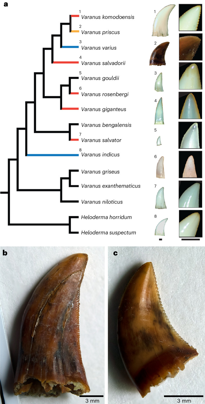
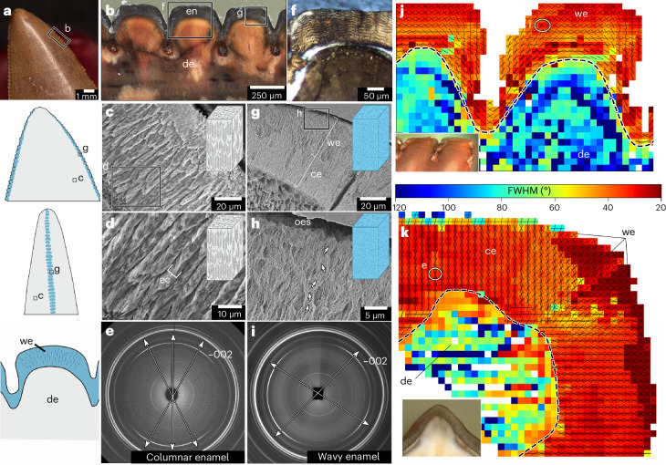
Komodo dragons (Varanus komodoensis) are the largest extant predatory lizards and their ziphodont (serrated, curved and blade-shaped) teeth make them valuable analogues for studying tooth structure, function and comparing with extinct ziphodont taxa, such as theropod dinosaurs. Like other ziphodont reptiles, V. komodoensis teeth possess only a thin coating of enamel that is nevertheless able to cope with the demands of their puncture-pull feeding. Using advanced chemical and structural imaging, we reveal that V. komodoensis teeth possess a unique adaptation for maintaining their cutting edges: orange, iron-enriched coatings on their tooth serrations and tips. Comparisons with other extant varanids and crocodylians revealed that iron sequestration is probably widespread in reptile enamels but it is most striking in V. komodoensis and closely related ziphodont species, suggesting a crucial role in supporting serrated teeth. Unfortunately, fossilization confounds our ability to consistently detect similar iron coatings in fossil teeth, including those of ziphodont dinosaurs. However, unlike V. komodoensis, some theropods possessed specialized enamel along their tooth serrations, resembling the wavy enamel found in herbivorous hadrosaurid dinosaurs. These discoveries illustrate unexpected and disparate specializations for maintaining ziphodont teeth in predatory reptiles.


Conflict of interest statement

The authors declare no competing interests.

Figures

Fig. 1
Fig. 1. Pigmented cutting edges in V. komodoensis teeth.
a, Lateral view of the skull of V. komodoensis (Natural History Museum, London, NHMUK 1934.9.2.1). b, Lingual view of a dentary tooth position showing several unerupted replacement teeth with orange pigmentation (American Museum of Natural History, AMNH 37912). c, Dorsal view of two erupted teeth from a fluid-preserved specimen (Zoological Society of London) showing pigmented cutting edges and apices. d, White light (WL) image of an erupted and unerupted tooth in the same specimen. e, Laser stimulated fluorescence (LSF) image of the same specimen, showing the pronounced serration pigmentation in both the erupted and unerupted teeth. f, Dorsal view of three left dentary teeth in NHMUK 1934.2.1 showing identical pigmentation on the tooth apices and mesial serrations. g, Lateral view of an isolated replacement tooth (Museum of Life Sciences, MoLS X263). h, Close-up of tooth apex in g showing the orange pigmentation along the tooth tip. i, Distal view of tooth serrations of MoLS X263 showing orange serrations and tooth apex. j, Polished thick section through mesial denticles of a tooth (Queensland Museum, Australia, J94036-2) showing orange pigmentation restricted to the enamel. k, SEM image of three mesial serrations of J94036-2. l, Close-up of serration enamel showing the bright coating. m, Close-up of the crown apex enamel showing the same nanocrystalline coating. Asterisks indicate pigmented regions. de, dentine; en, enamel; et, erupted tooth; gi, gingiva; ut, unerupted tooth.
Fig. 2
Fig. 2. Elemental and microstructural analyses of serration and tooth apex enamel in V. komodoensis.
a, Polished thick section of a tooth apex and distal serrations (MoLS X263). b, S-µXRF elemental map for iron along the tooth apex and serrations in MoLS X263. c,d, High-resolution S-µXRF map of iron along two apical serrations (c), compared with the calcium map of the same region (d), showing that iron is restricted to the outer enamel surface. e,f, High-resolution S-µXRF map of iron along the tip of the tooth (e), compared with the calcium map of the same region (f). g, High-resolution S-µXRF map of iron along a horizontal section through a distal serration in the same tooth. h, Wholeview image of horizontal section through MoLS X263. i, Higher resolution S-µXRF map of iron along the horizontal section through a mesial serration in MoLS X263. j, SEM–EDS map of iron from a thick section of a functional tooth (J94036-2) showing that the iron is most abundant in the coating overlying the enamel. k, SEM–EDS map of calcium from the same view as in j, showing reduced calcium signal in the outer coating. l, STEM–EDS map of iron from an FIB-milled portion of the outer enamel coating of J94036-2 (outer surface of tooth is towards the top). m, STEM–EDS map of calcium in the same region, showing lack of calcium in the iron-rich coating. n, STEM image of the interface between the iron-rich coating (above) and crystalline enamel (below) and iron-rich material interspersed between enamel crystallites (middle). In all elemental maps, brighter colours indicate higher counts. In the illustrations, red represents the pigmented regions, white is dentine and grey is enamel. am, amorphous region; cr, crystalline region; ecr, enamel crystallites; edj, enamel–dentine junction; Fe, iron.
Fig. 3
Fig. 3. Distribution of pigmented teeth within the genus Varanus.
a, Simplified phylogeny of Varanus species examined in this study (modified from ref. , position of V. priscus taken from ref. ). Red branches indicate taxa with obvious, consistently pigmented cutting edges. Black branches indicate those with no obvious pigmentation. Blue branches indicate taxa with occasional or inconsistently pigmented teeth. Orange branch indicates unknown level of pigmentation due to fossilization. Scale bars, 1 mm. b, Isolated tooth crown of the extinct V. priscus (SAMP 54739) showing no signs of pigmentation on the distal serrations. c, A second isolated crown assigned to V. priscus (SAMP 54739) showing no evidence of pigmentation.
Fig. 4
Fig. 4. Iron sequestration and pigmentation in crocodylian teeth.
a,b, Close-up of a posterior tooth of Tomistoma schlegelii under WL (a) and LSF (b), revealing a distinctive fluorescence pattern along the carina (asterisks). c, Close-up of the tooth tip of an anterior T. schlegelii tooth showing an orange carina under WL. d, Polished horizontal thick section through the carina of the tooth in c showing orange pigmentation (asterisks). e,f, LA-ICP-MS map of iron (e) and calcium (f), taken over a horizontal section through a carina from the tooth in a. g, Polished thick section of a caniniform tooth of A. mississippiensis cut parallel to the carinae, showing orange pigmentation along the outer enamel region (asterisks). h,i, LA-ICP-MS maps of iron (h) and calcium (i), from the enamel and dentine of the tooth in g. j, Welch test of hardness of pigmented and non-pigmented enamel in the same tooth measured using nano-indentation (n = 38 (non-pigmented) and 36 (pigmented) indents in one tooth). Central lines represent medians (3.33, 3.63), upper and lower bounds of boxes represent lower quartiles (3.14, 3.38) and upper quartiles (3.55, 4.06), minima (2.47, 2.68) and maxima (4.6, 5.63) for non-pigmented and pigmented enamel, respectively. P value (two-tailed) was 0.0058. k,l, S-µXRF maps of iron (k) and calcium (l) along the tip of a tooth section from a C. porosus tooth cut along the carina. m, Mesial view of the tooth sectioned and mapped in k and l. n, Section used in k and l, showing region of interest. o,p, S-µXRF maps of iron (o) and calcium (p) taken from a fossilized crocodylian tooth from Dinosaur Provinical Park (Canada) (UALVP 60550). q, Mesial view of UALVP 60550 before sectioning showing no evidence of pigmentation along the carina. r, Section used in o and p showing region of interest. In all elemental maps, brighter colours indicate higher counts. cn, carina.
Fig. 5
Fig. 5. Iron distribution within fossilized theropod tooth serrations.
a, Labiolingual view of a tooth of the tyrannosaurid Albertosaurus (NHMUK R12599). b,c, Distal view of NHMUK R12599 under WL (b) and LSF imaging (c), showing a lack of differential fluorescence between the serrations or the rest of the tooth. d, Labiolingual view of a dromaeosaurid tooth (UALVP 61165). e,f, Distal view of a dromaeosaurid tooth (UALVP 61165) under WL (e) and LSF imaging (f), showing no fluorescence difference on- or off-serration. g, Polished horizontal thick section through a distal serration of a tyrannosaurid tooth (UALVP 60554). h, LA-ICP-MS map of iron along the distal serration in g showing no evidence of iron sequestration along the serration enamel. i, Polished horizontal thick section through a distal serration of UALVP 61165. j, LA-ICP-MS map of iron along the same region as in g. k, Close-up view of serration enamel in a tyrannosaurid tooth (UALVP 60553). l, Serration enamel in a dromaeosaurid tooth (UALVP 61165). m, Polished thick section through distal serrations of a tyrannosaurid tooth (UALVP 60555). n, LA-ICP-MS map of iron along the serrations in UALVP 60555. o, Polished thick section through the distal serrations of a dromaeosaurid tooth (UALVP 61165). p, LA-ICP-MS map of iron along the serrations in UALVP 61165. In all elemental maps, brighter colours indicate higher counts.
Fig. 6
Fig. 6. Structural specializations of the serration enamel in tyrannosaurid teeth.
a, Close-up image of an isolated tyrannosaurid tooth (UALVP 60556), with illustrations of the distributions of columnar (grey) and wavy (blue) enamel based on structural analyses. b, Polished thick section through the distal serrations of UALVP 60555 showing positions of sections in subsequent analyses. c, SEM image of columnar enamel in a tyrannosaurid tooth in horizontal section (UALVP 60556). d, Higher magnification SEM image showing herringbone stacks of enamel crystallites within each column. e, S-µXRD-generated diffraction pattern taken through a region of columnar enamel in a tyrannosaurid tooth (UALVP 60554), showing three principal enamel crystallite orientations in the 002 reflection. f, Cross-polarized light image of serration enamel in a thin section of a tyrannosaurid tooth (UALVP 60398) showing wavy pattern in enamel. g, SEM image of enamel along a serration of a tyrannosaurid tooth in longitudinal section (UALVP 60557). h, Higher magnification image of the outer enamel surface showing the spiralled arrangements of serration enamel. i, S-µXRD-generated diffraction pattern taken through a region of wavy enamel in a tyrannosaurid tooth, showing two principal enamel crystallite orientations in the 002 reflection and a higher divergence angle between them compared with the columnar enamel (e). j, Composite apatite crystal texture map of two serrations in longitudinal section (UALVP 53472) derived from diffraction patterns. k, Composite texture map of enamel crystallites along a single serration in horizontal section (UALVP 60554). Lines within each pixel in j and k indicate principal enamel crystallite orientations. Hotter colours indicate more highly textured regions (lower full-width half maxima). Wavy enamel manifests as larger divergences between principal orientations and more textured regions along the serration. ce, columnar enamel; ec, enamel column; FWHM, full-width at half-maximum; oes, outer enamel surface; we, wavy enamel.

References

    1. Brink, K. S. & Reisz, R. R. Hidden dental diversity in the oldest terrestrial apex predator Dimetrodon. Nat. Commun.5, 3269 (2014). 10.1038/ncomms4269 - DOI - PubMed
    1. Whitney, M. R., LeBlanc, A. R. H., Reynolds, A. R. & Brink, K. S. Convergent dental adaptations in the serrations of hypercarnivorous synapsids and dinosaurs. Biol. Lett.16, 20200750 (2020). 10.1098/rsbl.2020.0750 - DOI - PMC - PubMed
    1. de Andrade, M. B., Young, M. T., Desojo, J. B. & Brusatte, S. L. The evolution of extreme hypercarnivory in Metriorhynchidae (Mesoeucrocodylia: Thalattosuchia) based on evidence from microscopic denticle morphology. J. Vertebr. Paleontol.30, 1451–1465 (2010).10.1080/02724634.2010.501442 - DOI
    1. Abler, W. L. The serrated teeth of tyrannosaurid dinosaurs and biting structures in other animals. Paleobiology18, 161–183 (1992).10.1017/S0094837300013956 - DOI
    1. Brink, K. S. et al. Developmental and evolutionary novelty in the serrated teeth of theropod dinosaurs. Sci. Rep.5, 12338 (2015). 10.1038/srep12338 - DOI - PMC - PubMed

Supplementary concepts