. 2024 Jun 24;16(6):e63037.
doi: 10.7759/cureus.63037. eCollection 2024 Jun.

Protean Drainage Patterns of the Left Renal Vein: A Cadaveric and Retrospective Clinical Study on the Surgical Implications and Technical Feasibility

Affiliations

Protean Drainage Patterns of the Left Renal Vein: A Cadaveric and Retrospective Clinical Study on the Surgical Implications and Technical Feasibility

Amit K Shreevastava et al. Cureus. .

Abstract

Background: The diverse drainage patterns of the left renal vein (LRV), often with asymptomatic congenital anomalies, present considerable challenges in renal and retroperitoneal surgical contexts. The potential for significant bleeding and subsequent renal compromise upon vascular injury highlights the need for increased surgical awareness.

Objective: This study investigates the LRV's variable anatomical drainage patterns and morphometry. It also evaluates the embryological factors contributing to these variations and discusses their surgical implications and technical considerations.

Methods: Anatomical dissections were conducted on 21 adult human cadavers within the Department of Anatomy. Concurrently, a retrospective analysis was conducted on 15 patients who underwent various retroperitoneal surgical interventions in the Urology Department. Demographic variables and intraoperative findings were recorded and analyzed.

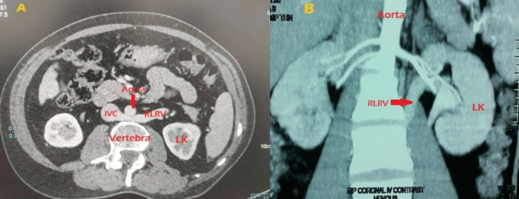
Results: Dissection analysis predominantly identified preaortic LRVs in 18 cadavers. Notable anatomical variations included a circumaortic left renal vein (CLRV), a delayed preaortic confluence of extrahilar duo LRVs, and an extrahilar tetramerous confluence with a retroiliac topography. The majority of LRVs usually end in the inferior vena cava. However, an extrahilar tetramerous variant had an unusual drainage pathway. Out of 15 cases, three (20%) had a retroaortic left renal vein (RLRV). One patient with a nonfunctioning kidney had type 1 RLRV, and another patient with pelvic ureteric junction obstruction had type 4 retroiliac left renal vein (RILRV). In both of these patients, symptoms were relieved after surgery. In a young patient with left varicocele and microscopic hematuria who had type 2 RLRV, symptoms resolved spontaneously after a few months.

Conclusion: A thorough understanding of the variable anatomical drainage patterns of the LRV is crucial for surgeons. Accurate preoperative identification can provide valuable insights, potentially leading to improved surgical outcomes in renal procedures.

Keywords: circumaortic; delayed confluence of veins; donor nephrectomy; inferior vena cava; left common iliac vein; left renal vein; renal transplant; retroaortic; variable anatomical drainage pattern.


Conflict of interest statement

Human subjects: All authors have confirmed that this study did not involve human participants or tissue. Animal subjects: All authors have confirmed that this study did not involve animal subjects or tissue. Conflicts of interest: In compliance with the ICMJE uniform disclosure form, all authors declare the following: Payment/services info: All authors have declared that no financial support was received from any organization for the submitted work. Financial relationships: All authors have declared that they have no financial relationships at present or within the previous three years with any organizations that might have an interest in the submitted work. Other relationships: All authors have declared that there are no other relationships or activities that could appear to have influenced the submitted work.

Figures

Figure 1
Figure 1. A delayed preaortic confluence of extrahilar double left renal veins (LRVs).
1) upper left hilar renal vein, 2) lower left hilar renal vein, 3) superior left extrahilar LRV, 4) inferior left extrahilar LRV, 5) left adrenal vein, 6) the main segment of a delayed confluence of LRV, 7) left ovarian vein, 8) main left renal artery, 9) accessory left renal artery, 10) left ureter, 11) right ureter, 12) inferior mesenteric artery, 13) right renal vein. LK: left kidney, RK: right kidney, IVC: inferior vena cava, AA: abdominal aorta, LRV: left renal vein, AG: adrenal gland, SMA: superior mesenteric artery
Figure 2
Figure 2. A circumaortic left renal vein (CLRV).
1) left hilar renal vein splitting to form a circumaortic left renal vein, 2) preaortic venous limb, 3) retroaortic venous limb, 4) retroaortic venous limb joining the IVC, 5) left testicular vein, 6) left adrenal vein, 7) left renal artery, 8) left ureter, 9) inferior mesenteric artery. LK: left kidney, RK: right kidney, AA: abdominal aorta, IVC: inferior vena cava, AG: adrenal gland, SMA: superior mesenteric artery. * Indicates the anastomosis of retroaortic venous limb and the hemiazygos vein.
Figure 3
Figure 3. An extrahilar tetramerous retroiliac left renal vein (LRV). The left kidney is ectopic.
1) upper hilar left renal vein, 2) lower hilar LRV, 3) left adrenal vein, 4) left testicular vein, 5) retroiliac LRV, 6) hemiazygos vein, 7) 4th lumbar vein, 8) splitting segment of retroiliac LRV draining into the inferior vena cava, 9) splitting segment of the retroiliac LRV draining into the left common iliac vein, 10) abdominal aorta, 11) superior mesenteric artery, 12) left renal artery, 13) left common iliac vein, 14) right common iliac vein, 15) left common iliac artery, 16) right common iliac artery, 17) left ureter, 18) right ureter, 19) right testicular vein. LRV: left renal vein, IVC: inferior vena cava, LK: left kidney, RK: right kidney
Figure 4
Figure 4. Lt PFK with RLRV type 1
A: CT angiogram showing the left renal vein draining into the IVC after passing posterior to the aorta. B: Reconstructed CT abdomen and pelvis image depicting the retroaortic course of the left renal vein. C: Lap retroperitoneoscopic image portraying the retroaortic course of the left renal vein (RALRV) draining into the IVC. D: Lap retroperitoneoscopic image with a dropped-down kidney laterally to display the retroaortic course of the left renal vein (RALRV) more vividly. PFK: poorly functioning kidney, RV: renal vein, RA: renal artery, IVC: inferior vena cava, RLRV: retroaortic left renal vein, GV: gonadal vein
Figure 5
Figure 5. Lt RILRV type IV (retroiliac renal vein) with PUJO, intrarenal pelvis, and left PFK
A: CT angiogram showing the oblique course of the left retroiliac renal vein (RIV) in a case of PUJ obstruction coursing posterior to the left common iliac artery. B: Laparoscopic transperitoneal image showing the course of three tributaries of the left renal vein (left suprarenal, lumbar vein, and parallel to the aorta trunk). C: Laparoscopic image showing the two tributaries of left renal vein emanating from straight parallel to the aorta trunk - gonadal vein and retroiliac trunk. PUJO: pelviureteral junction obstruction, PFK: poorly functioning kidney, GV: gonadal vein, LK: left kidney, RIV: retroiliac renal vein
Figure 6
Figure 6. Lt RLRV type II Lt varicocele with flank pain and microscopic hematuria
A: Axial CT abdomen image at the level of the left renal vein (RLRV) origin, showing its compression between the aorta and vertebra, leading to posterior nutcracker syndrome (NCS). B: Coronal CT abdomen and pelvis image depicting the oblique course of left renal vein (RLRV) posterior to aorta. RLRV: retroaortic left renal vein, IVC: inferior vena cava, LK: left kidney
Figure 7
Figure 7. A: Schematic representation of a circumaortic venous collar (renal collar) during the early embryonic period (transverse view). B: Schematic representation of a circumaortic venous collar (renal collar) during the early embryonic period (ventral view).
1) intersubcardinal anastomosis, 2) intersupracardinalanastomosis VSSCA: ventral subsupracardinal anastomosis, DSSCA: dorsal subsupracardinal anastomosis, RK: right kidney, LK: left kidney, AA: abdominal aorta
Figure 8
Figure 8. A: Schematic representation of degeneration of the retroaortic venous limb of circumaortic venous collar or renal collar (transverse view). 3: indicates the degeneration of intersupracardinal anastomosis, 4- obliteration of the left dorsal subsupracardinal anastomosis, 5- regression of the left supracardinal vein. B- Depicts a schematic representation of degeneration of the retroaortic venous limb of circumaortic venous collar or Renal collar (Ventral view). 3- indicates degeneration of intersupracardinal anastomosis, 4: obliteration of the left dorsal subsupracardinal anastomosis. Formation of the left renal vein (LRV).
LK: left kidney, RK: right kidney, AA: abdominal aorta, IVC: inferior vena cava
Figure 9
Figure 9. A) Schematic representation of the degeneration of the preaortic venous limb of circumaortic venous collar or renal collar (transverse view). 7: degeneration of intersubcardinal anastomosis, 8: degeneration of the left ventral subsupracardinal anastomosis, 9: regression of the left subcardinal vein. B) Schematic representation of the degeneration of the preaortic venous limb of the circumaortic venous collar or renal collar (ventral view). 6: formation of the retroaortic left renal vein. 7: degeneration of the intersubcardinal anastomosis, 8: degeneration of the left ventral subsupracardinal anastomosis. * indicates the regression of the remaining part of the left supracardinal vein.
LK: left kidney, RK: right kidney, AA: abdominal aorta, IVC: inferior vena cava

References

    1. Schoenwolf G, Bleyl S, Brauer P, et al. Philadelphia: Elsevier Health Sciences; 2021. Larsen’s human embryology.
    1. Sadler TW. Lippincott Williams & Wilkins; 2022. Langman's medical embryology.
    1. The clinical significance of a retroaortic left renal vein. Nam JK, Park SW, Lee SD, Chung MK. Korean J Urol. 2010;51:276–280. - PMC - PubMed
    1. Left renal vein variations. Satyapal KS, Kalideen JM, Haffejee AA, Singh B, Robbs JV. Surg Radiol Anat. 1999;21:77–81. - PubMed
    1. Evaluation of the frequency of left renal vein variations in computed tomography and its relationship with cancer development. Kuzan TY, Kuzan BN, Telli TA, Tüney D. Folia Morphol (Warsz) 2020;79:793–798. - PubMed

LinkOut - more resources