Permanent Severe Visual Field Defect Following Pre-eclampsia and Multiple Ophthalmological Pathologies: A Case Report
- PMID: 39050330
- PMCID: PMC11268793
- DOI: 10.7759/cureus.63052
Permanent Severe Visual Field Defect Following Pre-eclampsia and Multiple Ophthalmological Pathologies: A Case Report
Abstract
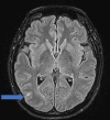
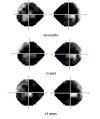
This clinical report discusses the interplay of various pathologies that may present similar clinical manifestations, with uncertainty about the distinct impact of each one of them. The patient is a 43-year-old young Asian female with no known medical conditions. She was 33 weeks pregnant when she was admitted for an urgent c-section because of preeclampsia with HELLP syndrome. While hospitalized, she complained about the visual field's loss. A comprehensive ophthalmological examination revealed a severe concentric visual field defect along with well-reduced visual acuity and impaired color vision. Her OCT revealed a bilateral serous macular detachment related to pre-eclampsia. A brain MRI revealed a microstroke at the temporo-parieto-occipital junction (TPO), although it did not fully account for the severity of the visual field deficit. Despite the macular pathology being resolved, the visual field remained deeply impacted. A thorough and complete investigation yielded negative results, leaving the cause of the patient's deficit unknown. The patient likely had a normal pressure glaucoma. Additionally, multifactorial bilateral microvascular ischemic neuropathy (caused especially by high myopia) has significantly affected her visual field. Furthermore, it is also probable that the patient had genetic neuropathy. Initial genetic testing was negative; however, due to the high suspicion of a genetic component, a retest was conducted, and the results were not conclusive. This case represents a highly unusual case of a profoundly affected visual field with no apparent identified cause. This is a notable example of the potential interaction of various local and systemic pathologies that can manifest with similar clinical presentations.
Keywords: glaucoma suspect; high myopia; optic nerve ischemia; pre-eclampsia; systemic lupus erythema; visual field defect.
Copyright © 2024, Abdouli et al.
Conflict of interest statement
Human subjects: Consent was obtained or waived by all participants in this study. Conflicts of interest: In compliance with the ICMJE uniform disclosure form, all authors declare the following: Payment/services info: All authors have declared that no financial support was received from any organization for the submitted work. Financial relationships: All authors have declared that they have no financial relationships at present or within the previous three years with any organizations that might have an interest in the submitted work. Other relationships: All authors have declared that there are no other relationships or activities that could appear to have influenced the submitted work.
Figures


References
Publication types
LinkOut - more resources
Full Text Sources
