Review
. 2024 Oct;116(4):948-962.
doi: 10.1002/cpt.3387. Epub 2024 Jul 25.

PharmVar GeneFocus: CYP2A6

Affiliations
Review

PharmVar GeneFocus: CYP2A6

Alec W R Langlois et al. Clin Pharmacol Ther. 2024 Oct.

Abstract

The Pharmacogene Variation Consortium (PharmVar) provides nomenclature for the human CYP2A gene locus containing the highly polymorphic CYP2A6 gene. CYP2A6 plays a role in the metabolism of nicotine and various drugs. Thus, genetic variation can substantially contribute to the function of this enzyme and associated efficacy and safety. This GeneFocus provides an overview of the clinical significance of CYP2A6, including its genetic variation and function. We also highlight and discuss caveats in the identification and characterization of allelic variation of this complex pharmacogene, a prerequisite for accurate genotype determination and prediction of phenotype status.


Conflict of interest statement

Conflicts of interests

C.N. and G.S are employed by PharmGenetix GmbH, a private laboratory providing PGx testing, reporting, and interpretation services. All other authors declared no competing interests for this work.

Figures

Figure 1
Figure 1. Overview of CYP2A gene locus and structural variation
The gene locus (top panel) spans approximately 252 kb and harbors three genes, CYP2A6 (blue), CYP2A7 (red) and CYP2A13 (green). The locus is shown as the reverse complement; as indicated by the colored arrows, CYP2A6 and CYP2A7 are both encoded on the negative strand while CYP2A13 is encoded on the positive strand. Numbered boxes depict exons. Deletion events with breakpoints downstream of CYP2A7 or within its 3’-UTR created a series of alleles lacking the entire CYP2A6 gene. Duplication events have been described for CYP2A6*1, which may contain CYP2A7-derived sequences as indicated by the red hashed lines. Three CYP2A7::CYP2A6 hybrids (*12, *34 and *47) have been identified to date with switches in intron 2, intron 4 and intron 8, respectively. The exact switch positions remain unknown due to high sequence homologies between the two genes. One CYP2A6::CYP2A7 hybrid (*53) has also been identified, which occurred in a duplication arrangement with an intact copy of CYP2A7. Additional and up-to-date information about SVs can be found in the Structural Variation document on the PharmVar CYP2A6 gene page .
Figure 2
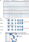
Figure 2. Overview of core alleles, suballeles, and the graphical Comparative Allele ViewEr (CAVE)
An excerpt of the CYP2A6 gene page is provided in panel (a). Grey bars show the CYP2A62*1, *15, *27 and *54 core allele definitions, whereas blue and white backgrounds signify suballeles. For CYP2A6*1, only selected suballeles are depicted, while all current suballeles are shown for *15, *27 and *54. Positions correspond to NG_008377.1 as the reference sequence (counting from the translation start). Star allele names, PharmVar ID (PVID), allele evidence levels, and references are shown for each allele. Legacy allele names are cross-referenced. rsIDs (where existing) are provided for each variant with direct link-out capability to dbSNP. Of note, CPIC has not assigned clinical allele function for CYP2A6 and therefore, function is shown as N/A for all star alleles. Panel (b) is a graphical representation of selected alleles with their respective core SNVs. Core SNVs are shared among all alleles listed under that star number and cause an amino acid change (CYP2A6*15, *24 and *26) or impact gene expression (*24 and *46); those known to impact function are shown in bolded black font and those with unknown or uncertain function are in grey font. The CYP2A7-derived 3’UTR conversion (which also impacts gene expression) is highlighted in red. Blue numbered boxes represent exons and untranslated regions are shown in light blue. Panel (c) represents the graphical output of the Comparative Allele ViewEr (CAVE) that allows easy visualization of which SNVs are i) unique to a star allele, e.g., 13G>A (rs28399434, G5R) is only found on *13 and 2134A>G is only found on *15, etc., or ii) occur on two or more star alleles, e.g., −48T>G is present on *9, *13, *15 and *50. The PharmVar symbols indicate which variants are unique to an allele and the function symbol signifies that the variant alters function.
Figure 3
Figure 3. NGS with paired-end short reads can produce misalignments due to high sequence identity between CYP2A6 and CYP2A7
Panel (a) visualizes the results of a simulation analysis in which 2×150 bp paired reads with average fragment sizes of 500 bp were generated from the CYP2A6 RefSeq, with an average coverage per position of 100 reads (CYP2A6 exons/introns are not drawn to scale); a second simulation used reads into which the 3’UTR conversion was introduced to simulate CYP2A6*46. For each position, the percentage of total reads that could not be disambiguated between CYP2A6 or CYP2A7, or which misaligned to CYP2A7 (in the case of the simulated CYP2A6*46), was determined. Misalignments were common in exon 5 and, when the 3’UTR conversion was present, exon 9. Misalignments in exon 9 are shown by position in a heatmap where the darkest red corresponds to >50% of misaligned reads. Panel (b) depicts a region from exon 8 to the 3’UTR (to scale) demonstrating how read misalignments can occur when NGS short reads have a mean fragment size of 500 bp. Read pairs are displayed as boxes with a connecting hashed black line. Blue and red lines indicate positions where CYP2A6 and CYP2A7 differ, i.e., represent informative positions, and the large blue and red block indicates the 3’UTR conversion region. Fragment size variation can produce misalignments when a read overlaps the 3’UTR conversion, as demonstrated by a hypothetical read pair shown above the CYP2A6*46 graph, which would produce “unambiguous” alignments to CYP2A7.
Figure 4
Figure 4. CYP2A6 haplotype characterization using single molecule sequencing
Panels (a) and (b) showcase two samples for which both haplotypes were initially found using WGS and subsequently further characterized with single molecule long read sequencing. CYP2A7-derived sequences are highlighted in red. SNVs in coding regions are in black font, and those in noncoding regions are shown in blue font. SNVs known to alter function are highlighted in larger font. The CYP2A6*9.002 allele in (a) represents a fully characterized sequence that was initially designated as *9B; several SNVs have since been added to the allele’s definition. The subject’s second allele represents a new CYP2A6*31 suballele, *31.003. The CYP2A6*17.002 and *54.001 alleles found in the subject shown in (b) are both novel. CYP2A6*54.001 has a 49-bp long region spanning parts of exon 3 and intron 3 (ex3/in3) that is CYP2A7-derived and listed as a conversion in the Structural Variation document. This region contains 11 SNVs, of which six contribute to five amino acid changes. CYP2A6*54.001 also has the CYP2D7-derived 3’UTR conversion. The top portions of panels (c) and (d) visualize the challenge of resolving CYP2A7 conversion regions with WGS short read sequencing. As shown in their respective bottom portions, these challenging regions can be resolved using single molecule long read sequencing. Screenshots of read alignments represent the sample depicted in panel (b).

References

    1. Hukkanen J, Jacob P, Benowitz NL. Metabolism and disposition kinetics of nicotine. Pharmacol Rev. Mar 2005;57(1):79–115. doi: 10.1124/pr.57.1.3 - DOI - PubMed
    1. Nagata K, Matsunaga T, Gillette J, Gelboin HV, Gonzalez FJ. Rat testosterone 7 alpha-hydroxylase. Isolation, sequence, and expression of cDNA and its developmental regulation and induction by 3-methylcholanthrene. J Biol Chem. Feb 25 1987;262(6):2787–93. - PubMed
    1. Yamano S, Tatsuno J, Gonzalez FJ. The CYP2A3 gene product catalyzes coumarin 7-hydroxylation in human liver microsomes. Biochemistry. Feb 6 1990;29(5):1322–9. doi: 10.1021/bi00457a031 - DOI - PubMed
    1. Fernandez-Salguero P, Hoffman SM, Cholerton S, et al. A genetic polymorphism in coumarin 7-hydroxylation: sequence of the human CYP2A genes and identification of variant CYP2A6 alleles. Am J Hum Genet. Sep 1995;57(3):651–60. - PMC - PubMed
    1. von Weymarn LB, Retzlaff C, Murphy SE. CYP2A6- and CYP2A13-catalyzed metabolism of the nicotine Δ5’(1’)iminium ion. J Pharmacol Exp Ther. Nov 2012;343(2):307–15. doi: 10.1124/jpet.112.195255 - DOI - PMC - PubMed

LinkOut - more resources