

Recovery of ambulation in small, nonbrachycephalic dogs after conservative management of acute thoracolumbar disk extrusion
- PMID: 39051966
- PMCID: PMC11423491
- DOI: 10.1111/jvim.17149
Recovery of ambulation in small, nonbrachycephalic dogs after conservative management of acute thoracolumbar disk extrusion
Abstract
Background: Currently, low-level evidence suggests loss of ambulation associated with acute thoracolumbar disk extrusion is best treated by decompressive spinal surgery. Conservative management can be successful, but the proportion of dogs that recover and the fate of herniated material are uncertain.
Objectives: Determine the proportion of nonambulatory dogs with conservatively treated acute thoracolumbar disk extrusion that recover ambulation and measure the change in spinal cord compression during the first 12 weeks after presentation.
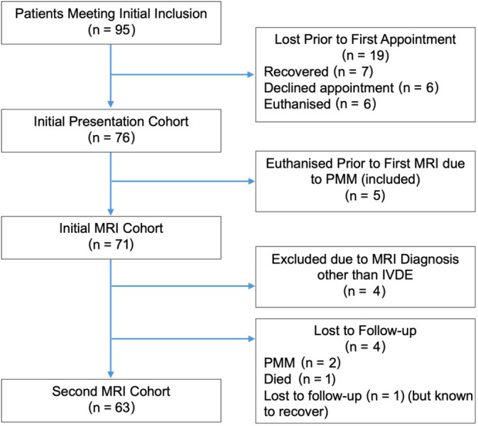
Animals: Seventy-two client-owned nonambulatory dogs with acute thoracolumbar intervertebral disk extrusion.
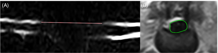
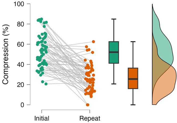
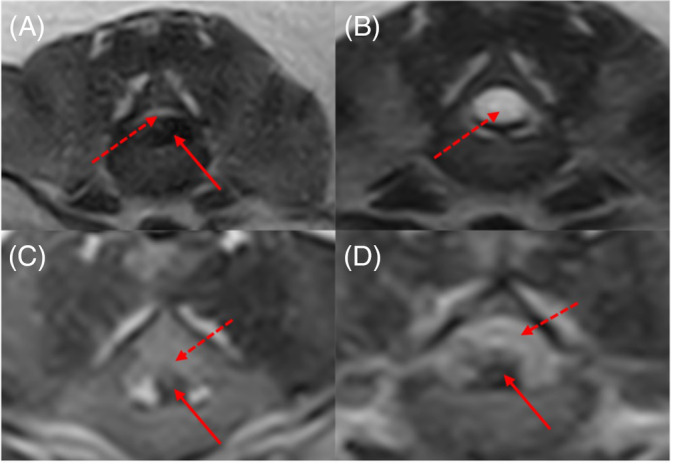
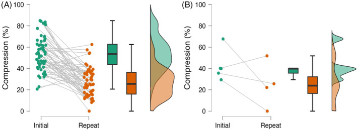
Methods: This is a prospective cohort study. Enrolled dogs underwent magnetic resonance imaging at presentation and owners were provided with conservative management recommendations. Imaging was repeated after 12 weeks. Recovery of ambulation was defined as 10 consecutive steps without falling. Spinal cord compression was determined from the cross-sectional area of the vertebral canal and extradural compressive material at the lesion epicenter. The association between recovery and change in compression over the 12-week observational period was examined.
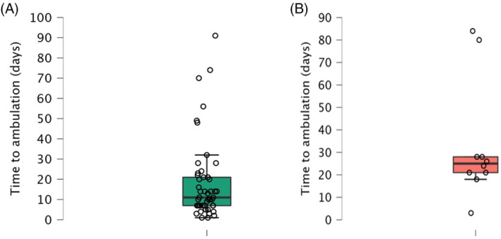
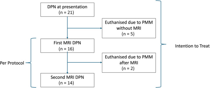
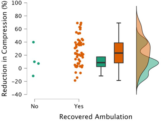
Results: Forty-nine of fifty-one (96%; 95% confidence interval [CI], 87%-99%) of deep pain-positive and 10/21 (48%; 95% CI, 28%-68%) of deep pain-negative dogs recovered ambulation within the 12-week period. The median time to ambulation was 11 and 25 days for deep pain-positive and -negative dogs, respectively. Reduction in spinal cord compression varied among individuals from minimal to complete and apparently was unrelated to the recovery of ambulation.
Conclusions and clinical importance: A high proportion of conservatively treated dogs recovered ambulation after conservative management of acute thoracolumbar disk herniation. Recovery was not dependent on the resolution of compression.
Keywords: compression; contusion; disk herniation; spinal cord injury.
© 2024 The Author(s). Journal of Veterinary Internal Medicine published by Wiley Periodicals LLC on behalf of American College of Veterinary Internal Medicine.
Conflict of interest statement
Authors declare no conflict of interest.
Figures
References
-
- Bergknut N, Egenvall A, Hagman R, et al. Incidence of intervertebral disk degeneration‐related diseases and associated mortality rates in dogs. J Am Vet Med Assoc. 2012;240(11):1300‐1309. - PubMed
-
- Fluehmann G, Doherr MG, Jaggy A. Canine neurological diseases in a referral hospital population between 1989 and 2000 in Switzerland. J Small Anim Pract. 2006;47(10):582‐587. - PubMed
-
- Hansen HJ. A pathologic‐anatomical study on disc degeneration in dog, with special reference to the so‐called enchondrosis intervertebralis. Acta Orthop Scand Suppl. 1952;11:1‐117. - PubMed
-
- Olsson SE. Observations concerning disc fenestration in dogs. Acta Orthop. 1951;20(4):349‐356. - PubMed
MeSH terms
Grants and funding
LinkOut - more resources
Full Text Sources
Medical
