. 2024 Aug 1;80(Pt 8):183-190.
doi: 10.1107/S2053230X24006915. Epub 2024 Jul 25.

Crystal structures of the 3C proteases from Coxsackievirus B3 and B4

Affiliations

Crystal structures of the 3C proteases from Coxsackievirus B3 and B4

Haihai Jiang et al. Acta Crystallogr F Struct Biol Commun. .

Abstract

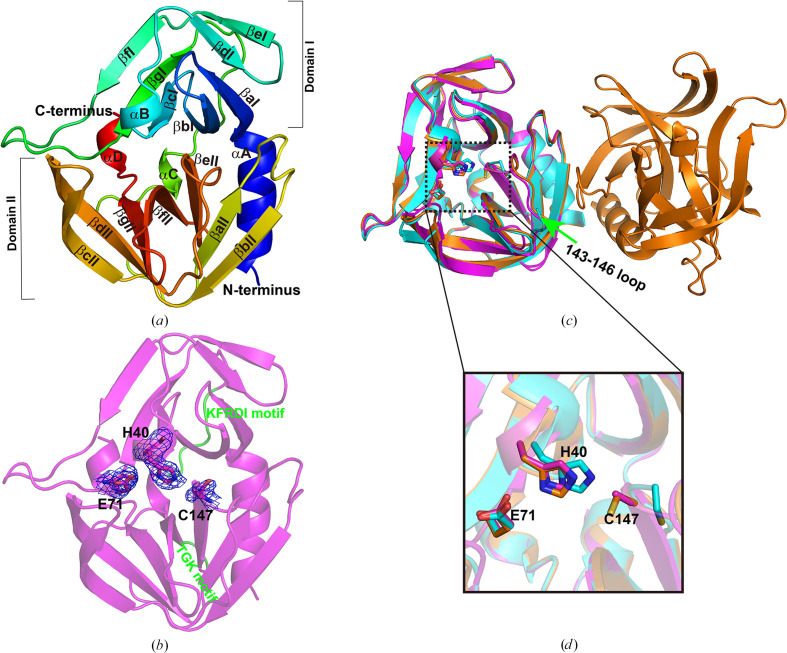
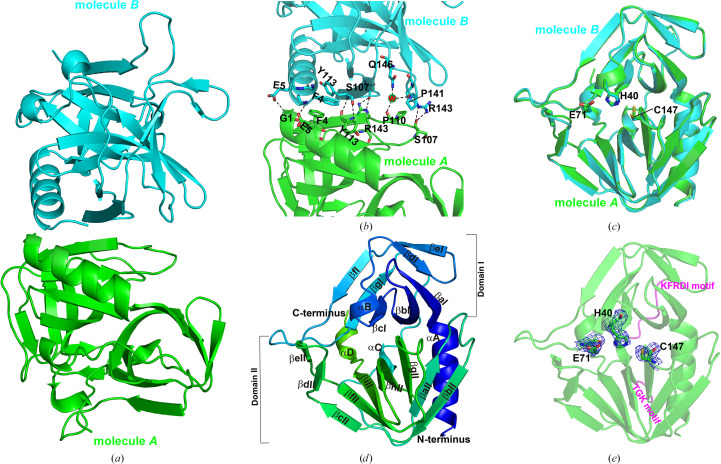
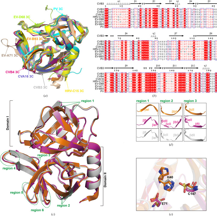
Enteroviruses cause a wide range of disorders with varying presentations and severities, and some enteroviruses have emerged as serious public health concerns. These include Coxsackievirus B3 (CVB3), an active causative agent of viral myocarditis, and Coxsackievirus B4 (CVB4), which may accelerate the progression of type 1 diabetes. The 3C proteases from CVB3 and CVB4 play important roles in the propagation of these viruses. In this study, the 3C proteases from CVB3 and CVB4 were expressed in Escherichia coli and purified by affinity chromatography and gel-filtration chromatography. The crystals of the CVB3 and CVB4 3C proteases diffracted to 2.10 and 2.01 Å resolution, respectively. The crystal structures were solved by the molecular-replacement method and contained a typical chymotrypsin-like fold and a conserved His40-Glu71-Cys147 catalytic triad. Comparison with the structures of 3C proteases from other enteroviruses revealed high similarity with minor differences, which will guide the design of 3C-targeting inhibitors with broad-spectrum properties.

Keywords: 3C proteases; CVB3; CVB4; Coxsackievirus; crystal structure.


Figures

Figure 1
Figure 1
Crystal structure of the 3C protease from CVB3. (a) Overall structure of the CVB3 3C protease. The structure is colored from blue at the N-terminus to red at the C-terminus. The α-helices in the two domains (I and II) are labeled αA–αD. The β-strands are labeled βaI–βgI in domain I and βaII–βgII in domain II according to their occurrence along the primary structure. (b) Catalytic triad and RNA-binding sites of the CVB3 3C protease. The 2FoFc electron densities for the catalytic residues (His40, Glu71 and Cys147) are contoured at 1σ. The RNA-binding motifs, KFRDI and TGK, are colored green. (c) Comparison of structures of the CVB3 3C protease. The structure solved in this study is colored magenta. The structures solved previously, named form I (PDB entry 2zty) and form II (PDB entry 2ztz), are colored cyan and orange, respectively. The loop consisting of residues 143–146 (the 143–146 loop) is indicated by a green arrow. (d) An enlarged view of the comparison of catalytic residues. The catalytic residues are shown as sticks. Electron density for Cys147 is missing in our structure.
Figure 2
Figure 2
Crystal structure of the 3C protease from CVB4. (a) Overall structure of the CVB4 3C protease. The two molecules (molecule A and molecule B) in the asymmetric unit of the CVB4 3C protease structure are shown in cartoon representation. Molecule B (cyan) is packed against the surface of molecule A (green). (b) A magnified view showing detailed crystal-packing interactions. The residues involved in crystal packing are shown as sticks. Hydrogen-bonding interactions are indicated as black dashed lines. W represents the water molecule that mediates hydrogen-bonding interactions. (c) Comparison of molecule A and molecule B in the CVB4 3C protease structure. The catalytic residues (His40, Glu71 and Cys147) are shown as sticks. (d) Overall structure of molecule A. The structure is colored from blue at the N-terminus to green at the C-terminus. Four α-helices in two domains (I and II) are marked αA–αD. A total of 16 β-strands are labeled (βaI–βgI in domain I and βaII–βgII in domain II) according to their occurrence along the primary structure. (e) Catalytic triad and RNA-binding sites of the CVB4 3C protease. The 2FoFc electron densities for the catalytic residues are contoured at 1σ. The RNA-binding motifs, KFRDI and TGK, are colored magenta.
Figure 3
Figure 3
Comparison of enteroviral 3C proteases with reported structures. (a) Structural alignment of 3C proteases. 3C proteases from poliovirus (PV, cyan, PDB entry 1l1n; Mosimann et al., 1997 ▸), EV-B93 (orange, PDB entry 3q3x; Costenaro et al., 2011 ▸), EV-D68 (lemon, PDB entry 3zv8; Tan et al., 2013 ▸), CVA16 (slate, PDB entry 3sj8; Lu et al., 2011 ▸), HRV-C15 (yellow, PDB entry 6ku7; Yuan et al., 2020 ▸), EV-A71 (wheat, PDB entry 3osy; Cui et al., 2011 ▸), CVB3 (gray, this study) and CVB4 (magenta, this study) are included in the comparison. Alignments of these 3C proteases yield r.m.s.d. values that demonstrate the similarity between these enzymes. (b) Multiple-sequence alignment of 3C proteases from different enteroviruses. The MEGA software (version 10.2.5) was used to perform the alignment. The sequence-alignment visualization was generated using ESPript 3.0. (c) Structural alignment of the CVB3 (gray, this study), CVB4 (magenta, this study) and EV-B93 (orange, PDB entry 3q3x) 3C proteases. Obvious local differences are labeled with green lines or dashed boxes. (d) An enlarged view of reigions 1–3 with obvious local differences. (e) An enlarged view of the catalytic triad of 3C proteases.

Similar articles

Cited by

References

    1. Alhazmi, A., Nekoua, M. P., Michaux, H., Sane, F., Halouani, A., Engelmann, I., Alidjinou, E. K., Martens, H., Jaidane, H., Geenen, V. & Hober, D. (2021). Microorganisms, 9, 1177. - PMC - PubMed
    1. Baggen, J., Thibaut, H. J., Strating, J. R. P. M. & van Kuppeveld, F. J. M. (2018). Nat. Rev. Microbiol.16, 368–381. - PubMed
    1. Barral, P. M., Sarkar, D., Fisher, P. B. & Racaniello, V. R. (2009). Virology, 391, 171–176. - PMC - PubMed
    1. Bergmann, E. M., Mosimann, S. C., Chernaia, M. M., Malcolm, B. A. & James, M. N. G. (1997). J. Virol.71, 2436–2448. - PMC - PubMed
    1. Birtley, J. R., Knox, S. R., Jaulent, A. M., Brick, P., Leatherbarrow, R. J. & Curry, S. (2005). J. Biol. Chem.280, 11520–11527. - PubMed

MeSH terms

LinkOut - more resources