Case Reports
. 2024 Sep 1;142(9):808-817.
doi: 10.1001/jamaophthalmol.2024.2385.

Severe Spaceflight-Associated Neuro-Ocular Syndrome in an Astronaut With 2 Predisposing Factors

Affiliations
Case Reports

Severe Spaceflight-Associated Neuro-Ocular Syndrome in an Astronaut With 2 Predisposing Factors

Tyson J Brunstetter et al. JAMA Ophthalmol. .

Abstract

Importance: Understanding potential predisposing factors associated with spaceflight-associated neuro-ocular syndrome (SANS) may influence its management.

Objective: To describe a severe case of SANS associated with 2 potentially predisposing factors.

Design, setting, and participants: Ocular testing of and blood collections from a female astronaut were completed preflight, inflight, and postflight in the setting of the International Space Station (ISS).

Exposure: Weightlessness throughout an approximately 6-month ISS mission. Mean carbon dioxide (CO2) partial pressure decreased from 2.6 to 1.3 mm Hg weeks before the astronaut's flight day (FD) 154 optical coherence tomography (OCT) session. In response to SANS, 4 B-vitamin supplements (vitamin B6, 100 mg; L-methylfolate, 5 mg; vitamin B12, 1000 μg; and riboflavin, 400 mg) were deployed, unpacked on FD153, consumed daily through FD169, and then discontinued due to gastrointestinal discomfort.

Main outcomes and measures: Refraction, distance visual acuity (DVA), optic nerve, and macular assessment on OCT.

Results: Cycloplegic refraction was -1.00 diopter in both eyes preflight and +0.50 - 0.25 × 015 in the right eye and +1.00 diopter in the left eye 3 days postflight. Uncorrected DVA was 20/30 OU preflight, 20/16 or better by FD90, and 20/15 OU 3 days postflight. Inflight peripapillary total retinal thickness (TRT) peaked between FD84 and FD126 (right eye, 401 μm preflight, 613 μm on FD84; left eye, 404 μm preflight, 636 μm on FD126), then decreased. Peripapillary choroidal folds, quantified by surface roughness, peaked at 12.7 μm in the right eye on FD154 and 15.0 μm in the left eye on FD126, then decreased. Mean choroidal thickness increased throughout the mission. Genetic analyses revealed 2 minor alleles for MTRR 66 and 2 major alleles for SHMT1 1420 (ie, 4 of 4 SANS risk alleles). One-week postflight, lumbar puncture opening pressure was normal, at 19.4 cm H2O.

Conclusions and relevance: To the authors' knowledge, no other report of SANS documented as large of a change in peripapillary TRT or hyperopic shift during a mission as in this astronaut, and this was only 1 of 4 astronauts to experience chorioretinal folds approaching the fovea. This case showed substantial inflight improvement greater than the sensitivity of the measure, possibly associated with B-vitamin supplementation and/or reduction in cabin CO2. However, as a single report, such improvement could be coincidental to these interventions, warranting further evaluation.


Conflict of interest statement

Conflict of Interest Disclosures: Dr Laurie reported receiving grants from NASA during the conduct of the study. Dr Lee reported receiving advisory board fees from Stoke, Amgen, and Viridian; speaker fees from Alexion; and consultant fees from AstraZeneca, Bristol Myers Squibb, NASA, and NFL. No other disclosures were reported.

Figures

Figure 1.
Figure 1.. Progression of a Retinal Pigment Epithelial Detachment (PED) in an Astronaut
A, Scanning laser ophthalmoscope image with vertical green arrow denoting location of B-scans shown in panels B through G. B, Preexisting PED shown with optical coherence tomography (OCT) on vertical B-scan, worsening during flight and improving postflight (C-G). Within 2 to 5 months of return, the PED was superseded by serous chorioretinopathy, with subsequent self-resolution. Subtle retinal pigment epithelial disruption remains at 26 months postflight (G). Chorioretinal folds are also visible inflight and postflight.
Figure 2.
Figure 2.. Change in Total Retinal Thickness (TRT), Mean Choroidal Thickness, and Surface Roughness Along With 24-Hour Mean Ambient Carbon Dioxide (CO2) Concentrations
A, Change in peripapillary TRT in the left and right eyes during and postflight. B, Change in average choroid thickness during and postflight. C, Change in surface roughness as an objective measure of choroidal folds inflight and postflight. D, Twenty-four–hour mean CO2 concentrations from the Columbus module (data from other modules similar and not presented). Average CO2 levels were 2.56 mm Hg until crew size decreased midmission. CO2 levels then averaged 1.3 mm Hg until additional astronauts arrived near the end of the mission. Vertical dotted line indicates when the crewmember took additional B vitamins, starting on flight day (FD) 153, 1 day before the optical coherence tomography scan collected on FD154, through FD169. Postflight collections in these graphs extend to approximately 15 months after flight. ISS indicates International Space Station.
Figure 3.
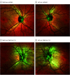
Figure 3.. Retinal Multicolor Imaging of the Right and Left Posterior Poles of a Spaceflight-Associated Neuro-Ocular Syndrome Case
A, Preflight image, right eye. B, Preflight image, left eye. C and D, Flight day (FD) 154 showing bilateral optic disc edema, optic nerve head protrusion (left eye greater than right eye), and chorioretinal folds, some of which enter the macula, in the right and left eyes, respectively.
Figure 4.
Figure 4.. Optical Coherence Tomography (OCT)–Derived Surface Topography Maps of the Posterior Pole of the Right and Left Eyes, Progressing From Preflight, Flight Day (FD) 63, FD183, and 150 Days After Return
A, Retinal internal limiting membrane topography showing bilateral optic disc edema and retinal folds. B, Bruch membrane topography showing bilateral choroidal folds. Dark circles represent the optic nerve head in each eye; blue indicates lower elevations; red indicates higher elevations.

References

    1. Mader TH, Gibson CR, Pass AF, et al. Optic disc edema, globe flattening, choroidal folds, and hyperopic shifts observed in astronauts after long-duration space flight. Ophthalmology. 2011;118(10):2058-2069. doi: 10.1016/j.ophtha.2011.06.021 - DOI - PubMed
    1. National Aeronautics and Space Administration . Risk of spaceflight-associated neuro-ocular syndrome (SANS). Accessed February 20, 2024. https://humanresearchroadmap.nasa.gov/evidence/reports/SANS.pdf
    1. Pardon LP, Greenwald SH, Ferguson CR, et al. Identification of factors associated with the development of optic disc edema during spaceflight. JAMA Ophthalmol. 2022;140(12):1193-1200. doi: 10.1001/jamaophthalmol.2022.4396 - DOI - PMC - PubMed
    1. Zwart SR, Gibson CR, Mader TH, et al. Vision changes after spaceflight are related to alterations in folate- and vitamin B-12–dependent 1-carbon metabolism. J Nutr. 2012;142(3):427-431. doi: 10.3945/jn.111.154245 - DOI - PubMed
    1. Zwart SR, Gregory JF, Zeisel SH, et al. Genotype, B-vitamin status, and androgens affect spaceflight-induced ophthalmic changes. FASEB J. 2016;30(1):141-148. doi: 10.1096/fj.15-278457 - DOI - PMC - PubMed

Publication types

Supplementary concepts