. 2024 Jul 25;19(7):e0307662.
doi: 10.1371/journal.pone.0307662. eCollection 2024.

Vitamin K2 sensitizes the efficacy of venetoclax in acute myeloid leukemia by targeting the NOXA-MCL-1 pathway

Affiliations

Vitamin K2 sensitizes the efficacy of venetoclax in acute myeloid leukemia by targeting the NOXA-MCL-1 pathway

Tetsuzo Tauchi et al. PLoS One. .

Abstract

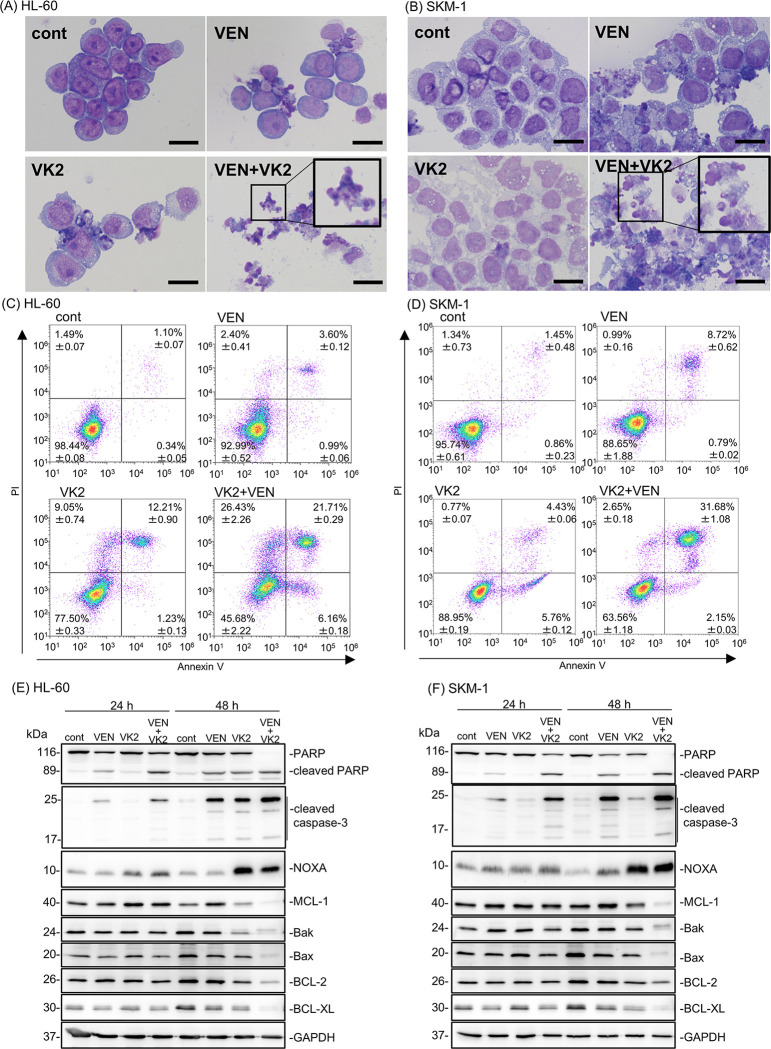
Promising outcomes have been reported in elder patients with acute myeloid leukemia (AML) using combined therapy of venetoclax (VEN) and azacytidine (AZA) in recent years. However, approximately one-third of patients appear to be refractory to this therapy. Vitamin K2 (VK2) shows apoptosis-inducing activity in AML cells, and daily oral VK2 (menaquinone-4, GlakayR) has been approved for patients with osteoporosis in Japan. We observed a high response rate to AZA plus VEN therapy, with no 8-week mortality in the newly diagnosed AML patients consuming daily VK2 in our hospital. The median age of the patients was 75.9 years (range 66-84) with high-risk features. Patients received AZA 75 mg/m2 on D1-7, VEN 400 mg on D1-28, and daily VK2 45 mg. The CR/CRi ratio was 94.7% (18/19), with a CR rate of 79%. Complete cytogenetic CR was achieved in 15 of 19 (79%) patients, and MRD negativity in 2 of 15 (13%) evaluable CR patients. Owing to the extremely high response rate in clinical settings, we further attempted to investigate the underlying mechanisms. The combination of VK2 and VEN synergistically induced apoptosis in all five AML cell lines tested. VK2, but not VEN, induced mitochondrial reactive oxygen species (ROS), leading to the transcriptional upregulation of NOXA, followed by MCL-1 repression. ROS scavengers repressed VK2 induced-NOXA expression and led to the cancellation of pronounced apoptosis and the downregulation of MCL-1 by VK2 plus VEN. Additionally, knockdown and knockout of NOXA resulted in abrogation of the MCL-1 repression as well as enhanced cytotoxicity by the two-drug combination, indicating that VK2 suppresses MCL-1 via ROS-mediated NOXA induction. These data suggest that the dual inhibition of BCL-2 by VEN and MCL-1 by VK2 is responsible for the remarkable clinical outcomes in our patients. Therefore, large-scale clinical trials are required.


Conflict of interest statement

The authors have declared that no competing interests exist.

Figures

Fig 1
Fig 1. Therapeutic outcome of 19 AML patients receiving VEN+AZA+VK2.
CR, complete response; CRi, CR with incomplete count recovery; MLFS, morphologic leukemia-free state; MRD, minimal residual disease; M, months after the initiation of AZA+VEN treatment.
Fig 2
Fig 2. Enhanced cell growth inhibition by combination treatment of VK2 and VEN in AML cell lines.
(A-E) AML cell lines (HL-60, SKM-1, THP-1, U-937, and MOLM-14) were treated with VK2 in the presence or absence of VEN at indicated concentrations for 48 h and 72 h. Upper: The viable cell number was assessed by CellTiter Blue assay. Data are presented as the mean ± SD. *p<0.05 vs. VEN 0 nM. Lower: The synergistic effect of VK2 and VEN combined treatment on AML cell proliferative inhibition was statistically analyzed using Combenefit software. Mapping of the synergy levels on the experimental combination dose-response surface. A higher score shown in denser blue indicates a stronger synergistic effect. n = 3.
Fig 3
Fig 3. Pronounced apoptosis induction by simultaneous treatment with VK2 and VEN along with up-regulation of NOXA and repression of MCL-1 in HL-60 and SKM-1 cells.
(A, B) HL-60 and SKM-1 cells were treated with either VK2 (10 μM for HL-60, 25 μM for SKM-1), VEN (25 nM for HL-60, 2.5 μM for SKM-1), or VK2 plus VEN for 48 h, and stained with May-Grunwald-Giemsa. Scale bar = 20 μm. (C, D) Cells were treated with either VK2 (10 μM for HL-60, 25 μM for SKM-1), VEN (25 nM for HL-60, 2.5 μM for SKM-1), or VK2 plus VEN for 48 h. Flow cytometry was performed with Annexin V and PI double staining. The number of each area indicates the percentage of cells. n = 3 (E, F) Cellular proteins were lysed, separated by SDS-PAGE, and immunoblotting was performed using indicated antibodies. Immunoblotting with anti-GAPDH mAb was performed as an internal loading control.
Fig 4
Fig 4. ROS production in response to VK2 and/or VEN in HL-60 and SKM-1 cells.
Cells were treated with VK2 (10 μM for HL-60, 25 μM for SKM-1) or VEN (25 nM for HL-60, 2.5 μM for SKM-1) for 24 and 48 h. (A, B) ROS production in whole cells was assessed by staining with dihydroethidium (DHE) and detected by fluorescence microscopy in HL-60 and SKM-1 cells 48 h after treatment. Scale bar = 100 μm. (C, D) Mitochondrial ROS levels were determined using flow cytometry after staining with MitoSox-Red. The cells treated with a mitochondrial uncoupler, CCCP (10 μM) were used as a positive control for mitochondrial ROS production. Data are presented as the mean ± SD. n = 3, *p<0.05 vs. cont. (E) The mitochondrial membrane potential was assessed by flow cytometry after TMRE staining. CCCP was used as the positive control. Data are presented as the mean ± SD. n = 3, *p<0.05 vs. cont. (F) The effect of ROS scavengers on the mitochondrial membrane potential was assessed by flow cytometry. Data are presented as the mean ± SD. n = 3, *p<0.05 vs. 0 mM NAC. (G, H) Cells were treated with VK2 (10 μM for HL-60, 25 μM for SKM-1) and/or VEN (25 nM for HL-60, 2.5 μM for SKM-1) in the presence of ROS scavengers, namely NAC, melatonin (MEL), and Trolox (TRO) at the indicated concentrations for 48 h. The number of viable cells was assessed using the CellTiter Blue assay. Data are presented as the mean ± SD. *p<0.05 vs. 0 mM NAC, MEL, or TRO. (I, J) Immunoblotting with anti-NOXA, anti-MCL-1, and anti-PARP mAbs. Immunoblotting with anti-GAPDH mAb was performed as an internal loading control.
Fig 5
Fig 5. Gene expressions of BCL-2 family members after treatment with VK2 and/or VEN in HL-60 and SKM-1 cells.
(A, B) HL-60 and SKM-1 cells were treated with VK2 (10 μM for HL-60, 25 μM for SKM-1) and/or VEN (25 nM for HL-60, 2.5 μM for SKM-1) for 24 h and 48 h. Gene expressions of the BCL-2 family members were assessed by real-time PCR. Data are presented as the mean ± SD. *p<0.05 vs. cont. n = 3. (C, D) Effect of NAC treatment on NOXA mRNA expression was assessed by real-time PCR with HL-60 and SKM-1 cells. Data are presented as the mean ± SD., *p<0.05 vs. cont, #p<0.05 v.s. NAC (-), n = 3.
Fig 6
Fig 6. Effect of NOXA-knockout in HL-60 and SKM-1 cells on the cytotoxicity and MCL-1 repression by VK2 and VEN combination treatment.
(A, B) Control and NOXA knockout (KO) HL-60 and SKM-1 cells were treated with VK2 with/without VEN at indicated concentrations, and the viable cell numbers were assessed by CellTiter Blue assay. Data are presented as the mean ± SD. *p<0.05 vs. VEN 0 nM. The synergistic effect of VK2 and VEN combined treatment on each cell proliferative inhibition was statistically analyzed using Combenefit software. (C, D) After treatment with VK2 with/without VEN for 48 h, cellular proteins were separated by SDS-PAGE and immunoblotted with anti-NOXA, anti-MCL-1, and anti-PARP Abs. Immunoblotting with anti-GAPDH mAb was performed as an internal control. (E) Proposal scheme of the molecular mechanism of VK2 for sensitization to VEN in AML cells. VEN-treatment specifically inhibits BCL-2 but not MCL-1 [36]. VK2-treatment induces mitochondrial ROS production leading to up-regulation of NOXA, which results in inhibition and/or repression of MCL-1 to relieve BAK inhibition. Thus, concomitant treatment with VEN plus VK2 results in simultaneous inhibition of BCL-2 and MCL-1. This appears to enhance BAK- and/or BAK/BAX-mediated apoptosis induction in AML cells.

References

    1. Diepstraten ST, Anderson MA, Czabotar PE, Lessene G, Strasser A, Kelly GL. The manipulation of apoptosis for cancer therapy using BH3-mimetic drugs. Nat Rev Cancer. 2022; 22(1): 45–64. doi: 10.1038/s41568-021-00407-4 . - DOI - PubMed
    1. Eichhorst B, Niemann CU, Kater AP, Fürstenau M, von Tresckow J, Zhang C, et al.. First-Line Venetoclax Combinations in Chronic Lymphocytic Leukemia. N Engl J Med. 2023; 388(19): 1739–1754. doi: 10.1056/NEJMoa2213093 . - DOI - PubMed
    1. Lasica M, Anderson MA. Review of Venetoclax in CLL, AML and Multiple Myeloma. J Pers Med. 2021; 11(6). doi: 10.3390/jpm11060463 . - DOI - PMC - PubMed
    1. Sullivan GP, Flanagan L, Rodrigues DA, T NC. The path to venetoclax resistance is paved with mutations, metabolism, and more. Sci Transl Med. 2022; 14(674): eabo6891. doi: 10.1126/scitranslmed.abo6891 . - DOI - PubMed
    1. Daver NG, Maiti A, Kadia TM, Vyas P, Majeti R, Wei AH, et al.. TP53-Mutated Myelodysplastic Syndrome and Acute Myeloid Leukemia: Biology, Current Therapy, and Future Directions. Cancer Discov. 2022; 12(11): 2516–2529. doi: 10.1158/2159-8290.CD-22-0332 . - DOI - PMC - PubMed

MeSH terms