. 2024 Aug 20;5(8):101658.
doi: 10.1016/j.xcrm.2024.101658. Epub 2024 Jul 24.

Myeloid cells coordinately induce glioma cell-intrinsic and cell-extrinsic pathways for chemoresistance via GP130 signaling

Affiliations

Myeloid cells coordinately induce glioma cell-intrinsic and cell-extrinsic pathways for chemoresistance via GP130 signaling

Jiying Cheng et al. Cell Rep Med. .

Abstract

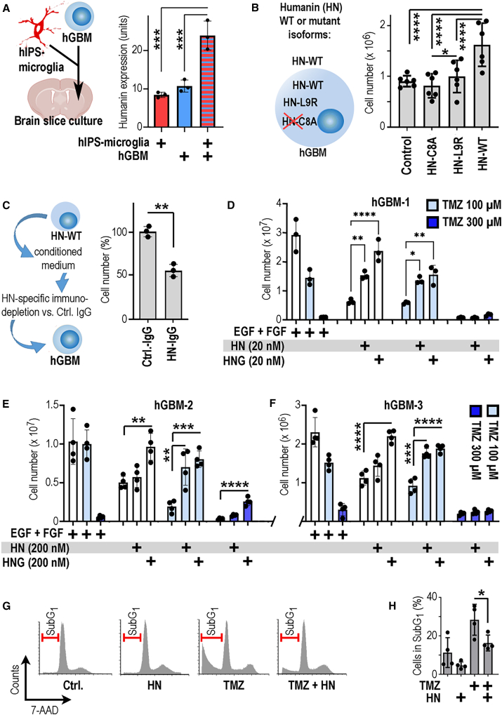
The DNA damage response (DDR) and the blood-tumor barrier (BTB) restrict chemotherapeutic success for primary brain tumors like glioblastomas (GBMs). Coherently, GBMs almost invariably relapse with fatal outcomes. Here, we show that the interaction of GBM and myeloid cells simultaneously induces chemoresistance on the genetic and vascular levels by activating GP130 receptor signaling, which can be addressed therapeutically. We provide data from transcriptomic and immunohistochemical screens with human brain material and pharmacological experiments with a humanized organotypic GBM model, proteomics, transcriptomics, and cell-based assays and report that nanomolar concentrations of the signaling peptide humanin promote temozolomide (TMZ) resistance through DDR activation. GBM mouse models recapitulating intratumoral humanin release show accelerated BTB formation. GP130 blockade attenuates both DDR activity and BTB formation, resulting in improved preclinical chemotherapeutic efficacy. Altogether, we describe an overarching mechanism for TMZ resistance and outline a translatable strategy with predictive markers to improve chemotherapy for GBMs.

Keywords: DDR; DNA damage response; GP130; IL6ST; TAM; blood-tumor barrier; chemotherapy; glioblastoma; humanin; temozolomide; tumor-associated myeloid cells.


Conflict of interest statement

Declaration of interests The authors declare no competing interests.

Figures

None
Graphical abstract
Figure 1
Figure 1
Humanin is strongly expressed in hGBMs (A) Myeloid cells purified from biopsies of epilepsy surgery (tumor free) or GBMs underwent transcriptomic profiling and bioinformatics analysis. (B) The mitochondrial ribosomal RNA-encoding gene MT-RNR2 is among the top-5 upregulated genes in GAMs. MT-RNR2 contains an open reading frame for the peptide humanin. (C) Confocal microscopy of GBMs immunolabeled for humanin and the myeloid cell marker Iba1; GAMs expressing humanin are indicated (arrowheads). (D) A single optical section of GAMs (arrow) and other intratumoral cells (double arrow) plus confocal cross hair inspection (insert). (E) Immunofluorescence labeling for humanin in GBMs and controls was quantified (dots indicating individual patient samples). (F) In IDH1-mutant (IDH1R132H), grade-IV astrocytomas, humanin expression is largely confined to GBM cells. The number of biological replicates is indicated (each dot in the graph indicates average data from one individual sample); error bars are presented as mean ± SDM. Statistical significance is shown as false discovery rate (FDR) in (A) and by t test (∗∗∗∗p < 0.0001) in (E); scales indicate 30 μm (C), 10 μm (D, F).
Figure 2
Figure 2
Humanin promotes GBM chemoresistance (A) hiPSC microglia or hGBMs were implanted (alone or in combination) into organ-cultured mouse brain slices; immunostained for humanin and staining was quantified. (B) Humanin (HN) exerts intra- and extracellular action, which can be inspected by distinct isoforms. The graph displays numbers of hGBMs expressing HN-WT, HN-C8A, HN-L9R, or unmanipulated controls. (C) Conditioned media from HN-WT GBMs were immunodepleted for HN (HN-IgG) or not (Ctrl.-IgG), control medium (Ctrl.-IgG) supported GBM cell expansion, but not HN-depleted medium. (D–F) hGBM-1, 2, or 3 was maintained under standard or under growth factor deprived conditions and partially supplemented with HN or the potent analog HNG. Partially, samples were exposed to temozolomide (TMZ) and cell numbers were quantified; note that nanomolar concentrations of humanin promote GBM chemoresistance. (G) hGBM1 cells were maintained in growth factor-free medium (Ctrl.), stimulated with 20 nM humanin (HN), 100 μM TMZ, or both HN and TMZ (HN + TMZ). Cell cycle profiles were obtained by flow cytometry; note that TMZ shifts hGBM1 cells into a sub-G1 fraction (indicative of apoptosis), which is rescued by HN (data from one representative experiment). (H) Quantification of the sub-G1 fraction (from the assay described in G; n = 4). The number of biological replicates is indicated (dots in graphs indicate data from individual experiments); all error bars are presented as mean ± SDM. Statistical significance was assessed by one-way ANOVA: ∗p < 0.05, ∗∗p < 0.01, ∗∗∗p < 0.001, ∗∗∗∗p < 0.0001.
Figure 3
Figure 3
GP130 is essential for humanin-induced chemoresistance (A) Quantitative reverse-transcription PCR (RT-PCR) for the humanin receptor subunit IL6ST (encoding GP130) was performed; note that IL6ST levels are much higher in humanin-sensitive than humanin-insensitive hGBMs. (B) hGBMs were stimulated with HN or HNG, partly sc144 was coapplied, which consistently abrogated the protumorigenic effect of HN and HNG (dashed line: controls without HN or sc144). (C) Expansion of hGBM1-HN-WT, HN-C8A, or HN-L9R cells, with or without sc144. (D) Humanin expression levels in brain slices with hiPSC-derived microglia and hGBM1 cells were attenuated after addition of sc144 (graphically summarized in E). The number of biological replicates is indicated (dots in graphs indicate data from individual experiments); all error bars are presented as mean ± SDM. Statistical significance is shown by one-way ANOVA in (A) and t test in (B–D): ∗∗p < 0.01; ∗∗∗∗p < 0.0001; NS, not significant.
Figure 4
Figure 4
GP130-mediated ERK signaling is required for humanin-induced chemoresistance (A) hGBM1 cells were stimulated with HNG or left untreated (0 min) and analyzed by western blotting. (B) Application of HN improved the viability of TMZ-treated hGBM-1, 2, or 3. Cotreatment with the ERK inhibitor ravoxertinib (RAV) consistently abrogated HN-induced chemoresistance. (C) Humanin-like peptide is expressed in the mouse colon (positive control; immunostaining partly counterstained with hematoxylin/eosin; H + E), but not in the mouse forebrain (D) or mouse gliomas (GL261; E). (F) Humanin was immunolabeled in orthotopic HN-WT GBMs (tumor is indicated by a dotted line; area pointed out by arrow is magnified). (G) HN-WT GBMs (as in H) received TMZ together with systemic application (i.p.) of the MEK (ERK pathway) inhibitor mirdanetinib (10 mg/kg, i.p.; n = 10 mice) or vehicle (n = 8 mice) and overall survival was quantified. (H) Schematic summary: extracellular humanin induces GP130-mediated chemoresistance in GBMs, which can be addressed therapeutically. The number of biological replicates is indicated (dots in graphs indicate data from individual experiments); all error bars are presented as mean ± SDM. Statistical significance is shown by one-way ANOVA in (B) or Mantel-Cox test (G): ∗p < 0.05, ∗∗p < 0.01, ∗∗∗p < 0.001. Scales indicate 200 μm (C) or 1 mm (D –F).
Figure 5
Figure 5
Humanin-induced chemoresistance requires ATR signaling (A) hGBM1 cells were stimulated with HN or vehicle (Ctrl.), underwent transcriptomics, and differentially expressed genes (DEGs) were analyzed by bioinformatics. (B) Experiments described in (A) were repeated with hGBM-1, 2, and 3 cells providing 12 consistent DEGs, of which several components assembled in a network. (C) HUS1 was associated with outcome in human GBMs. (D) In a myeloid-free brain sample, hGBMs have a basal level of HUS1 expression, which is upregulated by interaction with hiPSC microglia in a GP130-dependent manner. (E and F) Contribution of the ATR pathway to humanin-induced GBM expansion (E) and chemoresistance (F) was demonstrated with the ATR inhibitor AZ20. (G) Western blots showing expression levels of HUS1, ATR and beta-actin (loading control) and a readout for of ATR activation (pT1989) in hGBM1 cells treated with bovine serum albumin (control), TMZ, HN, or AZ20. (H) In summary, AZ20 does not cooperate with TMZ per se, but blocks HN-induced TMZ resistance. The number of biological replicates is indicated (dots in graphs indicate data from individual experiments); all error bars are presented as mean ± SDM. Statistical significance is shown as FDR in (A), one-way ANOVA (D, E), or two-way ANOVA (F): ∗p < 0.05; ∗∗∗p < 0.001; ∗∗∗∗p < 0.0001; NS, not significant.
Figure 6
Figure 6
Humanin-induced chemoresistance can be blocked therapeutically (A) Tumor size of orthotopic HN-WT or HN-C8a tumors was compared in mice receiving TMZ or vehicle (in animals with established tumor growth, 5x per week for 2 weeks; pre-defined endpoint was at 3 weeks). (B) Orthotopic hGBM1 was infused with HN (100 nM) or vehicle (artificial cerebrospinal fluid, aCSF) and i.p. injected with bazedoxifene-A (5 injections of BZA per week; 40 mg/kg; for 2 weeks) or vehicle; brains were labeled for HUS1; HUS1 expression was quantified. (C) Mice with established, orthotopic HN-WT tumors received TMZ (50 mg/kg) and were cotreated with vehicle or BZA (as in B); after 3 weeks, tumor size was quantified (dashed line: average data from untreated WT GBMs). (D) Mice with HN-WT GBMs received TMZ and were cotreated with vehicle or BZA (as in C); GBM samples were immunostained for active caspase-3 and immunolabeling was quantified (dashed line: average data from untreated WT GBMs). (E) Intratumoral vascularization and vessel diameter were compared in HN-WT or HN-C8a tumors receiving TMZ. (F) The HN-WT GBM mouse model was i.p. injected with TMZ and cotreated either with BZA or vehicle and the extent of intratumoral vascularization was compared. The number of biological replicates is indicated (dots in graphs indicate data from individual mice); all error bars are presented as mean ± SDM. Statistical significance is shown by one-way ANOVA (A, E), two-way ANOVA (B–D), or t test (F): ∗p < 0.05, ∗∗p < 0.01, ∗∗∗p < 0.001, ∗∗∗∗p < 0.0001; NS, not significant. Scale bars in (B, C) indicate 1 mm; scales in (D) represent 500 (overview) or 10 μm (magnified).
Figure 7
Figure 7
Humanin-mediated BTB formation is blunted by GP130 blockage increasing chemotherapeutic efficacy and survival (A) Quantification of vascular mural coverage on tumor vessels in orthotopic HN-WT or HN-C8a GBMs with or without TMZ (indicated by blue bars). (B and C) Vascular mural coverage of tumor vessels in TMZ-treated orthotopic HN-WT GBMs with or without BZA. (D) Endothelial cells and pericytes were purified from a transgenic GBM mouse model (GL261, n = 11; infused with humanin, HN, or aCSF, control) and analyzed by transcriptomics; GSEA indicated enrichment for angiogenic traits in HN-stimulated endothelia (as compared to aCSF). (E) Signaling pathways between endothelia and pericytes were analyzed from HN-infused versus control GBMs; in HN-infused GBMs, pericytes promote IL6ST (GP130) signaling in endothelia; in HN-infused GBMs, endothelia promote BMP signaling in pericytes. (F) In HN-WT GBMs, receiving TMZ cotreatment with BZA reduced pericyte (PDGFRB+) coverage of tumor vessels (CD31+; as compared to cotreatment with vehicle). (G) Intravenous application of 70 kDA dextran as a tracer for vessel tightness showed that BZA-treated gliomas had significantly increased leakage (across CD31+ vessels) into the tumor parenchyma, as compared to vehicle-treated mice. (H) In mice with orthotopic HN-WT GBMs, intracerebral infusion of sc144 (10 μM) during TMZ chemotherapy prolonged survival as compared to intracerebral infusion of vehicle (n = 12 per group). (I) Schematic summary: the BTB and DDR protect humanin-sensitive GBMs from TMZ. GP130 inhibitors reduce BTB tightness and blunt chemoresistance. Scale bars indicate 200 μm in (A) and 20 μm in (G). The number of biological replicates is indicated (dots in graphs indicate data from individual mice); all error bars are presented as mean ± SDM. Statistical significance is shown by one-way ANOVA (A), t test (F, G), or by Mantel-Cox test (F): ∗p < 0.05, ∗∗p < 0.01, ∗∗∗p < 0.001.

References

    1. Ostrom Q.T., Price M., Neff C., Cioffi G., Waite K.A., Kruchko C., Barnholtz-Sloan J.S. CBTRUS Statistical Report: Primary Brain and Other Central Nervous System Tumors Diagnosed in the United States in 2016-2020. Neuro Oncol. 2023;25:iv1–iv99. doi: 10.1093/neuonc/noad149. - DOI - PMC - PubMed
    1. Aldape K., Brindle K.M., Chesler L., Chopra R., Gajjar A., Gilbert M.R., Gottardo N., Gutmann D.H., Hargrave D., Holland E.C., et al. Challenges to curing primary brain tumours. Nat. Rev. Clin. Oncol. 2019;16:509–520. doi: 10.1038/s41571-019-0177-5. - DOI - PMC - PubMed
    1. Aasland D., Gotzinger L., Hauck L., Berte N., Meyer J., Effenberger M., Schneider S., Reuber E.E., Roos W.P., Tomicic M.T., et al. Temozolomide Induces Senescence and Repression of DNA Repair Pathways in Glioblastoma Cells via Activation of ATR-CHK1, p21, and NF-κB. Cancer Res. 2019;79:99–113. doi: 10.1158/0008-5472.CAN-18-1733. - DOI - PubMed
    1. Yan S., Michael W.M. TopBP1 and DNA polymerase alpha-mediated recruitment of the 9-1-1 complex to stalled replication forks: implications for a replication restart-based mechanism for ATR checkpoint activation. Cell Cycle. 2009;8:2877–2884. doi: 10.4161/cc.8.18.9485. - DOI - PubMed
    1. Parrilla-Castellar E.R., Arlander S.J., Karnitz L. Dial 9-1-1 for DNA damage: the Rad9-Hus1-Rad1 (9-1-1) clamp complex. DNA Repair. 2004;3:1009–1014. doi: 10.1016/j.dnarep.2004.03.032. - DOI - PubMed

MeSH terms