

Intrauterine device (IUD) migration to the fallopian tube: a rare location for a translocated IUD with no visceral injury
- PMID: 39054493
- PMCID: PMC11270885
- DOI: 10.1186/s40834-024-00278-8
Intrauterine device (IUD) migration to the fallopian tube: a rare location for a translocated IUD with no visceral injury
Abstract
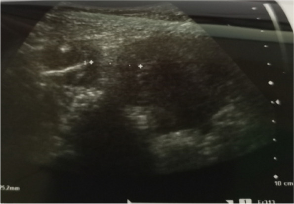
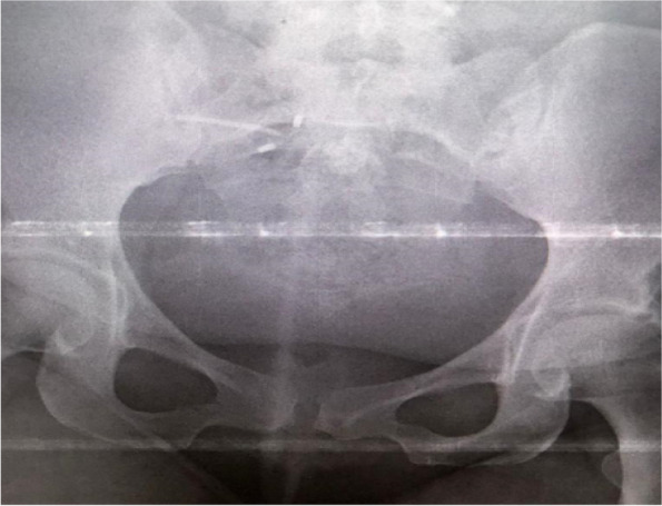
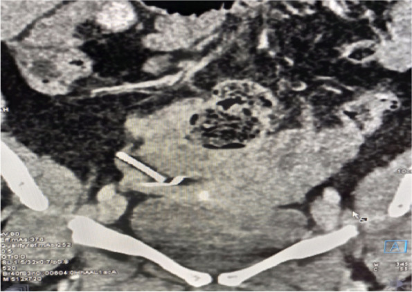
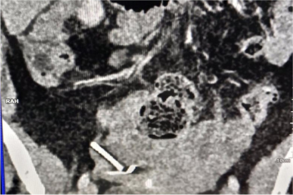
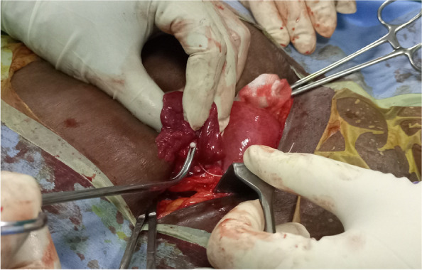
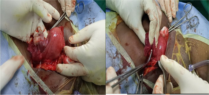
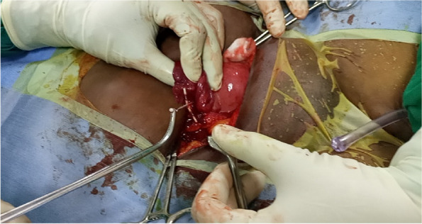
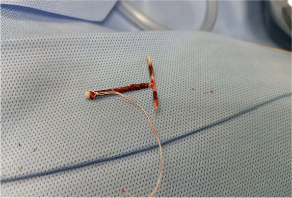
Background: Loss of Intra Uterine Device (IUD) following silent perforation of the uterus either during or after IUD insertion is an uncommon finding due to a lack of immediate follow-up. We report a rare case in which uterine perforation following the migration of IUD to the right fallopian tube without visceral injury. The patient presented with lower abdominal pain and pain during sex for one year since IUD insertion. On examination, we noted tenderness on the right suprapubic region and on speculum examination, no IUD thread was seen. A radiological pelvic examination showed an empty uterus without an IUD. Laparotomy and retrieval of migrated IUD was done followed by repair of perforated uterus.
Conclusion: Migrated IUD with silent uterine perforation without visceral injury is a distressing clinical condition both to the patient and the clinician. This case is reported to increase awareness in doing immediate vaginal examination and pelvic ultrasound post-IUD insertion.
Keywords: Chronic pelvic pain; Intrauterine devices; Migration; No visceral injury; Silent uterine perforation.
© 2024. The Author(s).
Conflict of interest statement
The authors declare no competing interests.
Figures
Similar articles
-
TRANSMIGRATION OF INTRA-UTERINE DEVICE, EXPLORATORY LAPAROTOMY, RETRIEVAL AND REPAIR OF PERFORATED UTERUS.Niger J Med. 2015 Jul-Sep;24(3):273-6. Niger J Med. 2015. PMID: 27487601
-
Intravesical migration of intrauterine device.J Urol. 1992 Jan;147(1):132-4. doi: 10.1016/s0022-5347(17)37159-8. J Urol. 1992. PMID: 1729505
-
Ileal penetration by a Multiload-Cu 375 intrauterine contraceptive device. A case report with review of the literature.Contraception. 1998 Nov;58(5):295-304. doi: 10.1016/s0010-7824(98)00116-4. Contraception. 1998. PMID: 9883385 Review.
-
Appendicitis caused by an intrauterine contraceptive device.Br J Surg. 1986 Nov;73(11):927-8. doi: 10.1002/bjs.1800731130. Br J Surg. 1986. PMID: 3790929
-
Complete and partial uterine perforation and embedding following insertion of intrauterine devices. II. Diagnostic methods, prevention, and management.Obstet Gynecol Surv. 1981 Aug;36(8):401-17. doi: 10.1097/00006254-198108000-00001. Obstet Gynecol Surv. 1981. PMID: 6455610 Review.
References
-
- Badu-Peprah A. The role of multimodality radiological imaging in extrauterine misplaced iucd: a case report. Afr J Reprod Health. 2020;24(4):212–7. - PubMed
-
- Elsheikh H, ElRefaei M, Abd Elfattah S. Assessment of misplaced intrauterine contraceptive devices by different imaging modalities: a cross-sectional study. Benha Med J. 2021;38:137–46.
LinkOut - more resources
Full Text Sources
